

The relationship between longitudinal serum leptin measures and measures of magnetic resonance imaging-assessed knee joint damage in a population of mid-life women
- PMID: 23576710
- PMCID: PMC3884071
- DOI: 10.1136/annrheumdis-2012-202685
The relationship between longitudinal serum leptin measures and measures of magnetic resonance imaging-assessed knee joint damage in a population of mid-life women
Abstract
Background and objective: Serum leptin measures are associated with radiographic knee osteoarthritis, but no studies have examined leptin levels with respect to different measures of knee joint damage from MRI.
Methods: Participants in the Michigan Study of Women's Health Across the Nation underwent bilateral knee MRIs at follow-up visit 11 for assessment of cartilage defects, bone marrow lesions, osteophytes, meniscal tears, synovitis and joint effusion. Serum leptin measures were available from baseline, follow-up visits 1 and 3-7.
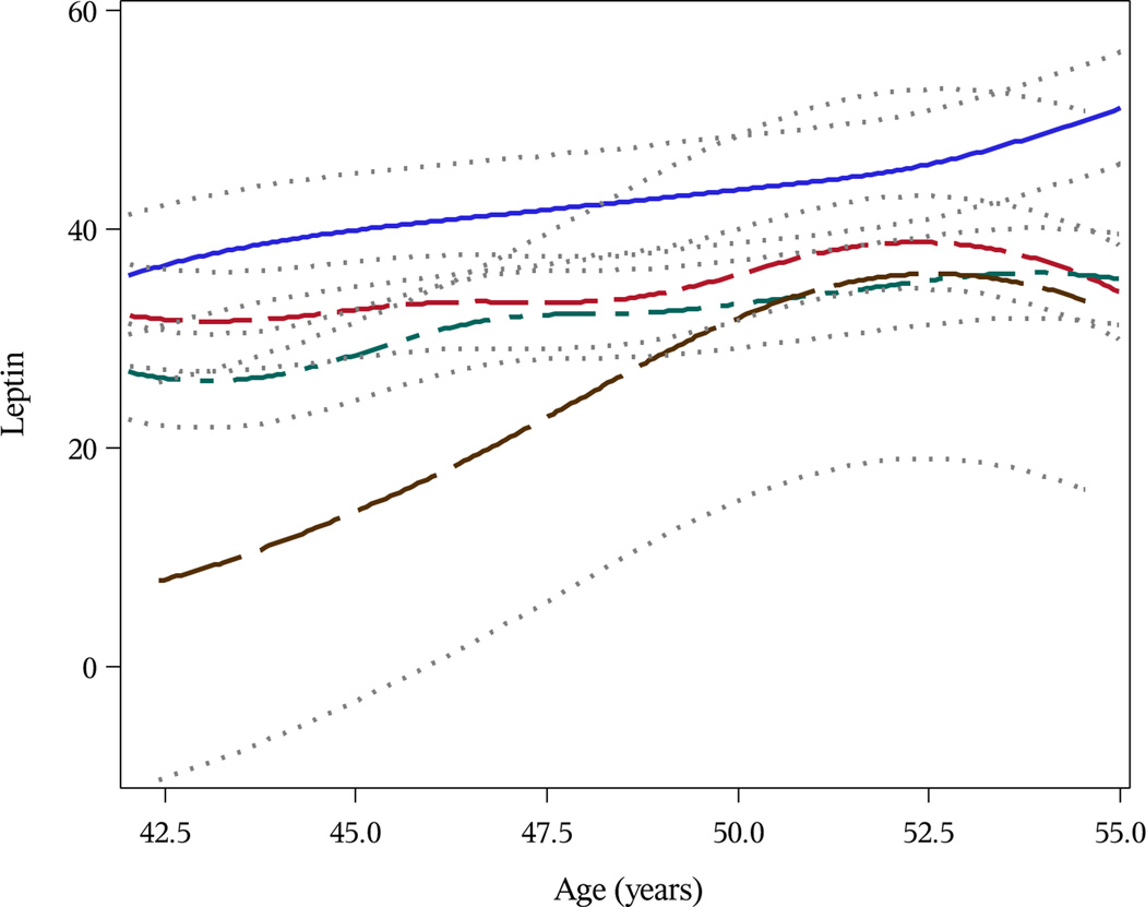
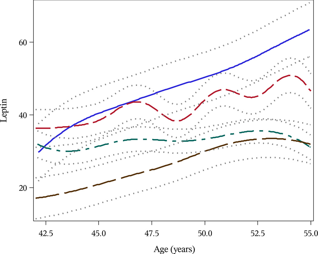
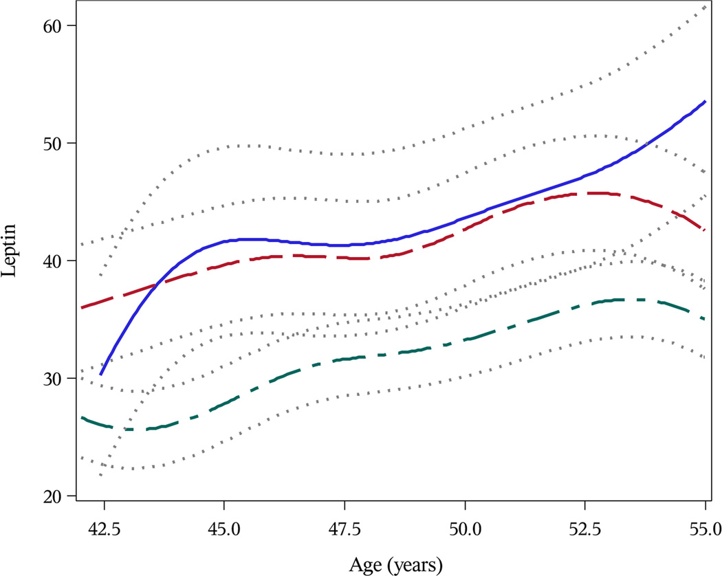
Results: Baseline serum leptin levels were associated with greater odds of having more severe knee joint damage at follow-up visit 11 after adjustment for age, smoking status, menopause status and body mass index residuals. The greatest effect was observed for osteophytes; a 5 ng/ml increase in baseline leptin was associated with 24% higher odds of having larger osteophytes (95% CI 1.17 to 1.32). Correlations with baseline serum leptin were greatest for MRI-assessed osteophytes (r=0.41), followed by effusion (r=0.32), synovitis (r=0.30), cartilage defects (r=0.28), bone marrow lesions (r=0.24) and meniscal abnormalities (r=0.21).
Conclusions: Leptin levels 10 years prior to MRI assessment were associated with the presence of cartilage defects, bone marrow lesions, osteophytes, meniscal tears, synovitis and effusion among a population of middle-aged women. Understanding the role that leptin plays in the joint degradation process is critical for development of more targeted interventions for osteoarthritis.
Keywords: Epidemiology; Knee Osteoarthritis; Osteoarthritis.
Conflict of interest statement
None of the authors have any financial conflicts of interests to declare.
Figures
Similar articles
-
Associations of anatomical measures from MRI with radiographically defined knee osteoarthritis score, pain, and physical functioning.J Bone Joint Surg Am. 2011 Feb 2;93(3):241-51. doi: 10.2106/JBJS.I.00667. J Bone Joint Surg Am. 2011. PMID: 21266638 Free PMC article.
-
Association of leptin levels with radiographic knee osteoarthritis among a cohort of midlife women.Arthritis Care Res (Hoboken). 2013 Jun;65(6):936-44. doi: 10.1002/acr.21922. Arthritis Care Res (Hoboken). 2013. PMID: 23281224 Free PMC article.
-
Associations between suprapatellar pouch effusion-synovitis, serum cartilage oligomeric matrix protein, high sensitivity C-reaction protein, knee symptom, and joint structural changes in patients with knee osteoarthritis.Clin Rheumatol. 2020 May;39(5):1663-1670. doi: 10.1007/s10067-019-04905-7. Epub 2020 Jan 3. Clin Rheumatol. 2020. PMID: 31897961
-
Tibial subchondral bone size and knee cartilage defects: relevance to knee osteoarthritis.Osteoarthritis Cartilage. 2007 May;15(5):479-86. doi: 10.1016/j.joca.2007.01.003. Epub 2007 Feb 8. Osteoarthritis Cartilage. 2007. PMID: 17291789 Review.
-
The reliability of a new scoring system for knee osteoarthritis MRI and the validity of bone marrow lesion assessment: BLOKS (Boston Leeds Osteoarthritis Knee Score).Ann Rheum Dis. 2008 Feb;67(2):206-11. doi: 10.1136/ard.2006.066183. Epub 2007 May 1. Ann Rheum Dis. 2008. PMID: 17472995 Review.
Cited by
-
The relationship between serum leptin level and disease activity and inflammatory markers in fibromyalgia patients.North Clin Istanb. 2018 Apr 16;5(2):102-108. doi: 10.14744/nci.2017.31644. eCollection 2018. North Clin Istanb. 2018. PMID: 30374474 Free PMC article.
-
The relationship between body composition and knee osteoarthritis in postmenopausal women.Turk J Phys Med Rehabil. 2018 Mar 3;64(2):121-125. doi: 10.5606/tftrd.2018.1496. eCollection 2018 Jun. Turk J Phys Med Rehabil. 2018. PMID: 31453501 Free PMC article.
-
Metabolic enrichment of omega-3 polyunsaturated fatty acids does not reduce the onset of idiopathic knee osteoarthritis in mice.Osteoarthritis Cartilage. 2014 Sep;22(9):1301-9. doi: 10.1016/j.joca.2014.06.033. Epub 2014 Jul 4. Osteoarthritis Cartilage. 2014. PMID: 25008209 Free PMC article.
-
Imaging-based measures of synovitis in knee osteoarthritis: A scoping review and narrative synthesis.Osteoarthr Cartil Open. 2025 Mar 24;7(2):100602. doi: 10.1016/j.ocarto.2025.100602. eCollection 2025 Jun. Osteoarthr Cartil Open. 2025. PMID: 40235523 Free PMC article. Review.
-
Emerging role of metabolic signaling in synovial joint remodeling and osteoarthritis.J Orthop Res. 2016 Dec;34(12):2048-2058. doi: 10.1002/jor.23420. Epub 2016 Sep 26. J Orthop Res. 2016. PMID: 27605370 Free PMC article. Review.
References
-
- Dillon CF, Rasch EK, Gu Q, et al. Prevalence of knee osteoarthritis in the United States: arthritis data from the Third National Health and Nutrition Examination Survey 1991–94. J Rheumatol. 2006;33:2271–2279. - PubMed
-
- Sowers M, Lachance L, Hochberg M, et al. Radiographically defined osteoarthritis of the hand and knee in young and middle-aged African American and Caucasian women. Osteoarthritis Cartilage. 2000;8:69–77. - PubMed
-
- Hootman JM, Helmick CG. Projections of US prevalence arthritis and associated activity limitation. Arthritis Rheum. 2006;54:226–229. - PubMed
-
- Flegal KM, Carroll MD, Kit BK, et al. Prevalence of obesity and trends in the distribution of body mass index among US adults, 1999–2010. JAMA. 2012;307:491–497. - PubMed
Publication types
MeSH terms
Substances
Grants and funding
LinkOut - more resources
Full Text Sources
Other Literature Sources
