

Inducible deletion of the Blimp-1 gene in adult epidermis causes granulocyte-dominated chronic skin inflammation in mice
- PMID: 23576729
- PMCID: PMC3631680
- DOI: 10.1073/pnas.1219462110
Inducible deletion of the Blimp-1 gene in adult epidermis causes granulocyte-dominated chronic skin inflammation in mice
Abstract
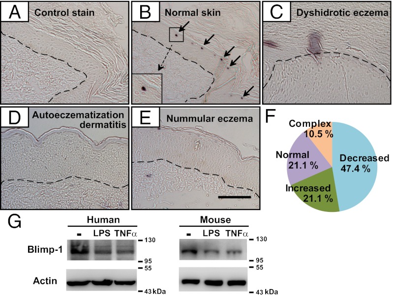
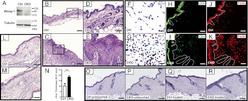
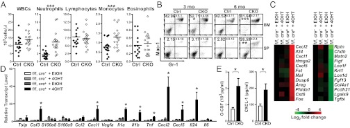
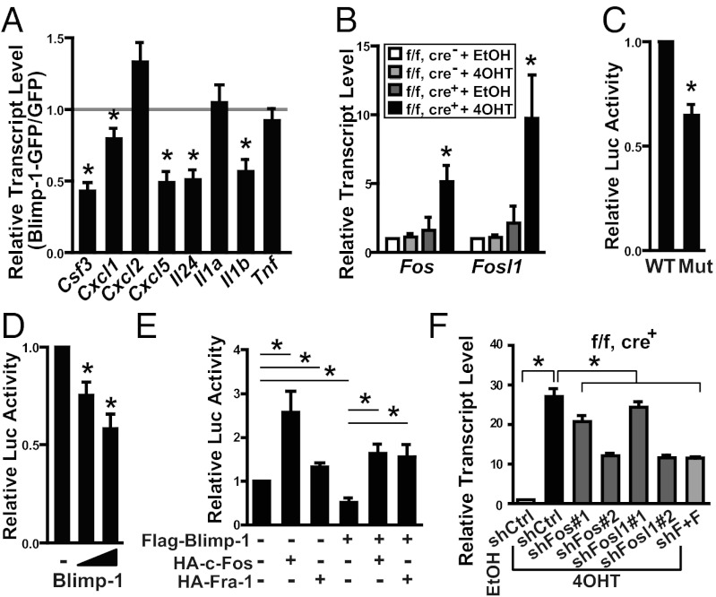
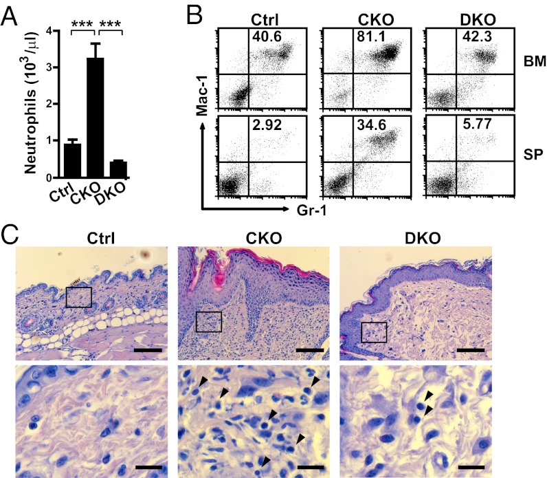
B lymphocyte-induced maturation protein-1 (Blimp-1) is a transcriptional repressor important for the differentiation and function of several types of immune cells. Because skin serves as a physical barrier and acts as an immune sentinel, we investigated whether Blimp-1 is involved in epidermal immune function. We show that Blimp-1 expression is reduced in skin lesions of some human eczema samples and in stimulated primary keratinocytes. Epidermal-specific deletion of PR domain containing 1, with ZNF domain (Prdm1), the gene encoding Blimp-1, in adult mice caused spontaneously inflamed skin characterized by massive dermal infiltration of neutrophils/macrophages and development of chronic inflammation associated with higher levels of cytokines/chemokines, including granulocyte colony-stimulating factor (G-CSF), and enhanced myelopoiesis in bone marrow. Deletion of Prdm1 in the epidermis of adult mice also led to stronger inflammatory reactions in a tape-stripping test and in a disease model of contact dermatitis. The elevated G-CSF produced by keratinocytes after deletion of Prdm1 in vitro was mediated by the transcriptional activation of FBJ osteosarcoma oncogene (Fos) and fos-like antigen 1 (Fosl1). Systemic increases in G-CSF contributed to the inflammatory responses, because deletion of the G-CSF gene [colony stimulating factor 3, (Csf3)] prevented neutrophilia and partially ameliorated the inflamed skin in Prdm1-deficient mice. Our findings indicate a previously unreported function for Blimp-1 in restraining steady-state epidermal barrier immunity.
Conflict of interest statement
The authors declare no conflict of interest.
Figures
Similar articles
-
Epidermal terminal differentiation depends on B lymphocyte-induced maturation protein-1.Proc Natl Acad Sci U S A. 2007 Sep 18;104(38):14988-93. doi: 10.1073/pnas.0707323104. Epub 2007 Sep 10. Proc Natl Acad Sci U S A. 2007. PMID: 17846422 Free PMC article.
-
CXCR2 ligands and G-CSF mediate PKCalpha-induced intraepidermal inflammation.J Clin Invest. 2006 Oct;116(10):2757-66. doi: 10.1172/JCI27514. Epub 2006 Sep 7. J Clin Invest. 2006. PMID: 16964312 Free PMC article.
-
Absence of the transcriptional repressor Blimp-1 in hematopoietic lineages reveals its role in dendritic cell homeostatic development and function.J Immunol. 2009 Dec 1;183(11):7039-46. doi: 10.4049/jimmunol.0901543. Epub 2009 Nov 13. J Immunol. 2009. PMID: 19915049
-
Mononuclear phagocyte regulation by the transcription factor Blimp-1 in health and disease.Immunology. 2020 Dec;161(4):303-313. doi: 10.1111/imm.13249. Epub 2020 Sep 27. Immunology. 2020. PMID: 32799350 Free PMC article. Review.
-
Radiation Dermatitis: Radiation-Induced Effects on the Structural and Immunological Barrier Function of the Epidermis.Int J Mol Sci. 2024 Mar 15;25(6):3320. doi: 10.3390/ijms25063320. Int J Mol Sci. 2024. PMID: 38542294 Free PMC article. Review.
Cited by
-
Compartmentalized Epidermal Activation of β-Catenin Differentially Affects Lineage Reprogramming and Underlies Tumor Heterogeneity.Cell Rep. 2016 Jan 12;14(2):269-81. doi: 10.1016/j.celrep.2015.12.041. Epub 2016 Jan 7. Cell Rep. 2016. PMID: 26771241 Free PMC article.
-
Dermal Blimp1 Acts Downstream of Epidermal TGFβ and Wnt/β-Catenin to Regulate Hair Follicle Formation and Growth.J Invest Dermatol. 2017 Nov;137(11):2270-2281. doi: 10.1016/j.jid.2017.06.015. Epub 2017 Jun 28. J Invest Dermatol. 2017. PMID: 28668474 Free PMC article.
-
Prdm1 Regulates Thymic Epithelial Function To Prevent Autoimmunity.J Immunol. 2017 Aug 15;199(4):1250-1260. doi: 10.4049/jimmunol.1600941. Epub 2017 Jul 12. J Immunol. 2017. PMID: 28701508 Free PMC article.
-
Mechanisms regulating skin immunity and inflammation.Nat Rev Immunol. 2014 May;14(5):289-301. doi: 10.1038/nri3646. Epub 2014 Apr 11. Nat Rev Immunol. 2014. PMID: 24722477 Review.
-
Expression of B lymphocyte-induced maturation protein 1 (Blimp-1) in keratinocyte and cytokine signalling drives human Th17 response in psoriasis.Arch Dermatol Res. 2023 Apr;315(3):481-490. doi: 10.1007/s00403-022-02379-3. Epub 2022 Aug 30. Arch Dermatol Res. 2023. PMID: 36042041
References
Publication types
MeSH terms
Substances
Associated data
- Actions
LinkOut - more resources
Full Text Sources
Other Literature Sources
Molecular Biology Databases
Research Materials
