. 2013 Apr 16;110(16):E1452-61.
doi: 10.1073/pnas.1219813110. Epub 2013 Apr 1.

Shaping organs by a wingless-int/Notch/nonmuscle myosin module which orients feather bud elongation

Affiliations

Shaping organs by a wingless-int/Notch/nonmuscle myosin module which orients feather bud elongation

Ang Li et al. Proc Natl Acad Sci U S A. .

Abstract

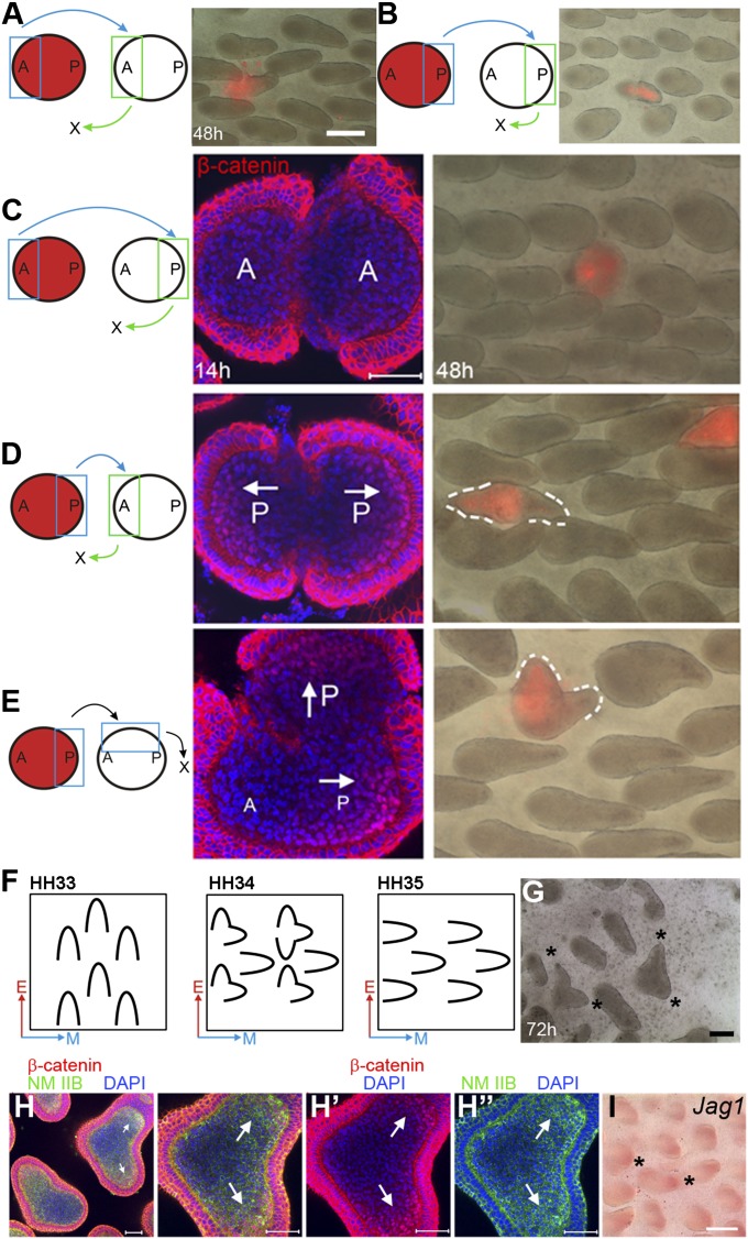
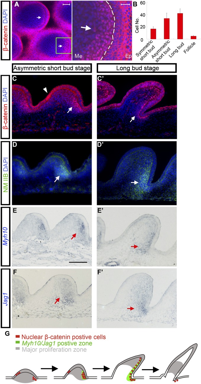
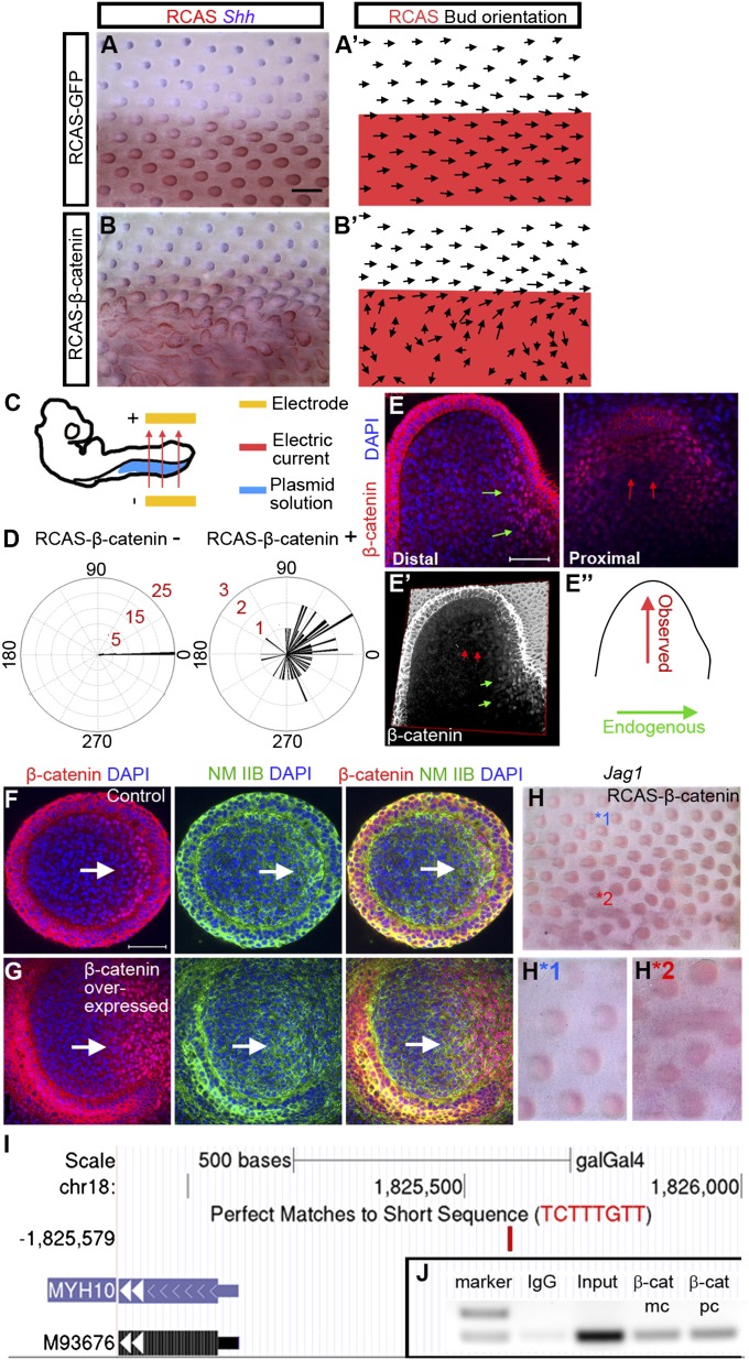
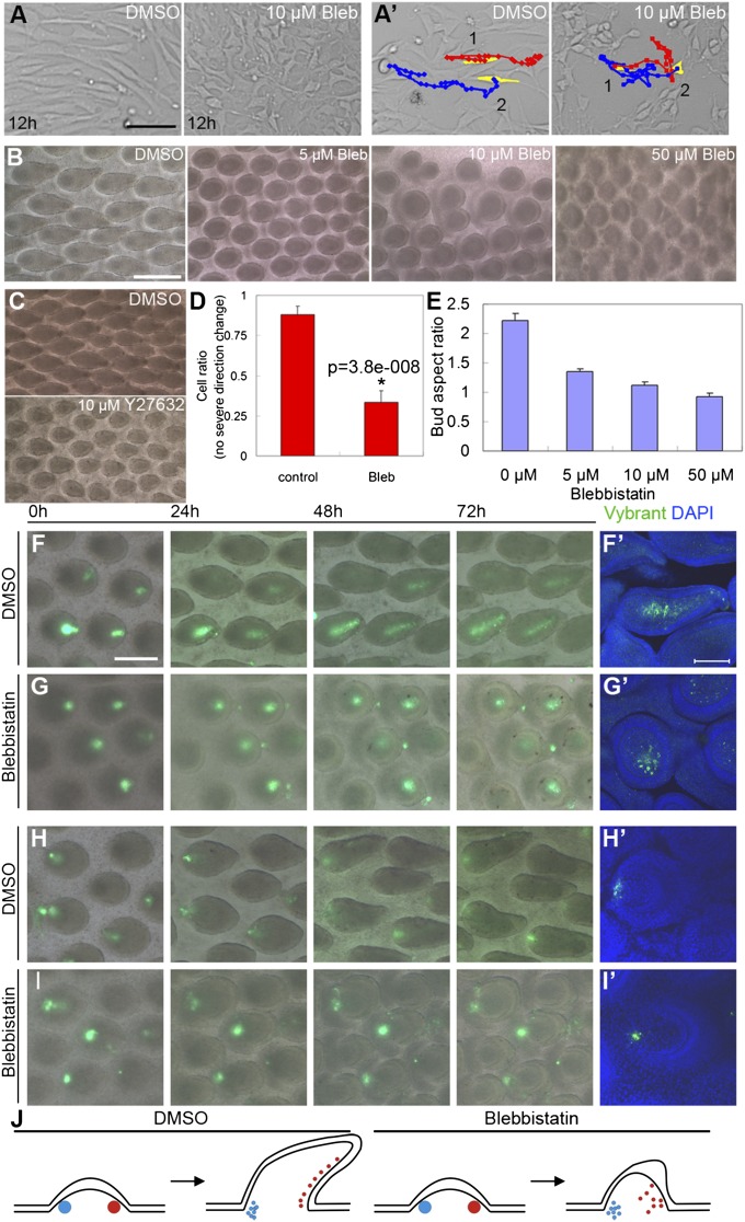
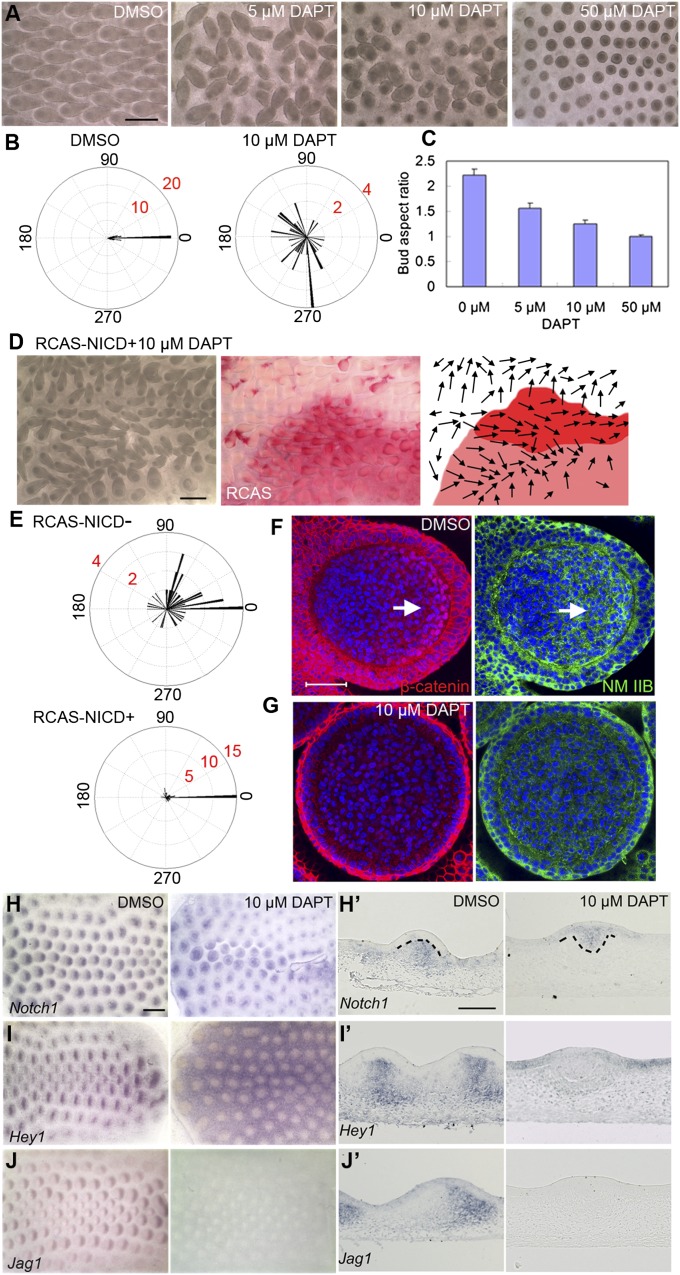
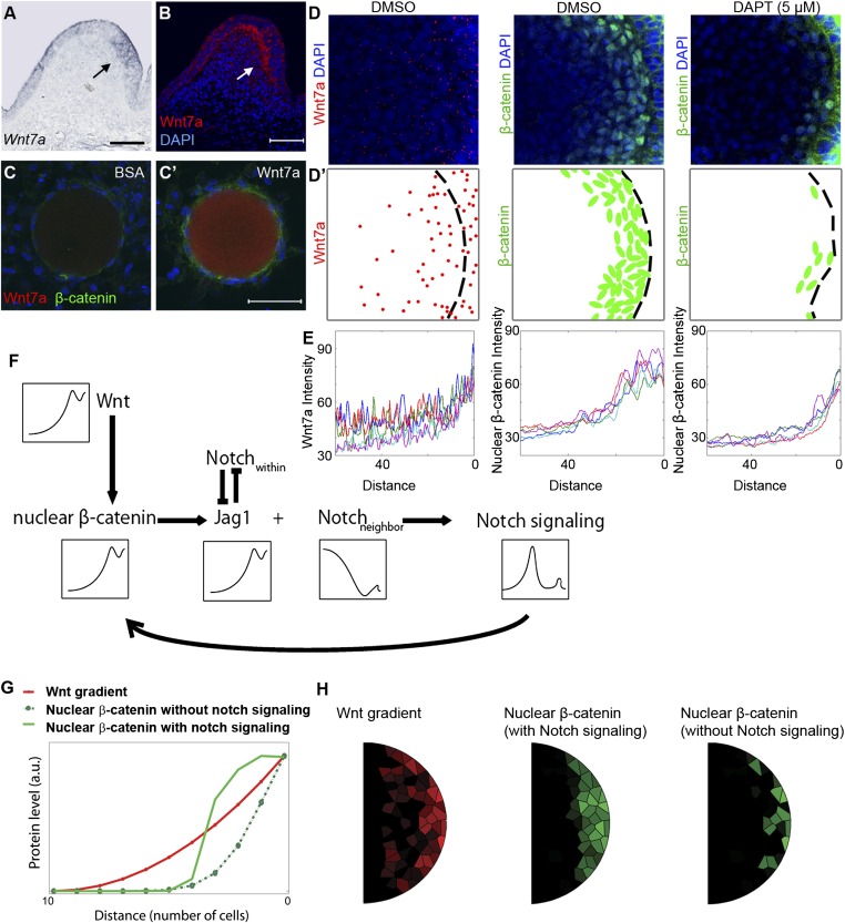
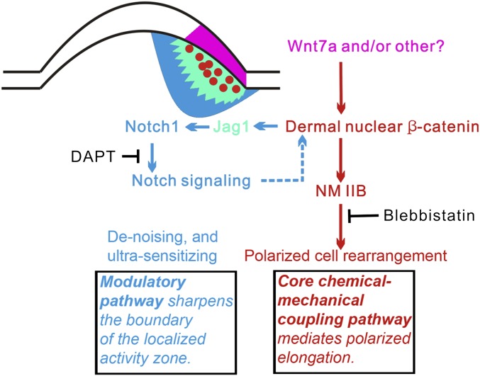
How organs are shaped to specific forms is a fundamental issue in developmental biology. To address this question, we used the repetitive, periodic pattern of feather morphogenesis on chicken skin as a model. Avian feathers within a single tract extend from dome-shaped primordia to thin conical structures with a common axis of orientation. From a systems biology perspective, the process is precise and robust. Using tissue transplantation assays, we demonstrate that a "zone of polarizing activity," localized in the posterior feather bud, is necessary and sufficient to mediate the directional elongation. This region contains a spatially well-defined nuclear β-catenin zone, which is induced by wingless-int (Wnt)7a protein diffusing in from posterior bud epithelium. Misexpressing nuclear β-catenin randomizes feather polarity. This dermal nuclear β-catenin zone, surrounded by Notch1 positive dermal cells, induces Jagged1. Inhibition of Notch signaling disrupts the spatial configuration of the nuclear β-catenin zone and leads to randomized feather polarity. Mathematical modeling predicts that lateral inhibition, mediated by Notch signaling, functions to reduce Wnt7a gradient variations and fluctuations to form the sharp boundary observed for the dermal β-catenin zone. This zone is also enriched for nonmuscle myosin IIB. Suppressing nonmuscle myosin IIB disrupts directional cell rearrangements and abolishes feather bud elongation. These data suggest that a unique molecular module involving chemical-mechanical coupling converts a pliable chemical gradient to a precise domain, ready for subsequent mechanical action, thus defining the position, boundary, and duration of localized morphogenetic activity that molds the shape of growing organs.


Conflict of interest statement

The authors declare no conflict of interest.

Figures

Fig. 1.
Fig. 1.
Exchanging different parts of feather buds indicates that the nuclear β-catenin positive dermis bears polarizing activity. This result is supported by epithelial–mesenchymal recombination and rotation experiments (EMRR). (AE) Exchanging different parts of feather buds. Chimeric bud morphology observed 48 h after surgery. Blue: transplant donor site. Green: transplant recipient site. (Red) DiI-labeled donor tissue. (Scale bar: 250 μm.) (A) Anterior-to-anterior transplantation. (B) Posterior-to-posterior transplantation. (C) Replacing posterior bud with anterior bud. (D) Replacing anterior bud with posterior bud. (E) Replacing left lateral bud with posterior bud. (CE) Confocal images of the corresponding chimeric buds stained for β-catenin 14 h after surgery. (F) Summary of EMRR experiments. A–P axis orientation for epithelium (red arrows) and dermis (blue arrows). (G) St. 34 chicken embryo dorsal skin cultured for 72 h after EMRR shows bifurcated orientation (*). (Scale bar: 1 mm.) (H) Same stage specimen cultured 20 h after EMRR. Arrows: nuclear β-catenin positive dermis and up-regulated nonmuscle myosin (NM) IIB. (H′) and (H″) show the β-catenin and NM IIB pattern, respectively. (Scale bar: 50 μm.) (I) Jag1 expression increases at the location of nuclear β-catenin positive dermis (*). (Scale bar: 500 μm.)
Fig. 2.
Fig. 2.
A zone of nuclear β-catenin positive cells is localized to posterior dermis during feather bud elongation. This zone has high levels of NM IIB and Jag1. (A) Confocal pictures of E8 chicken embryo dorsal skin feather buds at the asymmetric short bud stage show nuclear β-catenin positive dermis. Green boxes show the magnified region. Ep, epithelium; Me, mesenchyme. (Scale bar: 50 μm and 20 μm, respectively.) (B) Counts of dermal cells with nuclear accumulated β-catenin from sagittal sections of feather buds at the symmetric short bud stage, asymmetric short bud stage, long bud stage, and follicle stage. For each stage n = 12. (C) and (C′) β-catenin staining on sagittal sections of feather buds at asymmetric short bud and long bud stage, respectively. (Arrows) Nuclear β-catenin positive dermis. (Arrowheads) Nuclear β-catenin positive epithelium. (Scale bar: 50 μm.) (DF′) High-level NM IIB and Jag1 are detected in the nuclear β-catenin positive dermis. Myh10 encodes NM IIB heavy chain. (Scale bar: 100 μm.) (G) Schematic summary of the results.
Fig. 3.
Fig. 3.
Misexpression of stabilized β-catenin caused drastic feather misorientation and up-regulation of NM IIB and Jag1. (A) E9 chicken embryo with RCAS-GFP electroporated to its left side at E3. (blue, Shh in situ; red, RCAS staining) (n = 35). (B) E9 chicken embryo with RCAS-β-catenin electroporated to its left side at E3 (n = 72). (Scale bar: 500 μm.) (A′–B′) Schematic drawing of feather orientation and RCAS positive area. (C) E3 embryo electroporation setup. (D) Summary of feather bud orientation relative to the body A–P axis in RCAS-β-catenin negative (n = 45) and positive (n = 57) areas, respectively. (E) Confocal images at two different levels (proximal and distal) of a misoriented, β-catenin overexpressing feather bud. The distal view shows a nuclear β-catenin zone at the original posterior region (green arrows). The proximal view shows a second nuclear β-catenin zone in the new, reoriented posterior region (red arrows). (E′) 3D reconstruction of β-catenin staining pattern in E. (E″) Schematic representation of the original and new bud A–P axis. (F and G) Control and β-catenin overexpressed short feather buds stained with β-catenin, and NM IIB. (Scale bar: 50 μm.) (H) Jag1 whole mount in situ of RCAS β-catenin expressing skin. Enlargement of feather buds from a control (H*1) and RCAS β-catenin expressing (H*2) region in H. (I) Potential WRE located upstream of Myh10 (shown in University of California, Santa Cruz genome browser. M93676 is Myh10 accession code in GenBank). (J) ChIP-PCR result. β-cat mc, monoclonal β-catenin antibody; β-cat pc, polyclonal β-catenin antibody.
Fig. 4.
Fig. 4.
Blocking NM IIB function disrupted fibroblast migration patterns in vitro and inhibited feather bud elongation in vivo. (A) E7 chicken embryo dorsal skin fibroblasts transfected with RCAS-β-catenin to mimic posterior dermis were cultured 12 h in media containing DMSO or 10 μM Blebbistatin (Bleb), respectively. Notice the difference in cell shape. (A′) Most control cells moved in the direction of their long axis, whereas many Blebbistatin treated cells moved randomly. (Scale bar: 100 μm.) Red, blue lines show the movement trajectory of two cells artificially colored yellow at time 0. (B) Skin explants cultured 4 d with DMSO (control) and different doses of Blebbistatin, respectively. (Scale bar: 500 μm.) (C) Inhibition of bud elongation by Blebbistatin could be phenocopied by treatment with Y27632 (ROCK inhibitor). (D) Statistics of cell rearrangements for A′. For details please see Statistics. (E) Feather bud aspect ratio (length/width) at different Blebbistatin concentrations (n = 20 for each treatment condition). (FI) Time-course pictures of Vybrant dye labeled posterior (F and G) and anterior (H and I) bud dermis in control and Blebbistatin-treated buds. (Scale bar: 250 μm.) (F′–I′) Confocal images of feather buds from the corresponding 72-h explants. (Scale bar: 100 μm.) (J) Schematic representing cell tracking results for the anterior (blue) and posterior (red) dermis.
Fig. 5.
Fig. 5.
Inhibition of Notch signaling by DAPT causes drastic feather misorientation and dose-dependent inhibition of bud elongation. Meanwhile DBZ nuclear β-catenin and NM IIB level are dramatically decreased. (A) E7 skin explants cultured for 4 d with DMSO or different doses of DAPT. (B) Summary of feather bud orientation relative to the body A–P axis upon DMSO (n = 40) or 10 μM DAPT (n = 40) treatment, respectively. (C) Feather buds’ aspect ratio at different DAPT concentrations (n = 20 for each concentration). (D) E8 chicken embryo dorsal skin (electroporated with RCAS-NICD at E3) cultured with 10 μM DAPT for 72 h. Red signal shows RCAS positive area. Arrows in the schematic drawing represent feather bud orientation. (E) Divergence of feather bud orientations from the body A–P axis in the RCAS-NICD negative and positive area, respectively (n = 44 for each condition). (F and G) Confocal image of a feather bud on E7 skin explant cultured for 1.5 d with DMSO and DAPT (10 μM) in media, respectively. (Arrow) nuclear β-catenin positive cells. (Scale bar: 50 μm.) (HJ) Whole-mount in situ hybridization of DMSO- and DAPT-treated skin explants (after 40 h in culture) with Notch1, Hey1, Jag1 probes, respectively. (Scale bar: 500 μm.) (H′–J′) Section in situ hybridization of DMSO- and DAPT-treated skin explants with the three probes, respectively. (Scale bar: 100 μm.)
Fig. 6.
Fig. 6.
Mathematical modeling of Wnt–Notch cross-talk simulates the change from a noisy, gradual Wnt gradient to a definitive threshold Wnt response. (A) Section in situ hybridization for Wnt7a (arrow) in feather buds. (B) Fluorescent staining of Wnt7a. (C and C′) Effect of BSA and Wnt7a (0.2 M) soaked beads on E7 chicken skin dermis cultured for 24 h. (Scale bar: 50 μm.) (D) Wnt7a and β-catenin staining in control and 5 μM DAPT-treated feather buds (compact confocal Z-stack pictures). (D′) Schematic summary of D. (E) Wnt7a, β-catenin signal intensity measured from five feather buds for each condition (distance is calculated from the posterior epithelial–dermal boundary to the center of the bud). (F) Wnt–Notch cross-talk relationships used for mathematical modeling. (G) Simulation (1D) shows Wnt–Notch cross-talk can help nuclear β-catenin form an ultrasensitive response to Wnt ligand. (H) Simulation (2D) results.
Fig. 7.
Fig. 7.
Summary of the core and modulatory molecular modules for polarized elongation during feather bud morphogenesis. Purple, Wnt7a and/or other Wnt molecules from posterior epithelium induce β-catenin nuclear accumulation in DBZ cells (red spots); yellow, Jag1 positive zone; blue, Notch1 positive zone; red arrows, nuclear β-catenin–Notch feedback loop; green arrows, nuclear-β-catenin–induced directional cell rearrangement; dashed lines, unknown mechanism.

Similar articles

Cited by

References

    1. Slack JM. Origin of stem cells in organogenesis. Science. 2008;322(5907):1498–1501. - PubMed
    1. Chuong CM, Randall VA, Widelitz RB, Wu P, Jiang TX. Physiological regeneration of skin appendages and implications for regenerative medicine. Physiology (Bethesda) 2012;27(2):61–72. - PMC - PubMed
    1. Wu P, Jiang TX, Suksaweang S, Widelitz RB, Chuong CM. Molecular shaping of the beak. Science. 2004;305(5689):1465–1466. - PMC - PubMed
    1. Chodankar R, et al. Shift of localized growth zones contributes to skin appendage morphogenesis: Role of the Wnt/beta-catenin pathway. J Invest Dermatol. 2003;120(1):20–26. - PMC - PubMed
    1. Yu M, Wu P, Widelitz RB, Chuong CM. The morphogenesis of feathers. Nature. 2002;420(6913):308–312. - PMC - PubMed

Publication types

LinkOut - more resources