. 2013 Apr 16;110(16):6370-5.
doi: 10.1073/pnas.1300135110. Epub 2013 Apr 1.

Characterization of mechanical and biochemical properties of developing embryonic tendon

Affiliations

Characterization of mechanical and biochemical properties of developing embryonic tendon

Joseph E Marturano et al. Proc Natl Acad Sci U S A. .

Abstract

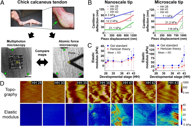
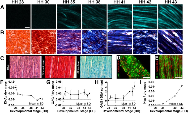
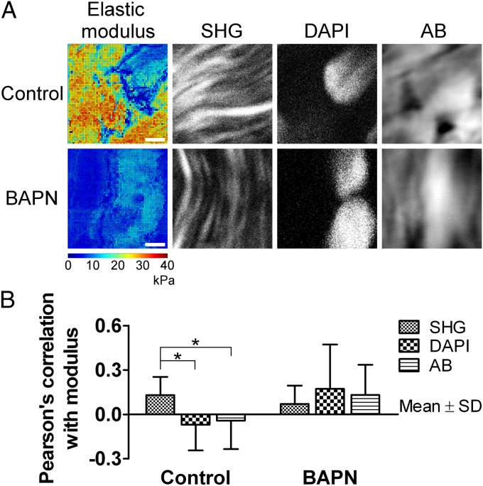
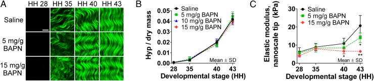
Tendons have uniquely high tensile strength, critical to their function to transfer force from muscle to bone. When injured, their innate healing response results in aberrant matrix organization and functional properties. Efforts to regenerate tendon are challenged by limited understanding of its normal development. Consequently, there are few known markers to assess tendon formation and parameters to design tissue engineering scaffolds. We profiled mechanical and biological properties of embryonic tendon and demonstrated functional properties of developing tendon are not wholly reflected by protein expression and tissue morphology. Using force volume-atomic force microscopy, we found that nano- and microscale tendon elastic moduli increase nonlinearly and become increasingly spatially heterogeneous during embryonic development. When we analyzed potential biochemical contributors to modulus, we found statistically significant but weak correlation between elastic modulus and collagen content, and no correlation with DNA or glycosaminoglycan content, indicating there are additional contributors to mechanical properties. To investigate collagen cross-linking as a potential contributor, we inhibited lysyl oxidase-mediated collagen cross-linking, which significantly reduced tendon elastic modulus without affecting collagen morphology or DNA, glycosaminoglycan, and collagen content. This suggests that lysyl oxidase-mediated cross-linking plays a significant role in the development of embryonic tendon functional properties and demonstrates that changes in cross-links alter mechanical properties without affecting matrix content and organization. Taken together, these data demonstrate the importance of functional markers to assess tendon development and provide a profile of tenogenic mechanical properties that may be implemented in tissue engineering scaffold design to mechanoregulate new tendon regeneration.


Conflict of interest statement

The authors declare no conflict of interest.

Figures

Fig. 1.
Fig. 1.
FV-AFM results. (A, Upper) HH 43 limb with calcaneus tendon highlighted; the sectioning plane is shown in green. (Lower) Cryosection of tendon visualized with multiphoton microscopy (SHG; Left) and brightfield microscopy with the AFM probe in view (Right). Yellow boxes show the AFM-probed area. Verhoeff stain (green arrows) was used to mark the same regions for multiphoton microscopy and FV-AFM measurements. (Scale bar, 50 μm.) (B) Force-displacement AFM curves of tendon from HH 28–43. Slopes of linear regions were used to calculate modulus. (C) Tendon elastic modulus measured via FV-AFM as a function of developmental stage, calculated with agarose gel standards and Hertzian theory. Modulus increased nonlinearly during development, with the greatest increases between HH 28 and 30 and after HH 38 (n = 5). (D) FV-AFM topography and nanoscale elastic modulus maps of embryonic tendon from HH 28–43. Both showed increasing heterogeneity during development. (Scale bar, 2 μm.)
Fig. 2.
Fig. 2.
Morphological and biochemical characterization. (A) SHG imaging showed increases in collagen density and organization during tendon development. (Scale bar, 2 μm.) (B) Trichrome staining showed a decrease in cellularity (red) and increase in collagen content (blue) with development. (Scale bar, 20 μm.) (C) HH 43 tendon sections show high density of cell nuclei (H&E), aligned collagen (Picrosirius red), and GAG deposits (Alcian blue). (Scale bar, 25 μm.) (D) Cell nuclei (green) and GAG deposits (red) were similar in shape and size but spatially distinct in HH 43 tendon. (Scale bar, 25 μm.) (E) GAG deposits (red) appeared closely associated with collagen fibers (green). (Scale bar, 20 μm.) From HH 28–43, DNA-to-dry mass content decreased by 67% (F; n = 3); GAG-to-dry mass content remained relatively constant (G; n = 3); and GAG-to-DNA ratio (H; n = 3) and Hyp-to-dry mass ratio (I; n = 3), representative of collagen content, increased dramatically.
Fig. 3.
Fig. 3.
Spatial correlation of nanoscale modulus with collagen fibers (SHG), cell nuclei (DAPI), and GAGs (Alcian blue). Collagen fibers seemed more spatially correlated to elastic modulus than cell nuclei or GAGs when normally cross-linked. (A) Comparison of modulus maps (color images) with microscopy (black and white images) of the same areas. In control tissue, there appeared to be correlation between high-modulus regions and collagen fibers, and between low-modulus regions and cell nuclei and GAGs. After BAPN treatment, average modulus was reduced and areas of GAGs and cell nuclei appeared to be more correlated with modulus. (Scale bars, 2 μm.) (B) Pearson’s correlation coefficient between FV-AFM modulus maps and multiphoton microscopy (n = 10). In control samples, collagen fibers were weakly but significantly more correlated than cell nuclei or GAGs (*P < 0.05). This effect was lost with BAPN treatment (P = 0.59).
Fig. 4.
Fig. 4.
Effects of BAPN treatment to inhibit LOX activity. (A) SHG images showed no apparent differences in collagen microstructure with BAPN treatment compared with a saline control. (Scale bar, 10 μm.) (B) BAPN treatment also had no effect on Hyp content, representative of collagen content, during development (P ≥ 0.51; n = 3). (C) BAPN treatment significantly reduced tendon elastic modulus measured with a nanoscale tip by 38% at HH 40 (*P < 0.05) and by 68% at HH 43 (**P < 0.001; n = 5).

References

    1. Butler DL, Awad HA. Perspectives on cell and collagen composites for tendon repair. Clin Orthop Relat Res. 1999;(367) Suppl:S324–S332. - PubMed
    1. Woo SL, Abramowitch SD, Kilger R, Liang R. Biomechanics of knee ligaments: Injury, healing, and repair. J Biomech. 2006;39(1):1–20. - PubMed
    1. AAOS . United States Bone and Joint Decade: The Burden of Musculoskeletal Diseases in the United States. Rosemont, IL: American Academy of Orthopaedic Surgeons; 2008.
    1. Kardon G. Muscle and tendon morphogenesis in the avian hind limb. Development. 1998;125(20):4019–4032. - PubMed
    1. Kuo CK, Petersen BC, Tuan RS. Spatiotemporal protein distribution of TGF-betas, their receptors, and extracellular matrix molecules during embryonic tendon development. Dev Dyn. 2008;237(5):1477–1489. - PMC - PubMed

Publication types