Review
. 2013 Mar;15(1):53-65.
doi: 10.31887/DCNS.2013.15.1/esibille.

Molecular aging of the brain, neuroplasticity, and vulnerability to depression and other brain-related disorders

Affiliations
Review

Molecular aging of the brain, neuroplasticity, and vulnerability to depression and other brain-related disorders

Etienne Sibille. Dialogues Clin Neurosci. 2013 Mar.

Abstract

The increased risk for neurodegenerative and neuropsychiatric disorders associated with extended lifespan has long suggested mechanistic links between chronological age and brain-related disorders, including depression, Recent characterizations of age-dependent gene expression changes now show that aging of the human brain engages a specific set of biological pathways along a continuous lifelong trajectory, and that the same genes that are associated with normal brain aging are also frequently and similarly implicated in depression and other brain-related disorders. These correlative observations suggest a model of age-by-disease molecular interactions, in which brain aging promotes biological changes associated with diseases, and additional environmental factors and genetic variability contribute to defining disease risk or resiliency trajectories. Here we review the characteristic features of brain aging in terms of changes in gene function over time, and then focus on evidence supporting accelerated molecular aging in depression. This proposed age-by-disease biological interaction model addresses the current gap in research between "normal" brain aging and its connection to late-life diseases. The implications of this model are profound, as it provides an investigational framework for identifying critical moderating factors, outlines opportunities for early interventions or preventions, and may form the basis for a dimensional definition of diseases that goes beyond the current categorical system.

El mayor riesgo para los trastornos neurodegenerativos y neuropsiquiátricos asociados con el aumento de la expectativa de vida ha sugerido hace tiempo asociaciones mecanicistas entre la edad cronológica y los trastornos cerebrates, incluyendo la depresión. Recientes caracterizaciones en los cambios en la expresión génica dependiente de la edad han mostrado que el envejecimiento del cerebro humano requiere de un conjunto específico de vías biológicas en un continuo a través del curso de toda la vida, y que los mismos genes que están asociados con el envejecimiento normal se implican frecuentemente y de igual forma en la depresión como en otros trastornos cerebrates. Estas sucesivas observaciones sugieren un modelo de interacciones moleculares de enfermedades por la edad, en las cuales el envejecimiento cerebral promueve cambios biológicos asociados con las enfermedades, y en forma adicional los factores ambientales y la variabilidad genética contribuyen a definir el riesgo de enfermar o de tener una evolución con resiliencia. En este artículo se revisan las caracteristicas distintivas del envejecimiento cerebral en términos de cambios en la función génica a lo largo del tiempo, y luego se enfoca en la evidencia que sustenta un acelerado envejecimiento molecular en la depresión. Este modelo que propone la interacción biológica de enfermedades por la edad Ie da una dirección a la actual brecha en la investigación entre el envejecimiento cerebral “normal” y su conexión con enfermedades del final de la vida. Las sugerencías de este modelo son profundas y proporcionan un marco de investigación para identificar factores moderadores criticos, delinear oportunidades para intervenciones o prevención precoces y poder formar las bases para una definición dimensional de las enfermedades que vaya más allá del actual sistema de categoríes.

L'augmentation du risque de troubles neurodégénératifs et neuropsychiatriques associé à l'allongement de la durée de vie évoque depuis longtemps l'existence de liens mécanistes entre l'âge chronologique et les troubles cérébraux, dont la dépression. Des mises en évidence récentes de modifications d'expression des gènes en fonction de l'âge suggèrent maintenant que le vieillissement cérébral humain met enjeu un ensemble spécifique de voies biologiques sur une trajectoire continue tout au long de la vie. Or, les gènes associés au vieillissement normal du cerveau sont aussi impliqués de façon fréquente et identique dans la dépression ainsi que dans d'autres troubles cérébraux. Ces observations proposent un modèle d'interaction moléculaire âge-maladie, dans lequel le vieillissement cérébral favorise des modifications biologiques associeés aux maladies et dans lequel des facteurs environnementaux supplémentaires et la variabilité génétique contribuent à définir le risque pathologique ou celui des trajectoires de résilience. Nous passons en revue ici les traits caractéristiques du vieillissement cérébral en termes de modification de la fonction des gènes au cours du temps. Puis nous nous interessons aux arguments en favour de l'accélération du vieillissement moléculaire dans la dépression. Cette proposition de modèle d'interaction biologique âge-maladie aborde le décalage actuel dans la recherche entre le vieillissement cérébral normal et ses connexions aux maladies de la vieillesse. Les implications de ce modèle sont importantes, en termes de cadre d'investigation pour l'identification des facteurs de modération déterminants, d'opportunités pour une prévention ou un traitement précoce et de création d'une définition dimensionnelle des maladies allant au-delà du système catégoriel actuel.

Keywords: age; brain molecular aging; depression; neurological disorder; neuroplasticity; psychiatric.


Figures
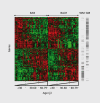
Figure 1.
Figure 1.. Continuous and progressive gene expression changes in human prefrontal cortex. Age-dependent changes for a core set of exemplary genes (n=588) are presented together for two regions of the prefrontal cortex (Brodmann area (BA9), dorsolateral prefrontal cortex; BA47, orbital ventral prefrontal cortex). Each gene is represented by a row, each array, or brain area per subject, by a column. Samples are organized left to right by brain area and increasing age. Green and red bars indicate decreased and increased gene expression, respectively, versus the averaged signal for these genes across all samples. For example, a horizontal row going from red to green indicates a gene for which expression decreases with age in that brain area. Genes are organized along the y-axis according to similarities in expression profiles across age. This study illustrated several points: (i) similar numbers of genes are downregulated or upregulated throughout the lifetime; (ii) gene changes display continuous and progressive trajectories throughout adult life; (iii) profiles of age-dependent changes are conserved across the two areas; and (iv) downregulated genes are mostly expressed in neurons, whereas upregulated genes include most glial-enriched and some neuronal-enriched genes, as indicated on the right hand of the figure. Columns to the right indicate the distribution of genes with glial-(white matter [WM]- enriched) or neuronal-enriched (gray matter [GM]-enriched) signals. Adapted from ref 7: Erraji-Benchekroun L, Underwood MD, Arango V, et al. Molecular aging in human prefrontal cortex is selective and continuous throughout adult life. Biol Psychiatry. 2005;57:549-558. Copyright © Elsevier 2005
Figure 2.
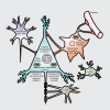
Figure 2.. Age-dependent biological changes in neurons and glia. Known age-related cellular phenotypes are highlighted for neurons and glia. Blue, pyramidal cells; Purple, interneurons; Orange, astrocyte; Green, microglia; Brown, oligodendrocyte. Not shown are changes in brain white matter track and blood vessel integrity. Many neuronal phenotypes (such as DNA damage) occur in neuron and glia. In parentheses are single representative examples (amongst many) of age-regulated gene expression changes, which may contribute to the particular cellular phenotypes. CRF, Corticotropin-releasing hormone; CALB-1, calbindin 1; SOD2, superoxide dismutase 2; BCL-2, B-cell CLL/lymphoma 2; DRD1, Dopamine receptor D1; SYN2, synapsin II; GFAP, glial fibrillary acidic protein; NF-KB, nuclear factor kappa B; CNP, 2',3'-cyclic nucleotide 3' phosphodiesterase; MHC, myosin heavy chain. Adapted from ref 19: Glorioso C, Sibille E. Between destiny and disease: genetics and molecular pathways of human central nervous system aging. Prog Neurobiol. 2011;93:165-181. Copyright © Pergamon Press 2011
Figure 3.
Figure 3.. Dendritic inhibition, a biological module at the intersection of age and psychiatric disorders. A) Excitatory pyramidal neurons (PYR) are regulated by different types of inhibitory γ-aminobutyric acid (GABA) neurons. Somatostatin (SST)-, neuropeptide Y (NPY)- and cortistatin (CORT)-positive GABA neurons (red) target PYR distal dendrites. Parvalbumin- (PV) and cholecystokinin- (CCK) positive GABA neurons target PYR cell body and axon initial segment (blue). Calretinin-(CR) positive GABA neurons (green) regulate other GABA neurons. B) Markers of interneurons that target PYR dendrites show decreased expression with age, and great effect or statistical significance of changes in subjects with major depression. C) Brain-derived neurotropic factor (BDNF) expression, measured by quantitative polymerase chain reaction (qPCR), is significantly and inversely correlated with chronological age in control and depressed subjects. Values are in arbitrary units of qPCR signal intensity. Respective to age-matched control subjects, subjects with major depression display greater BDNF downregulation (-22%; P<0.05). D) Age-regulation of BDNF- and depression-related genes. The average relative age effects for each individual subject are shown for the set of BDNF-related genes that display significant depression-related effects. Microarray-based gene expression values were normalized to the group means of each gene and averaged per subject. The BDNF-related gene set was split based on the effect of age on those genes in control subjects (top panel, age-upregulated; bottom panel, age-downregulated). The results show that age effects are systematically in the same direction, and of greater effect sizes in subjects with major depression (red squares) compared with controls (blue circles). Values are Pearson correlation factors. *, P<0.05; **, P<0.01 . Data in B is from Erraji et al, Glorioso et al, and Guilloux et al. Figures C and D are adapted from ref 18: Douillard-Guilloux G, Guilloux JP, Lewis DA, Sibille E. Anticipated brain molecular aging in Major Depression. Am J Geriatr Psychiatry. 2012. Oct 31 [epub ahead of print]. Copyright © American Psychiatry Press 2012
Figure 4.
Figure 4.. A proposed age-by-disease molecular interaction model. The graph depicts the age-dependent change in expression that is frequently observed for genes that are otherwise implicated in brain-related disorders (a decrease is shown here). Progression below a threshold (horizontal red line) marks the onset of disease symptoms. Changes in the trajectory of agerelated changes in expression of disease-related genes (Y-axis) determine the age (X-axis), or even if, an individual develops disease symptoms (vertical red arrows). Per this model, modulators (black arrows), genetic or environmental, place subjects on an “at risk” or protected trajectory for developing symptoms or brain-related disorders LLD (late-life depression). Adapted from ref 8: Glorioso C, Oh S, Douillard GG, Sibille E. Brain molecular aging, promotion of neurological disease and modulation by Sirtuin5 longevity gene polymorphism. Neurobiol Dis. 2011;41:279-90. Copyright © Blackwell Science 2011; ref 19: Glorioso C, Sibille E. Between destiny and disease:genetics and molecular pathways of human central nervous system aging. Prog Neurobiol. 2011;93:165-181. Copyright © Pergamon Press 2011

References

    1. Vaupel JW. Biodemography of human ageing. Nature. 2010;464:536–542. - PMC - PubMed
    1. Fiske A., Wetherell JL., Gatz M. Depression in older adults. Annu Rev Clin Psychol. 2009;5:363–389. - PMC - PubMed
    1. Belmaker RH., Again G. Major depressive disorder. N Engl J Med. 2008;358:55–68. - PubMed
    1. Wolkowitz OM., Reus VI., Mellon SH. Of sound mind and body: depression, disease, and accelerated aging. Dialogues Clin Neurosci. 2011;13:25–39. - PMC - PubMed
    1. Carroll BJ. Ageing, stress and the brain. Novartis Found Symp. 2002;242:26–36;discussion 36-45. - PubMed

Publication types

Substances