Liposclerosing myxofibrous tumor (LSMFT), a study of 33 patients: should it be a distinct entity?
- PMID: 23576919
- PMCID: PMC3565412
Liposclerosing myxofibrous tumor (LSMFT), a study of 33 patients: should it be a distinct entity?
Abstract
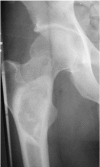
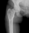
Liposclerosing myxoid fibrous tumor (LSMFT) is a recently described bone lesion site specific to the proximal femur. We have studied the radiographs and clinical features of 33 patients with this disorder. Histologic material was available for study in 18 of these patients. Histologic study revealed that 12 of these 18 had an underlying fibrous dysplasia and these had an underlying intraosseous lipoma. Because of this histologic evidence as well as the radiographic spectrum of the other lesions, we conclude that LSMFT is not a specific lesion and use of the term should be discontinued.
Figures


References
-
- Flanagan AM, Delaney D, O’Donnell PO. The benefits of molecular pathology in the diagnosis of musculoskeletal disease: Part I of a two-part review: soft tissue tumors. Skeletal Radiol. 2010;39(2):105–15. - PubMed
-
- Flanagan AM, Delaney D, O’Donnell PO. Benefits of molecular pathology in the diagnosis of musculoskeletal disease: part II of a two-part review: bone tumors and metabolic disorders. Skeletal Radiol. 2010;39(3):213–24. - PubMed
-
- Argani P, Aulmann S, Illei PB, Netto GJ, Ro J, Cho H, Dogan S, Ladanyi M, Martignoni G, Goldblum JR, Weiss SW. A Distinctive Subset of PEComas harbors TFE3 Gene Fusions. Am J Surg Pathol. 2010;34(10):1395–406. - PubMed
-
- Ragsdale BD, Sweet DE, Vinh TN. Radiology as Gross Pathology in Evaluating Chondroid Lesions. Hum Pathol. 1989;20(10):930–51. - PubMed
-
- Ragsdale BD. Polymorphic Fibro-osseous Lesions of Bone: An Almost Site-Specific Diagnostic Problem of the Proximal Femur. Hum Pathol. 1993;24(5):505–12. - PubMed
MeSH terms
LinkOut - more resources
Full Text Sources
