Review
. 2012:32:189-95.

SAPHO syndrome--a pictorial assay

Affiliations
Review

SAPHO syndrome--a pictorial assay

Lokesh Khanna et al. Iowa Orthop J. 2012.

Abstract

SAPHO (synovitis, acne, pustulosis, hyperostosis and osteitis) syndrome is a distinct clinical entity representing involvement of the musculoskeletal and dermatologic systems. It is well known to rheumatologists because of characteristic skin manifestations and polyarthropathy. However, few reports exist in the orthopaedic literature. It is important to be aware of sAPHO syndrome as it can mimic some of the more common disease entities such as infection, tumor, and other inflammatory arthropathies. Anterior chest wall pain centered at sternoclavicular and sternocostal joints is an important and characteristic clinical finding which can point to its diagnosis. A patient may undergo different diagnostic tests and invasive procedures such as biopsies before a diagnosis is made. Imaging can be helpful by offering a detailed evaluation of the abnormalities. More importantly it helps in revealing subclinical foci of involvement due to the polyostotic nature of the disease. The treatment is mostly nonsurgical. NSAIDS are the first line agents. However multiple new agents are being used for refractory cases. Surgery is reserved to treat complications.


Figures

Figure 1
Figure 1. Hyperostosis and osteitis of the left sternoclavicular and sternocostal joints.
35 year old female presented with chronic recurring jaw pain after tooth extraction. (a) CT sagittal reformatted image of the left mandible showing hyperostosis of the ramus and body. (b) Chest x-ray, (c) CT chest and (d) CT 3d reformatted images show hyperostosis and sclerosis involving the manubrium and proximal end of first rib on the left side. Involvement of sternocostal, sternoclavicular and manubriosternal joints is highly diagnostic of SAPHO syndrome. CT is much more sensitive in depicting the lesions as compared to radiographs. (e) Bone scan showing increased uptake in left sternoclavicular region. Scintigraphy is particularly useful in revealing subclinical areas of involvement. (f) MRI of the chest, axial T2 fat-saturated image show edema in the marrow and adjacent soft tissue edema at anterior end of the left first rib and adjacent manubrium. Water sensitive sequences on MR show high sensitivity and are helpful in detecting subclinical foci. (g) Follow up MRI shows reduced marrow edema suggestive of response to NSAIDS and methotrexate. (h) However CT chest a year later showed progression of abnormalities.
Figure 2
Figure 2. Multifocal spondylodiskitis and sacroiliitis confused for metastatic disease.
50 year old man presented with trauma and back pain. (a) Radiograph of the lumbar spine showed sclerotic areas in multiple vertebral bodies. These were initially suspected to be sclerotic metastases. (b) A bone scan obtained showed high uptake in the sacroiliac joints and lumbar spine. (c) A sagittal reformatted image of the lumbar spine showed sclerotic lesions and erosions centered on the corners of vertebral bodies and adjacent end plates. No associated soft tissue mass was seen. Corner erosions are one of the characteristic features of SAPHO. (d) to (f) MRI of lumbar spine including sagittal T1, T2 and T1 fat-sat contrast images showed corner lesions to be hypointense on T1, hyperintense on T2 with contrast enhancement. There is no abnormal signal in disc or any associated fluid col ection which rules out an infectious etiology. (g) MRI Pelvis axial T2 fat suppressed image centered at SI joints showed edema around the bilateral sacroiliac joints, more severe on right side.
Figure 3
Figure 3. Spondylodiskitis and sacroiliitis in SAPHO syndrome.
46 year old female presented to the emergency treatment center with acute chest pain and had a chest CT done to rule out pulmonary embolism. (a, b) There were multiple areas of hyperostosis and sclerosis in the manubrium, clavicle and first rib on the left side. (c) The sagittal reformatted image of the thoracic spine showed sclerotic lesions in the adjacent vertebral bodies. Disc spaces are well preserved. The spine is the second most common site of involvement in children and adults. These were earlier diagnosed as suspicious for metastatic disease. The patient had no history of cancer and further investigations revealed no focus of a primary tumor. (d) The Ferguson view of the sacroiliac joints and (e) a CT pelvis showed sclerosis and erosions in the bilateral sacroiliac joints, more severe on left side. Sacroiliac involvement in SAPHO is either unilateral or bilateral asymmetric, mainly involving the iliac side of the SI joint. This patient was also found to have palmar and plantar pustulosis.
Figure 4
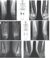
Figure 4. Hyperostosis and osteitis involving the long bones in SAPHO syndrome.
21 year old female presenting with pain in both legs. (a) Radiograph of both knees showed bony expansion and new bone formation (hyperostosis) with sclerosis. Long bone involvement is more common in pediatric patients. (b) Bone Scan showed increased uptake in both the distal femur diaphysis. (c) and (d) MRI showed thick periosteal reaction along with marrow edema(osteitis) in both femora. (e) to (h) follow up imaging studies after an year including radiographs of both knees, a bone scan and MRI of both femora respectively; showed significantly decreased disease activity after anti-TNF factor treatment.

References

    1. Windom RE, Sandford JP, Ziff M. Acne conglobata and arthritis. Arthritis Rheum. 1961;4:632–635. - PubMed
    1. Chamot A, Benhamou CL, Kahn MF, et al. Le syndrome acne pustulose hyperostose osteite (SA-PHO) Rev Rhum. 1987;54:187–196. - PubMed
    1. Sonozaki H, Mitsui H, Miyanaga Y, et al. Clinical features of 53 cases with pustulotic arthro-osteitis. Ann Rheum Dis. 1981;40(6):547–53. Dec. - PMC - PubMed
    1. Jang KA, Sung KJ, Moon KC, Koh JK, Choi JH. Four cases of pustulotic arthro-osteitis. J Dermatol. 1998;25(3):201–4. March. - PubMed
    1. Hyodoh K, Sugimoto H. Pustulotic arthro-osteitis: defining the radiologic spectrum of the disease. Semin Musculoskelet Radiol. 2001;5(2):89–93. Jun. - PubMed

MeSH terms

LinkOut - more resources