Review
. 2013 Apr 9:7:58.
doi: 10.3389/fncir.2013.00058. eCollection 2013.

Regulation of zebrafish sleep and arousal states: current and prospective approaches

Affiliations
Review

Regulation of zebrafish sleep and arousal states: current and prospective approaches

Cindy N Chiu et al. Front Neural Circuits. .

Abstract

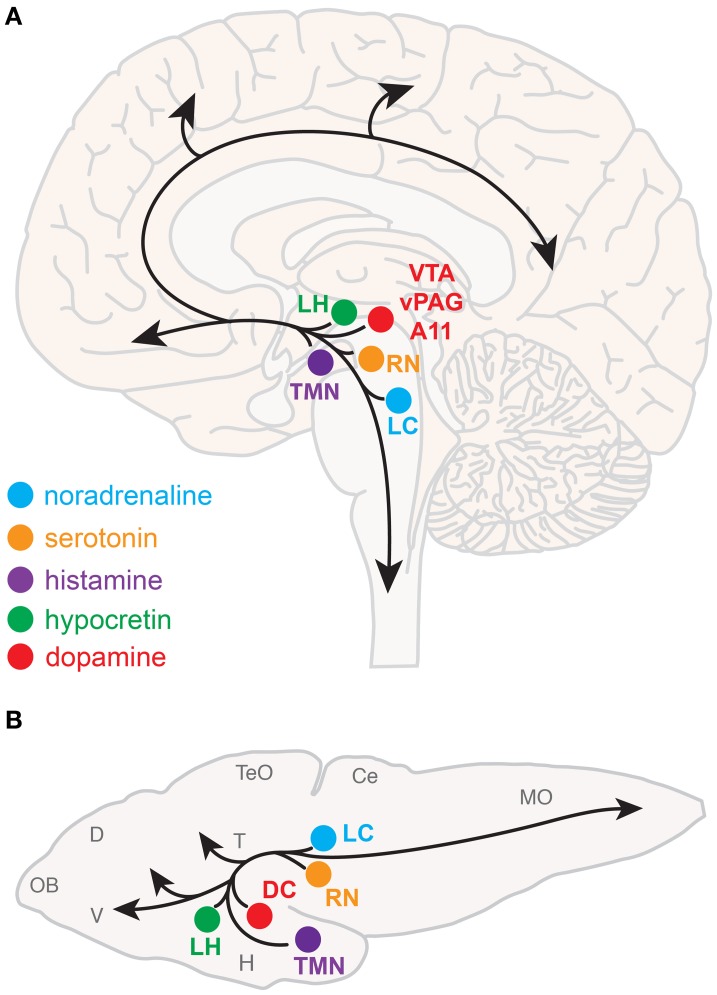
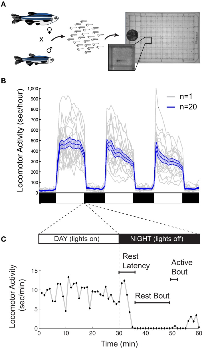
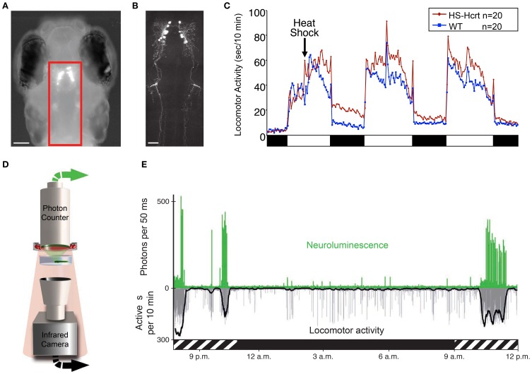
Every day, we shift among various states of sleep and arousal to meet the many demands of our bodies and environment. A central puzzle in neurobiology is how the brain controls these behavioral states, which are essential to an animal's well-being and survival. Mammalian models have predominated sleep and arousal research, although in the past decade, invertebrate models have made significant contributions to our understanding of the genetic underpinnings of behavioral states. More recently, the zebrafish has emerged as a promising model system for sleep and arousal research. Here we review experimental evidence that the zebrafish, a diurnal vertebrate, exhibits fundamental behavioral and neurochemical characteristics of mammalian sleep and arousal. We also propose how specific advantages of the zebrafish can be harnessed to advance the field. These include tractable genetics to identify and manipulate molecular and cellular regulators of behavioral states, optical transparency to facilitate in vivo observation of neural structure and function, and amenability to high-throughput drug screens to discover novel therapies for neurological disorders.

Keywords: arousal; hypocretin; neuromodulator; orexin; sleep; zebrafish.


Figures

Figure 1
Figure 1
Neuromodulatory systems that promote arousal in vertebrates. The approximate locations of key neuromodulatory regions are shown for human (A) and larval zebrafish (B) brains. Arrows indicate ascending projections that increase forebrain excitation and descending projections that increase muscle tone and sensorimotor function. Abbreviations of neuromodulatory regions: LC, locus coeruleus; RN, raphe nuclei; VTA, ventral tegmental area; vPAG, ventral periaqueductal gray; A11, mammalian dopamine cell group A11; DC, dopaminergic diencephalic cluster; TMN, tuberomammillary nucleus; LH, lateral hypothalamus. Abbreviations of larval zebrafish brain anatomy: OB, olfactory bulb; D, dorsal telencephalon; V, ventral telencephalon; TeO, optic tectum; H, hypothalamus; T, thalamus; Ce, cerebellum; MO, medulla oblongata. Note human and larval zebrafish brains are not depicted to scale.
Figure 2
Figure 2
Monitoring larval zebrafish sleep and wake behaviors. (A) Zebrafish larval locomotor activity assay. Individual zebrafish larva are placed in each well of a 96-well-plate on the 5th day of development. The plate is placed in a temperature-controlled chamber that is illuminated by white lights during the day and is continuously illuminated by infrared lights. The larvae are monitored by an infrared camera and the locomotor activity of each larva is recorded by a computer. (B) Representative locomotor activity data for each of 20 individual wild-type larvae (gray traces) and their mean locomotor activity (blue trace, ±standard error of the mean) is shown. Black and white bars indicate day and night, respectively. Larvae are more active during the day than at night, although there is considerable variability among individuals. (C) An example of typical larval zebrafish behavior at the end of the day is shown. A rest bout is defined as a period of at least 1 min of inactivity, which is associated with an increase in arousal threshold (Prober et al., 2006). Rest latency indicates the time between lights off at night and initiation of the first rest bout. Figure modified from Prober et al. (2006).
Figure 3
Figure 3
Zebrafish hypocretin is associated with arousal. (A) Dorsal view of a 4 dpf zebrafish larva that expresses GFP-Aequorin (GA) specifically in Hcrt neurons. (B) Two-photon z-projection image of the boxed area in (A). Scale bars represent 100 μm (A) and 50 μm (B). (C) Overexpression of Hcrt using a heat shock-inducible promoter (HS-Hcrt) increases locomotor activity. The mean locomotor activity of 20 HS-Hcrt larvae and 20 of their wild-type siblings is shown. The spike in activity during the afternoon of the 2nd and 3rd days of the experiment resulted from the addition of water to offset evaporation. (D) The GA assay. A large-area photon-counting photomultiplier tube is placed above a transparent behavior chamber in which a zebrafish larva that expresses GA in specific neurons is allowed to freely swim. The larva is imaged using infrared (IR) lights and an IR camera that is placed below the recording chamber. Spectral separation between GA neuroluminescence and the IR illumination allows the simultaneous recording of GA neuroluminescence and larval behavior. (E) Activity of Hcrt neurons during natural behavior. Data for a representative 4 dpf larva is shown. The larva exhibited periods of increased spontaneous locomotor activity (lower trace, thick line indicates 10 min running average) during the subjective day (hatched bar below graph) and little activity during the subjective night (black bar below graph). Most neuroluminescence signals produced by Hcrt neurons (upper trace) coincide with periods of robust locomotor activity during the subjective day, suggesting that Hcrt neuron activity is associated with arousal. Figure modified from Prober et al. (2006), Naumann et al. (2010).

Similar articles

Cited by

References

    1. Adamantidis A., de Lecea L. (2009). The hypocretins as sensors for metabolism and arousal. J. Physiol. 587, 33–40 10.1113/jphysiol.2008.164400 - DOI - PMC - PubMed
    1. Ahrens M. B., Li J. M., Orger M. B., Robson D. N., Schier A. F., Engert F., et al. (2012). Brain-wide neuronal dynamics during motor adaptation in zebrafish. Nature 485, 471–477 10.1038/nature11057 - DOI - PMC - PubMed
    1. Akerboom J., Chen T.-W., Wardill T. J., Tian L., Marvin J. S., Mutlu S., et al. (2012). Optimization of a GCaMP calcium indicator for neural activity imaging. J. Neurosci. 32, 13819–13840 10.1523/JNEUROSCI.2601-12.2012 - DOI - PMC - PubMed
    1. Alexander G. M., Rogan S. C., Abbas A. I., Armbruster B. N., Pei Y., Allen J. A., et al. (2009). Remote control of neuronal activity in transgenic mice expressing evolved G protein-coupled receptors. Neuron 63, 27–39 10.1016/j.neuron.2009.06.014 - DOI - PMC - PubMed
    1. Allada R., Siegel J. M. (2008). Unearthing the phylogenetic roots of sleep. Curr. Biol. 18, R670–R679 10.1016/j.cub.2008.06.033 - DOI - PMC - PubMed

Publication types

LinkOut - more resources