. 2013:2013:613864.
doi: 10.1155/2013/613864. Epub 2013 Mar 7.

Nuclear control of the inflammatory response in mammals by peroxisome proliferator-activated receptors

Affiliations

Nuclear control of the inflammatory response in mammals by peroxisome proliferator-activated receptors

Stéphane Mandard et al. PPAR Res. 2013.

Abstract

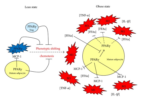
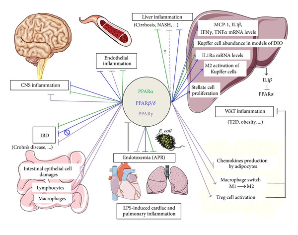
Peroxisome proliferator-activated receptors (PPARs) are ligand-activated transcription factors that play pivotal roles in the regulation of a very large number of biological processes including inflammation. Using specific examples, this paper focuses on the interplay between PPARs and innate immunity/inflammation and, when possible, compares it among species. We focus on recent discoveries establishing how inflammation and PPARs interact in the context of obesity-induced inflammation and type 2 diabetes, mostly in mouse and humans. We illustrate that PPAR γ ability to alleviate obesity-associated inflammation raises an interesting pharmacologic potential. In the light of recent findings, the protective role of PPAR α and PPAR β / δ against the hepatic inflammatory response is also addressed. While PPARs agonists are well-established agents that can treat numerous inflammatory issues in rodents and humans, surprisingly very little has been described in other species. We therefore also review the implication of PPARs in inflammatory bowel disease; acute-phase response; and central, cardiac, and endothelial inflammation and compare it along different species (mainly mouse, rat, human, and pig). In the light of the data available in the literature, there is no doubt that more studies concerning the impact of PPAR ligands in livestock should be undertaken because it may finally raise unconsidered health and sanitary benefits.


Figures

Figure 1
Figure 1
Contribution of the anti-inflammatory roles of PPARγ in the onset of WAT inflammation in the context of obesity and insulin resistance. In the lean state, PPARγ activity maintains homeostasis in mature adipocytes in preventing the secretion of chemokines such as MCP-1. In addition, alternatively activated macrophages (M2) and Treg cells are resident leukocytes in WAT coordinating numerous biological activities such as stimulating angiogenesis and cleaning of dead cells. The role of PPARγ in these cells is to prevent classical activation of macrophages and local inflammation to develop. When obesity is reached, mature adipocytes are exposed to excessive concentrations of free fatty acids (FFAs), which decrease Pparγ expression. In consequence, insulin sensitivity is also decreased in adipocytes, which elevates even more local FFAs concentrations as adipocytes are no longer able to properly store fatty acids and lipolysis also becomes activated. Furthermore, these FFAs activate macrophages shifting into an M1 phenotype, promoting the release of proinflammatory cytokines such as TNF-α and IL-1β. Secondly, as PPARγ transrepressional activity is decreased, adipocytes secrete high concentrations of chemokines (MCP-1), further promoting the recruitment of macrophages. Occurrence of this feed forward amplification loop between adipocytes and macrophages eventually leads to the elevation of local inflammation, further exacerbating local insulin resistance, which will turn systemic in the long term. MCP-1: monocyte chemoattractant protein-1; treg cells: regulatory T cells; FFA: free fatty acids; PPARγ: peroxisome proliferator-activated receptor γ; TNF-α: tumor necrosis factor-alpha; IL-1β: Interleukin-1 beta.
Figure 2
Figure 2
Representative illustration of PPAR main targets in inflammatory diseases. PPARα mostly displays anti-inflammatory properties in the context of liver inflammation. Its reported liver targets are hepatocytes and Küppfer cells [131]. IL-1β produced by Küppfer cells potently suppresses Pparα expression and activity via NF-κB–dependent inhibition of PPARα promoter activity [160]. Besides downregulating gene expression of proinflammatory mediators such as Mcp-1, Tnf-α, Ifn-γ, IL-1β, and PPARα also directly controls expression of IL1-ra in liver [131, 163]. Küppfer cell activation is also dependent on PPARβ/δ, which also targets stellate cells and therefore prevents liver fibrosis [75, 138]. In addition, PPARβ/δ has well-established anti-inflammatory properties in diseases associated with CNS inflammation. In CNS, PPARβ/δ has also proven anti-inflammatory properties in neurons, glial cells, and astrocytes [–202]. PPARγ anti-inflammatory properties are mainly illustrated in T2D and IBD. PPARγ serves as the molecular target of the insulin-sensitizing TZD drugs and plays a key role in T2D, adipogenesis and obesity. In WAT, mature adipocytes, Treg cells and macrophages have been identified as key cellular targets for PPARγ [66, 75, 116, 117]. Macrophage-specific deletion of PPARγ leads to specific reduction in alternatively activated macrophages (M2 state) in WAT leading to local inflammation [110]. Moreover, Treg-cell-specific deletion of Pparγ was shown to reduce the abundance of Treg cells in WAT resulting in the increase of WAT infiltration by proinflammatory macrophages (M1) and monocytes [116, 117]. In IBD, PPARγ acts in intestinal epithelial cells, macrophages and lymphocytes [–192]. Note that endotoxemia represses the mRNA expression level of Ppars (see black bar) [–155]. Furthermore, multiple lines of evidence indicated that PPARγ is very important in endothelial cells, because it inhibits the in situ production of proinflammatory molecules such as vascular adhesion molecule-1 (VCAM-1), intercellular adhesion molecule-1 (ICAM-1) and MCP-1 [, –228]. Similar conclusions were also drawn for the PPARα and PPARβ/δ isotypes [, –220]. Finally, PPARs display protective effets against endotoxemia [166, 167, 169, 236]. NASH: nonalcoholic steatoHepatitis; T2D: type-2 diabetes; CNS: central nervous system; Treg cells: Foxp3+ CD4+ regulatory T cells; DIO: diet-induced-obesity; APR: acute phase response; green lines: action of PPARα; blue lines: action of PPARβ/δ; purple lines: action of PPARγ; ?: Some PPARγ-independent effects of PPARγ activators have been proposed [146, 147]; : pharmacological activation of PPARβ/δ did not protect against dextran sulfate sodium-induced colitis pointing towards a ligand-independent anti-inflammatory effect of PPARβ/δ [180].

Similar articles

Cited by

References

    1. Kersten S, Mandard S, Escher P, et al. The peroxisome proliferator-activated receptor α regulates amino acid metabolism. FASEB Journal. 2001;15(11):1971–1978. - PubMed
    1. Nadra K, Anghel SI, Joye E, et al. Differentiation of trophoblast giant cells and their metabolic functions are dependent on peroxisome proliferator-activated receptor β/δ . Molecular and Cellular Biology. 2006;26(8):3266–3281. - PMC - PubMed
    1. Tan NS, Icre G, Montagner A, Bordier-Ten Heggeler B, Wahli W, Michalik L. The nuclear hormone receptor peroxisome proliferator-activated receptor β/δ potentiates cell chemotactism, polarization, and migration. Molecular and Cellular Biology. 2007;27(20):7161–7175. - PMC - PubMed
    1. Genolet R, Kersten S, Braissant O, et al. Promoter rearrangements cause species-specific hepatic regulation of the glyoxylate reductase/hydroxypyruvate reductase gene by the peroxisome proliferator-activated receptor α . Journal of Biological Chemistry. 2005;280(25):24143–24152. - PubMed
    1. Mandard S, Stienstra R, Escher P, et al. Glycogen synthase 2 is a novel target gene of peroxisome proliferator-activated receptors. Cellular and Molecular Life Sciences. 2007;64(9):1145–1157. - PMC - PubMed