. 2013:2013:956404.
doi: 10.1155/2013/956404. Epub 2013 Mar 13.

Differential cytotoxicity responses by dog and rat hepatocytes to phospholipogenic treatments

Affiliations

Differential cytotoxicity responses by dog and rat hepatocytes to phospholipogenic treatments

James K Morelli et al. J Toxicol. 2013.

Abstract

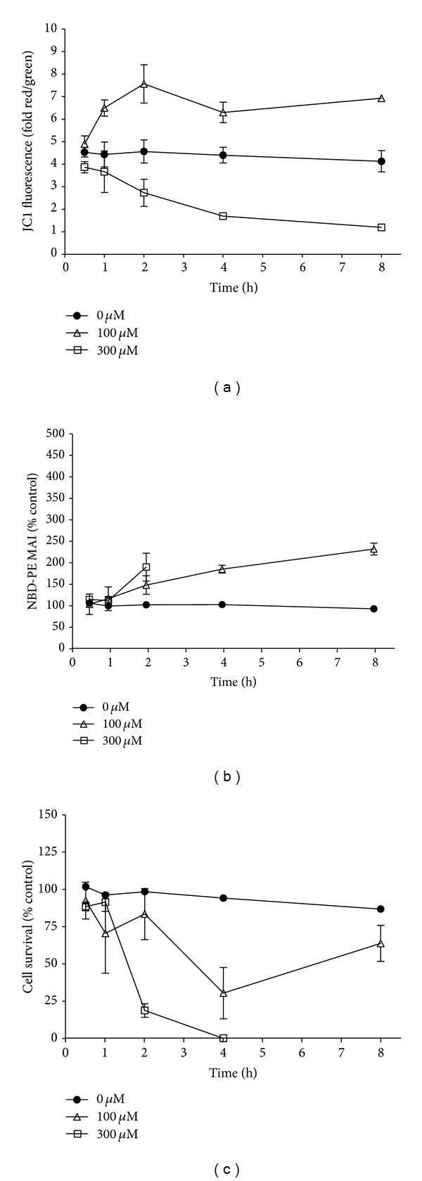
Dog and rat hepatocytes were treated with phospholipogenics to identify the more sensitive species and to determine whether lysosomal or mitochondrial changes were the primary cause of cytotoxicity. Endpoints included cell death, lysosome membrane integrity, mitochondrial membrane polarization, and fluorescent phospholipid (NBD-PE). Dog cells exhibited lower survival IC50 values than did rat cells with all phospholipogenic treatments and exhibited a lower capacity to accumulate NBD-PE in 4 of 5 phospholipogenic test conditions. The lysosomal modulator Bafilomycin A1 (Baf) rescued dog cells from cytotoxicity caused by 3 phospholipogenic 5HT1b antagonists and hydroxychloroquine, but not fluoxetine, and rescued rat cells from hydroxychloroquine and NMTMB, a 5HT1b antagonist. Following NMTMB treatment, rat mitochondrial membrane hyperpolarization was observed at modestly cytotoxic concentrations and depolarization at the highest concentration. At the highest test concentration, lysosomal loss of acridine orange occurred by 30 min, mitochondrial polarity changes by 1 hr, and NBD-PE accumulation by 2 hr, respectively. Baf shifted mitochondrial polarity from a depolarized state to a hyperpolarized state. These data demonstrate that (a) dog hepatocytes were generally less capable of mounting an adaptive, protective phospholipidotic response than rat hepatocytes, (b) effects on mitochondria and survival were preventable by lysosomal protection, and (c) destabilizing changes in both organelles are involved causally in cytotoxicity.


Figures

Figure 1
Figure 1
Chemical structures of select compounds used in this study.
Figure 2
Figure 2
(a) Differential effect of NMTMB on survival and NBD-PE accumulation in dog and rat hepatocytes. Cells were treated with compound in NBD-PE containing media. After 24 h cells were fixed with formaldehyde/Hoechst, and both survival and NBD-PE accumulation were quantified using the Cellomics Arrayscan instrument (as described in Section 2). Expressed as % control ±SD for 3 replicates from single isolations. Dog and rat hepatocytes were isolated for this experiment on the same day. (b) Differential effect of NMTMB on accumulation of NBD-PE in rat and dog hepatocytes. Cells were treated with NMTMB in NBD-PE containing media. After 24 h cells were fixed with formaldehyde/Hoechst and imaged using the Cellomics Arrayscan instrument. Image exposures were identical in all experiments (0.5 sec); background levels of NBD-PE accumulation were similar between dog and rat hepatocytes (approximately 50 mean average intensity units). Some artifactual accumulation of NBD-PE in dog preparations is apparent in the control sample. Dog and rat hepatocytes were isolated for this experiment on the same day.
Figure 3
Figure 3
Differential effects of phospholipogenic test articles on accumulation of the fluorescent phospholipid NBD-PE on dog and rat hepatocytes. Cells were treated with known phospholipogenic or nonphospholipogenic compounds in NBD-PE containing media. After 24 h cells were fixed with formaldehyde/hoechst, and NBD-PE accumulation was quantified using the Cellomics Arrayscan instrument. Image exposures were identical in all experiments (0.5 sec); background levels of NBD-PE accumulation were similar between dog and rat hepatocytes (approximately 50 mean average intensity units). Accumulation is expressed as % VC (vehicle control) at listed concentrations. Three replicates for each cell isolate were employed per experiment. Isolations occurred on separate days. The data are represented as an average of individual experiments where N = 1-2 (where histograms do not have error bars) or an average of 3 independent isolates ±SEM.
Figure 4
Figure 4
Effects of lysosomal modulator Baf (30 nM) cotreatments on 24 h survival of phospholipogenic-treated rat and dog hepatocytes. Cell survival was measured using the Cellomics Arrayscan (see Section 2). The data are expressed as an average of 3 replicates (±SD) for a single cell isolate. Statistically significant (P < 0.05, t-test) differences between dog phospholipogenic controls and modulator treatments occurred at the four top concentrations for NMTMB, top three concentrations for hydroxychloroquine, and top 2 concentrations for compounds A and B, and are illustrated by “ *”. For rat, statistically significant differences between phospholipogenic control and modulator treatments occurred at the top 3 concentrations for NMTMB and hydroxychloroquine and are illustrated by “ **”.
Figure 5
Figure 5
Effect of NMTMB on cell survival, NBD-PE accumulation, and mitochondrial polarization (JC-1 fluorescence) over 8 hours. Rat hepatocytes were treated with NMTMB for 30 minutes, 1 h, 2 h, 4 h, or 8 h. The data are expressed as an average of 3 (±SD) replicates from a single cell isolate. A statistically significant difference in JC-1 and NBD-PE levels between corresponding control replicates and that for NMTMB conditions was achieved by 1 h and 2 h, respectively (P < 0.05, t-test). As there was minimal survival observed for the higher test concentration beyond 2 h, NBDPE levels are not presented beyond 2 h.
Figure 6
Figure 6
Baf (30 nM) cotreatment shifted the mitochondrial polarization state of rat hepatocytes treated with NMTMB over an 8-hour time course. Rat hepatocytes were treated with NMTMB for 30 minutes, 1 h, 2 h, 4 h, 6 h or 8 h. Mitochondrial polarization status was monitored using JC-1 fluorescence on a Molecular Devices Flexstation instrument. The data are expressed as an average of 3 replicates (±SD) from a single cell isolate. Statistically significant differences between control replicates and that for all NMTMB conditions was achieved by 1 h (P < 0.05, t-test).
Figure 7
Figure 7
NMTMB affected acridine orange (AO) staining of lysosomes of rat hepatocytes over an 8-hour time course. AO was added to hepatocyte media for 10 minutes, removed, and replaced with media containing compound. Treatment with NMTMB proceeded for 30 minutes, 1 h, 2 h, 4 h or 8 h. AO staining was monitored using the Cellomics Arrayscan instrument. The data are expressed as an average of 3 replicates (±SD) from a single cell isolate. A statistically significant difference between control replicates and that for the NMTMB high test concentration is achieved by 30 min (P < 0.05, t-test) where marked cytotoxicity is evident by 4 hr (see Figure 5). Images from the Cellomics at the 30-minute time point are presented in (b).

Similar articles

References

    1. Reasor MJ, Hastings KL, Ulrich RG. Drug-induced phospholipidosis: issues and future directions. Expert Opinion on Drug Safety. 2006;5(4):567–583. - PubMed
    1. Daniel WA, Wójcikowski J. The role of lysosomes in the cellular distribution of thioridazine and potential drug interactions. Toxicology and Applied Pharmacology. 1999;158(2):115–124. - PubMed
    1. Daniel WA, Wójcikowski J, Palucha A. Intracellular distribution of psychotropic drugs in the grey and white matter of the brain: the role of lysosomal trapping. British Journal of Pharmacology. 2001;134(4):807–814. - PMC - PubMed
    1. Shacka JJ, Klocke BJ, Shibata M, et al. Bafilomycin A1 inhibits chloroquine-induced death of cerebellar granule neurons. Molecular Pharmacology. 2006;69(4):1125–1136. - PubMed
    1. Kornhuber J, Henkel AW, Groemer TW, et al. Lipophilic cationic drugs increase the permeability of lysosomal membranes in a cell culture system. Journal of Cellular Physiology. 2010;224(1):152–164. - PubMed

LinkOut - more resources