

Association between P(16INK4a) promoter methylation and non-small cell lung cancer: a meta-analysis
- PMID: 23577085
- PMCID: PMC3618325
- DOI: 10.1371/journal.pone.0060107
Association between P(16INK4a) promoter methylation and non-small cell lung cancer: a meta-analysis
Abstract
Background: Aberrant methylation of CpG islands acquired in tumor cells in promoter regions plays an important role in carcinogenesis. Accumulated evidence demonstrates P(16INK4a) gene promoter hypermethylation is involved in non-small cell lung carcinoma (NSCLC), indicating it may be a potential biomarker for this disease. The aim of this study is to evaluate the frequency of P(16INK4a) gene promoter methylation between cancer tissue and autologous controls by summarizing published studies.
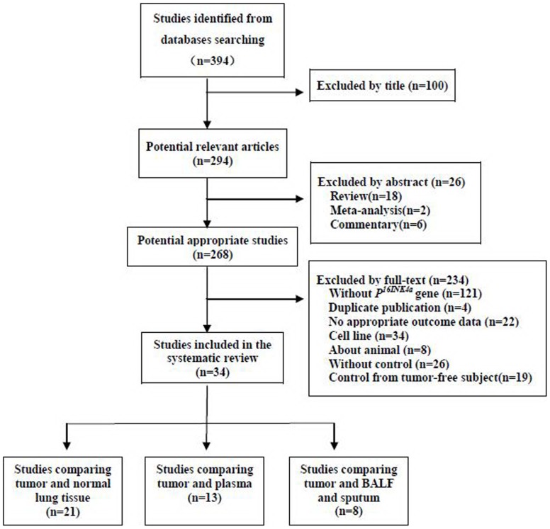
Methods: By searching Medline, EMBSE and CNKI databases, the open published studies about P(16INK4a) gene promoter methylation and NSCLC were identified using a systematic search strategy. The pooled odds of P(16INK4A) promoter methylation in lung cancer tissue versus autologous controls were calculated by meta-analysis method.
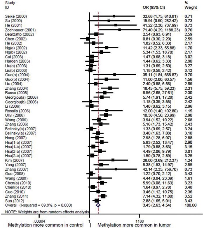
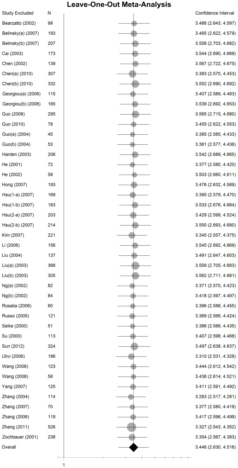
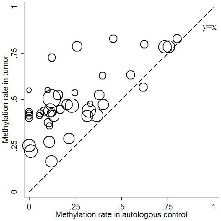
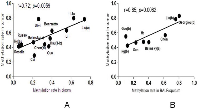
Results: Thirty-four studies, including 2 652 NSCLC patients with 5 175 samples were included in this meta-analysis. Generally, the frequency of P(16INK4A) promoter methylation ranged from 17% to 80% (median 44%) in the lung cancer tissue and 0 to 80% (median 15%) in the autologous controls, which indicated the methylation frequency in cancer tissue was much higher than that in autologous samples. We also find a strong and significant correlation between tumor tissue and autologous controls of P(16INK4A) promoter methylation frequency across studies (Correlation coefficient 0.71, 95% CI:0.51-0.83, P<0.0001). And the pooled odds ratio of P(16INK4A) promoter methylation in cancer tissue was 3.45 (95% CI: 2.63-4.54) compared to controls under random-effect model.
Conclusion: Frequency of P(16INK4a) promoter methylation in cancer tissue was much higher than that in autologous controls, indicating promoter methylation plays an important role in carcinogenesis of the NSCLC. Strong and significant correlation between tumor tissue and autologous samples of P(16INK4A) promoter methylation demonstrated a promising biomarker for NSCLC.
Conflict of interest statement
Figures
Similar articles
-
A meta-analysis of the relationship between RARβ gene promoter methylation and non-small cell lung cancer.PLoS One. 2014 May 5;9(5):e96163. doi: 10.1371/journal.pone.0096163. eCollection 2014. PLoS One. 2014. PMID: 24796328 Free PMC article. Review.
-
Does adenomatous polyposis coli gene promoter 1A methylation increase non-small cell lung cancer risk? A meta-analysis.Thorac Cancer. 2017 Sep;8(5):410-416. doi: 10.1111/1759-7714.12450. Epub 2017 May 12. Thorac Cancer. 2017. PMID: 28497891 Free PMC article.
-
[A meta-analysis of Association between MGMT gene promoter methylation and non-small cell lung cancer].Zhongguo Fei Ai Za Zhi. 2014 Aug 20;17(8):601-5. doi: 10.3779/j.issn.1009-3419.2014.08.04. Zhongguo Fei Ai Za Zhi. 2014. PMID: 25130966 Free PMC article. Chinese.
-
Bronchial lavage P 16INK4A gene promoter methylation and lung cancer diagnosis: A meta-analysis.Indian J Cancer. 2015 Dec;52 Suppl 2:e96-8. doi: 10.4103/0019-509X.172522. Indian J Cancer. 2015. PMID: 26728683
-
Quantitative assessment of the diagnostic role of FHIT promoter methylation in non-small cell lung cancer.Oncotarget. 2017 Jan 24;8(4):6845-6856. doi: 10.18632/oncotarget.14256. Oncotarget. 2017. PMID: 28036263 Free PMC article. Review.
Cited by
-
Quantitative assessment of the diagnostic role of APC promoter methylation in non-small cell lung cancer.Clin Epigenetics. 2014 Mar 24;6(1):5. doi: 10.1186/1868-7083-6-5. Clin Epigenetics. 2014. PMID: 24661338 Free PMC article.
-
A meta-analysis of the relationship between RARβ gene promoter methylation and non-small cell lung cancer.PLoS One. 2014 May 5;9(5):e96163. doi: 10.1371/journal.pone.0096163. eCollection 2014. PLoS One. 2014. PMID: 24796328 Free PMC article. Review.
-
Role of p14ARF and p15INK4B promoter methylation in patients with lung cancer: a systematic meta-analysis.Onco Targets Ther. 2016 Nov 11;9:6977-6985. doi: 10.2147/OTT.S117161. eCollection 2016. Onco Targets Ther. 2016. PMID: 27956841 Free PMC article.
-
Aberrant methylation of CDH13 can be a diagnostic biomarker for lung adenocarcinoma.J Cancer. 2016 Nov 25;7(15):2280-2289. doi: 10.7150/jca.15758. eCollection 2016. J Cancer. 2016. PMID: 27994665 Free PMC article.
-
P16INK4a gene promoter methylation as a biomarker for the diagnosis of non-small cell lung cancer: An updated meta-analysis.Thorac Cancer. 2018 Aug;9(8):1032-1040. doi: 10.1111/1759-7714.12783. Epub 2018 Jun 21. Thorac Cancer. 2018. PMID: 29927090 Free PMC article.
References
-
- Jemal A, Bray F (2011) Center MM, Ferlay J, Ward E, et al (2011) Global cancer statistics. CA Cancer J Clin 61(2): 69–90. - PubMed
-
- Risch A, Plass C (2008) Lung cancer epigenetics and genetics. Int J Cancer 123(1): 1–7. - PubMed
-
- Schiller JH, Harrington D, Belani CP, Langer C, Sandler A, et al. (2002) Comparison of four chemotherapy regimens for advanced non-small-cell lung cancer. N Engl J Med 346(2): 92–98. - PubMed
Publication types
MeSH terms
Substances
LinkOut - more resources
Full Text Sources
Other Literature Sources
Medical
