. 2013;8(4):e60813.
doi: 10.1371/journal.pone.0060813. Epub 2013 Apr 5.

Birbeck granule-like "organized smooth endoplasmic reticulum" resulting from the expression of a cytoplasmic YFP-tagged langerin

Affiliations

Birbeck granule-like "organized smooth endoplasmic reticulum" resulting from the expression of a cytoplasmic YFP-tagged langerin

Cédric Lenormand et al. PLoS One. 2013.

Abstract

Langerin is required for the biogenesis of Birbeck granules (BGs), the characteristic organelles of Langerhans cells. We previously used a Langerin-YFP fusion protein having a C-terminal luminal YFP tag to dynamically decipher the molecular and cellular processes which accompany the traffic of Langerin. In order to elucidate the interactions of Langerin with its trafficking effectors and their structural impact on the biogenesis of BGs, we generated a YFP-Langerin chimera with an N-terminal, cytosolic YFP tag. This latter fusion protein induced the formation of YFP-positive large puncta. Live cell imaging coupled to a fluorescence recovery after photobleaching approach showed that this coalescence of proteins in newly formed compartments was static. In contrast, the YFP-positive structures present in the pericentriolar region of cells expressing Langerin-YFP chimera, displayed fluorescent recovery characteristics compatible with active membrane exchanges. Using correlative light-electron microscopy we showed that the coalescent structures represented highly organized stacks of membranes with a pentalaminar architecture typical of BGs. Continuities between these organelles and the rough endoplasmic reticulum allowed us to identify the stacks of membranes as a form of "Organized Smooth Endoplasmic Reticulum" (OSER), with distinct molecular and physiological properties. The involvement of homotypic interactions between cytoplasmic YFP molecules was demonstrated using an A206K variant of YFP, which restored most of the Langerin traffic and BG characteristics observed in Langerhans cells. Mutation of the carbohydrate recognition domain also blocked the formation of OSER. Hence, a "double-lock" mechanism governs the behavior of YFP-Langerin, where asymmetric homodimerization of the YFP tag and homotypic interactions between the lectin domains of Langerin molecules participate in its retention and the subsequent formation of BG-like OSER. These observations confirm that BG-like structures appear wherever Langerin accumulates and confirm that membrane trafficking effectors dictate their physiology and, illustrate the importance of molecular interactions in the architecture of intracellular membranes.


Conflict of interest statement

Competing Interests: Ben Lich and David Wall are employed by FEI; own shares in the company and FEI holds patents where Ben Lich is one of the inventors. J. Salamero is heading the 9th Nikon Imaging Center on the behalf of his own academic institution (Roper Scientific S.A.S is a corporate partner of the NIC). While the authors constantly receive support from private companies Nikon S.A. and Roper Scientific S.A.S, this does not concern any activities such as employment, consultancy, patents, products in development etc. The authors simply acknowledge these companies who make it possible to use their microscopes for free in an open core facility, for academic research teams. This does not alter the authors' adherence to all the PLOS ONE policies on sharing data and materials.

Figures

Figure 1
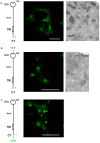
Figure 1. Impact of the position of the YFP tag on the distribution of Langerin.
(A) Transfected M10 melanoma cells stably expressing wild type Langerin (M10-22E cells) were fixed, immunolabeled with the anti-CD207 mAb DCGM4 and analyzed by confocal microscopy (left), or processed for electron microscopy (right). (B) Transfected M10 cells stably expressing Lang-YFP were fixed and processed for confocal microscopy analysis of the YFP distribution (left), or for ultrastructural analysis (right). (A and B) Arrows indicate the position of BGs. (C) Transfected M10 cells stably expressing YFP-Lang were fixed and processed for confocal microscopy analysis of the YFP distribution (left). A higher magnification of a region of interest is shown on the right. Scale bars, 50 µm.
Figure 2
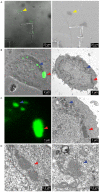
Figure 2. Correlative light-electron microscopy identifies the YFP+ puncta as stacks of BG-like membranes.
M10-YFP-Lang cells were grown on pre-patterned Aclar® culture supports and the ultrastructure of the YFP+ puncta was determined by CLEM. (A) A cell of interest was located by bright field microscopy (left panel) and readily retrieved under the electron microscope (right panel) with the help of the still apparent “7” mark. (B and C) Higher magnifications of the same cell in bright field fluorescent microscopy (left panels) and electron microscopy (right panels), where the fluorescent puncta (blue and red arrowheads) appear as stacks of BG-like membranes (better seen in (D) at a still higher magnification).
Figure 3
Figure 3. Continuity of the BG-like structures with the rough ER.
Transfected M10 cells stably expressing YFP-Lang were processed for electron microscopy. M10 cells stably expressing YFP-Lang were processed for FIB/SEM. The surface of the block was ion-milled and serial images were acquired. A 3D reconstruction was then obtained from the image stack after manual segmentation. (A) A stack of BG-like membranes and sacs of the rough ER in the same plane with (B) manual segmentation of the different objects of interest. The BG-like structures appear in orange, yellow and red and the ER in green. (C and D) A 3D reconstruction demonstrating that continuity (blue segments) exists between the BG-like membranes and the ER (two different angles of view, see also Video S1). Scale bars: 500 nm.
Figure 4
Figure 4. FRAP analysis of the mobilities of Langerin/YFP chimeras.
(A) Selected regions of M10 cells stably expressing either Lang-YFP (upper panels) or YFP-Lang (middle and lower panels) were bleached (arrows) and the fluorescence in single z-sections was recorded approximately every 1.6 s. (B) The fluorescence in the bleached area was quantified and plotted against time after correction for the change in total fluorescence. At least 10 cells were analyzed for each plot; error bars indicate the standard error of the mean. Fi and Fm designate the immobile and mobile fractions of the Langerin/YFP chimeras, respectively.
Figure 5
Figure 5. A monomerizing substitution in YFP impairs the formation of BG-like OSER and restores BG dynamics.
(A) M10 cells stably expressing mYFP-Lang were fixed and the YFP distribution was examined by confocal microscopy. Overlays of 5–10 z-sections from different representative cells are depicted. Left and middle panels: images representative of the majority of cells, with a pericentriolar concentration of fluorescence and small vesicles dispersed across the cytoplasm. Right panel: YFP+ puncta present only in a minority of cells. Scale bars: 25 µm. (B) FRAP experiments were carried out as in Fig. 5B . Left panel: to study the fluorescence recovery in the pericentriolar region, 16 cells were analyzed for each plot. Right panel: to study the fluorescence recovery in enlarged vesicles, 8 cells were analyzed for each plot. Error bars represent the standard error of the mean. Fi and Fm indicate the immobile and mobile fractions of mYFP-Lang molecules, respectively. (C) Soluble membrane extracts (30 µg) of M10 cells stably expressing transgenic Lang-YFP, YFP-Lang or mYFP-Lang were digested or not (NT) with PNGaseF (F) or endoglycosidase Hf (H) and separated by 7.5% SDS-PAGE. Fusion proteins were revealed by western blotting with an anti-GFP antibody. R and S indicate endoglycosidase Hf-resistant and sensitive species, respectively. Untransfected cells (WT) were used as a control.
Figure 6
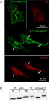
Figure 6. Mutation of the calcium binding domain does not block OSER formation, but restores the dynamic transport of YFP-Langerin mutant.
(A) M10 cells stably expressing YFP-LangE293A were fixed, stained with anti-calnexin and anti-Bip antibodies and analyzed by confocal microscopy. (B) EnodH resistance of YFP-LangE293A molecules was analyzed as described in Fig. 5C.
Figure 7
Figure 7. Schematic view of the proposed mechanism of formation of BG-like OSER.
The ER lumen (L) is depicted in pale gray and the cytoplasm (C) in white. Homotypic interactions between the CRD domains of Langerin (C-terminal, intra-luminal position, dark gray stars) are responsible for the luminal zipping of Langerin-enriched ER membranes, while homotypic interactions between YFP molecules (N-terminal, cytoplasmic position, green circles) are involved in the stacking of BG-like membranes. This “two-lock mechanism” could plausibly explain the high rigidity and immobility of these structures.

Similar articles

Cited by

References

    1. Shimomura O, Johnson FH, Saiga Y (1962) Extraction, purification and properties of aequorin, a bioluminescent protein from the luminous hydromedusan, Aequorea. J Cell Comp Physiol 59: 223–239. - PubMed
    1. Prasher DC, Eckenrode VK, Ward WW, Prendergast FG, Cormier MJ (1992) Primary structure of the Aequorea victoria green-fluorescent protein. Gene 111: 229–233. - PubMed
    1. Snapp EL (2009) Fluorescent proteins: a cell biologist's user guide. Trends Cell Biol 19: 649–655. - PMC - PubMed
    1. Zacharias DA, Violin JD, Newton AC, Tsien RY (2002) Partitioning of lipid-modified monomeric GFPs into membrane microdomains of live cells. Science 296: 913–916. - PubMed
    1. Snapp EL, Hegde RS, Francolini M, Lombardo F, Colombo S, et al. (2003) Formation of stacked ER cisternae by low affinity protein interactions. J Cell Biol 163: 257–269. - PMC - PubMed

Publication types

MeSH terms

LinkOut - more resources