. 2013;8(4):e60859.
doi: 10.1371/journal.pone.0060859. Epub 2013 Apr 8.

Overexpression of full-length centrobin rescues limb malformation but not male fertility of the hypodactylous (hd) rats

Affiliations

Overexpression of full-length centrobin rescues limb malformation but not male fertility of the hypodactylous (hd) rats

František Liška et al. PLoS One. 2013.

Abstract

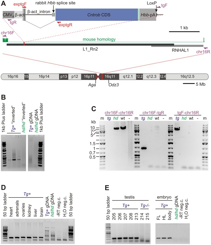
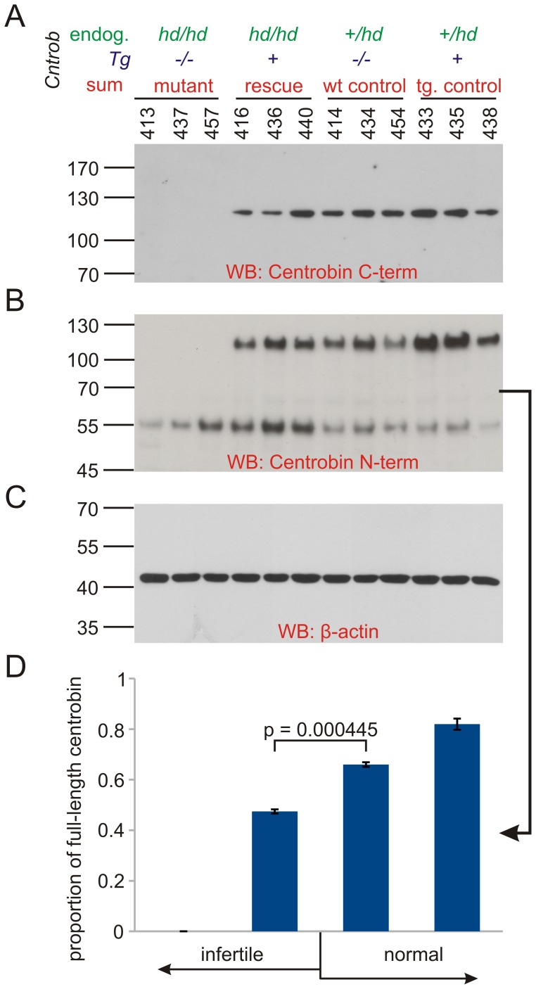
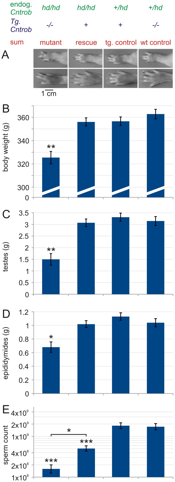
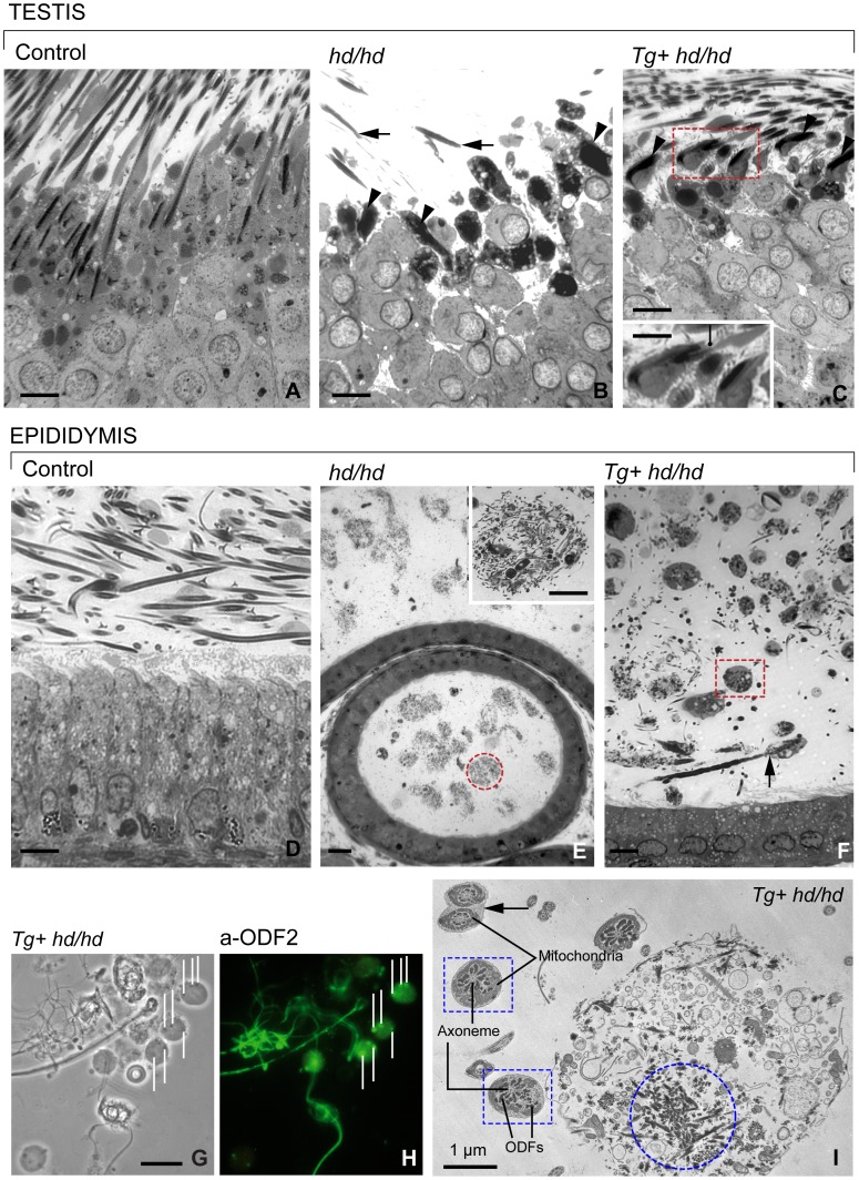
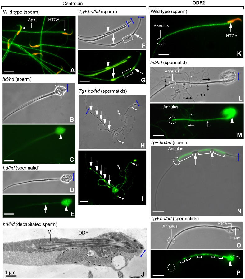
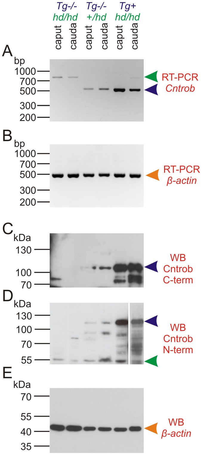
Rat hypodactyly (hd) mutation is characterized by abnormal spermatogenesis and sperm decapitation, limb malformation (missing digits II and III) and growth retardation. We have previously reported centrobin (centrosome BRCA2-interacting protein) truncation at the C-terminus in the hd mutant. Here, we report data from a transgenic rescue experiment carried out to determine a role of centrobin in pathogenesis of hd. The transgenic construct, consisting of full-length-coding cDNA linked to a ubiquitous strong promoter/enhancer combination, was inserted to chromosome 16 into a LINE repeat. No known gene is present in the vicinity of the insertion site. Transgenic centrobin was expressed in all tissues tested, including testis. Transgenic animals show normal body weight and limb morphology as well as average weight of testis and epididymis. Yet, abnormal spermatogenesis and sperm decapitation persisted in the transgenic animals. Western blotting showed the coexistence of full-length and truncated or partially degraded centrobin in sperm of the rescued transgenic animals. Immunocytochemistry showed a buildup of centrobin and ODF2 (outer dense fiber 2) at the sperm decapitation site in the hd mutant and rescued transgenic rats. Additional findings included bulge-like formations and thread-like focal dissociations along the sperm flagellum and the organization of multiple whorls of truncated sperm flagella in the epididymal lumen. We conclude that centrobin is essential for normal patterning of the limb autopod. Centrobin may be required for stabilizing the attachment of the sperm head to flagellum and for maintaining the structural integrity of the sperm flagellum. We postulate that the presence of truncated centrobin, coexisting with full-length centrobin, together with incorrect timing of transgenic centrobin expression may hamper the rescue of fertility in hd male rats.


Conflict of interest statement

Competing Interests: The authors have declared that no competing interests exist.

Figures

Figure 1
Figure 1. Transgenic construct, its localization and expression.
A Top: Transgenic construct contains CMVIE enhancer, chicken β-actin promoter together with first noncoding exon and intron with rabbit β-globin acceptor splice site, coding sequence and rabbit β-globin 3’UTR and polyA signal. Violet arrows represent PCR primers used to amplify the insertion sites (see B). Red arrows depict primers used to assess expression of the transgene (see D and E). A Middle: structure of the insertion site region showed in detail (genome assembly Rn3.4, chr16∶43998593–44005224). Green: homology with the mouse (above zero in the nonrepetitive flanking sequence), grey rectangles: repetitive sequences; the insertion site is red. A Bottom: Rat chromosome 16. Red line: insertion site; purple: centromere. The approximate positions of the genes flanking the transgene are shown. B: Inversion PCR amplifying a fragment of chromosome 16 only in the transgenic animals. C: Long range PCR confirming insertion of the transgene to chromosome 16. Expected product sizes were (for wild-type and transgenic respectively): chr16F-chr16R 6632 bp and 11898 bp (impossible to be amplified with the PCR system employed); chr16F-tgR 944 bp (only transgenic); tgF-chr16R 6080 bp (only transgenic). D: Expression of the transgene in somatic tissues by RT-PCR. E: Expression of the transgene in testis by RT-PCR. hd/hd or hd = hypodactylous mutants, transgene negative; Tg (Tg+) = transgene positive; wt = wild-type; ““ in C: water negative control; “−” RT in D and E, reverse transcriptase dropout negative control. Numbers of individual animals according to our internal system for animal identification are given in panel E.
Figure 2
Figure 2. Detection of centrobin in testis by Western blotting.
A: Western blot using centrobin C-terminal antibody that recognizes both endogenous and transgenic wild-type centrobin. hd-allele specific truncated form cannot be detected. Lack of centrobin in mutants is supplemented to approximately normal level in the rescued males. B: Centrobin N-terminal antibody confirms presence of wild-type protein and the hd-specific truncated protein (lower amount in heterozygote controls). C: We used β-actin as a loading control. D: Relative amount of full-length centrobin as determined from densitometry of the Western blot in B. The proportion is different among groups with full-length centrobin (ANOVA p = 10−5), post-hoc comparison by Tukey’s test shows significant difference between the adjacent infertile and fertile groups (p = 4.45 10−4). Numbers labeling the lanes identify individual animals.
Figure 3
Figure 3. Limb phenotype, growth retardation rescue and improvement of reproductive phenotype.
A: Autopods of mutants compared to transgenic rescue and controls. Left forelimb (top) and hind limb (bottom). Anterior to posterior axis from top to bottom. Normal hind limb has 5 toes, normal forelimb has 4 fingers and a rudimentary thumb. Note missing digit II and hypoplastic digit III in mutants only. B: Body weight. Transgenic rescue rats are indistinguishable from controls, mutants are significantly smaller (post-hoc P = 0.0039 in comparison to rescued males). C: Testis weight (both testes together). Transgenic rescued rats are indistinguishable from controls, mutants are significantly smaller (post-hoc P = 0.0018 in comparison to rescued males). D: Epididymis weight (both epididymides) Transgenic rescue rats are indistinguishable from controls, mutants are significantly smaller (post-hoc P = 0.0297 in comparison to rescued males). E: Sperm count (cauda epididymidis of both sides). Rescued males have more than 3 times more spermatozoa compared to mutants (post-hoc P = 0.00114). However, control males have at least 4 times more spermatozoa compared to rescued males (post hoc P = 0.000816 for rescued males compared to wild-type males). Mutants n = 3, rescued n = 7, transgenic controls n = 6, nontransgenic (wild-type = WT) controls n = 5. One-way ANOVA, with post hoc Tukey test for unequal N.
Figure 4
Figure 4. Characteristics of spermiation and sperm maturation in control, hd/hd mutant and rescued Tg+ hd/hd rats.
A: Histologic section of a control seminiferous epithelium showing the release of mature spermatids during stage IX of spermatogenesis. B: Histologic section of a hd/hd mutant displaying abnormally shaped heads (arrowheads) and severed flagella (arrows) of mature spermatids seen in a similar spermatogenic stage IX. Note that round spermatids display normal structure. C: Histologic section of a rescued Tg+ hd/hd rat showing heads (arrowheads) separated from the flagella seen in a similar spermatogenic stage IX. The inset illustrates a thin cytoplasmic bridge (pointer) connecting the spermatid head to its developing flagellum. The number of decapitated mature spermatids released at spermiation is larger in the rescued rat as compared to the mutant. Round spermatids display normal features. D: Histologic section of the tail region of the epididymis of a control rat showing well-developed sperm in the lumen and the epididymal epithelium with normal characteristics. E: In the hd/hd mutant, the epididymal lumen contains multiple compact whorls (dashed circle) each consisting of entangled flagella seen in the inset at higher magnification. The height of epididymal epithelium is reduced. F: In the rescued Tg+ hd/hd rat epididymis, flagellar whorls coexist with numerous spherical bodies (dashed box). The arrow indicates a decapitated sperm. The height of epididymal epithelium is reduced. G–H: Phase contrast microscopy (panel G) and immunofluorescent localization of ODF2 (panel H) in decapitated sperm and spherical bodies harvested from the epididymal cauda of a rescued Tg+ hd/hd rat. The pointers in panel H indicate immunoreactive ODF2. I: Electron microscopy of a spherical body and sperm flagella in the epididymal lumen of a Tg+ hd/hd rat. The dashed circle indicates aggregates of outer dense fibers in the spherical body equivalent to those seen in panels G and H. The dashed boxes indicate cross-sections of sperm flagella (middle piece) each with an intact axoneme and surrounded by fragmented outer dense fibers (ODFs). The arrow indicates two fused sperm flagella (principal piece). Scale bar in all panels and inset is 5 µm.
Figure 5
Figure 5. Immunofluorescence localization of centrobin (A, C, E, G and I; N-terminal antibody) and ODF2 (K, M, O and Q) in spermatids and/or epididymal sperm from control, hd/hd mutant and rescued Tg+ hd/hd rats. Panels B, D, F, H, L and P are phase-contrast microscopy.
Panel O is a phase-contrast microscopy-immunofluorescence merge. The blue bar indicates the decapitation region. A: Centrobin localization in wild-type control epididymal sperm. The location of the immunoreactive acroplaxome (Apx) and head-tail coupling apparatus (HTCA) is indicated. Note the regular immunoreactive centrobin banding pattern along the flagellum. B–C: Decapitated sperm with fused flagella (opposing arrows). Centrobin immunoreactivity predominates at the HTCA region (arrowhead). D–E: Decapitated spermatid. The arrowhead indicates substantial centrobin-containing material at the HTCA region. The arrow points to a developing bulge of the flagellum. F–G: The arrows denotes the presence of centrobin in the flagellar bulges. The dashed box indicates a thread-like dispersion at the inter-bulge linker. H–I: The arrows show the linear arrangement of centrobin-containing bulges along the spermatid flagellum. The single-crossed arrow identifies immunoreactive spermatid flagella of reduced diameter. The double-crossed arrows point to the centrobin-stained severed end of the thin, also immunoreactive, decapitated spermatid flagella. J: Transmission electron microscopy of a decapitated sperm. The dotted line indicates a large deposit of proteinaceous material at the decapitated end. The material is presumably equivalent to the accumulation of centrobin and ODF2 (and other proteins) detected by immunocytochemistry. The localization of outer dense fibers (ODF) and mitochondria (Mi) is indicated. K: Wild type control epididymal sperm stained with ODF2 antibody. The location of the HTCA and annulus is indicated. L–M: The arrowhead indicates a large deposit of ODF2 at the HTCA region and extending along the middle piece of the flagellum (single-crossed arrow) up to the annulus. The double-crossed arrows point to densities at the decapitated end of several developing spermatid flagella. The arrows denote less intense ODF2 immunoreactive spermatid thin flagella contrasting with the more intense staining of the thicker flagellum (single-crossed arrow) present in the field. N: Merged phase-contrast-fluorescence microscopy images displaying ODF2 immunoreactivity along segments (brackets) linking non-immunoreactive bulges (arrows) of a decapitated sperm flagellum. ODF2 immunoreactivity extends up to the annulus. O–P: Mature spermatids with a persistent attached head can be seen. A significant ODF2 deposit is visualized at the HTCA region (arrowhead). The brackets indicate a less intense staining of ODF2 in regions presumed to become bulges. Scale bar in phase-contrast and fluorescence microscopy panels: 10 µm.
Figure 6
Figure 6. Centrobin expression in epididymal sperm.
A: RT-PCR with primers flanking the junction between exon 10 and 11. Mutants show products with higher molecular weight due to the insertion of an endogenous retrovirus . (green arrowhead). Wild-type product (blue arrowhead) can be amplified both from endogenous locus transcripts as well as from the transgenic transcripts. The “mutant” product can be also seen in the rescued males, although during PCR, the product can be masked by preferential amplification of the smaller molecular weight amplicon coming from the transgene. B: β-actin is shown as endogenous control. Robust expression of centrobin can be seen in sperm both from caput (less mature) and cauda (more mature) epididymis. C: Western blot with an antibody against C-terminal part of centrobin. There is no signal in mutants because they lack the C-terminal portion of centrobin. Note a lower molecular weight species of centrobin from from caput and cauda epididymidis sperm of rescued rats that may correspond to proteolytic processing or degradation. This can be seen in the wild-type sperm too with higher exposition time (not shown). D: Western blot with an N-terminal-specific antibody (middle panel) shows similar results as seen with the C-terminal antibody, 55 kDa mutant truncated protein is also identifiable (green arrowhead). Note: the last lane had to be developed separately due to excess signal. E: β-actin served as the loading control.

Similar articles

Cited by

References

    1. Boivin J, Bunting L, Collins JA, Nygren KG (2007) International estimates of infertility prevalence and treatment-seeking: potential need and demand for infertility medical care. Hum Reprod 22: 1506–1512. - PubMed
    1. Matzuk MM, Lamb DJ (2008) The biology of infertility: research advances and clinical challenges. Nat Med 14: 1197–1213. - PMC - PubMed
    1. Liška F (2003) Selected genetic aspects of male infertility–what animal models tell us. Folia Biol (Praha) 49: 129–141. - PubMed
    1. Manandhar G, Simerly C, Schatten G (2000) Centrosome reduction during mammalian spermiogenesis. Curr Top Dev Biol 49: 343–363. - PubMed
    1. Woolley DM, Fawcett DW (1973) The degeneration and disappearance of the centrioles during the development of the rat spermatozoon. Anat Rec 177: 289–301. - PubMed

Publication types

LinkOut - more resources