. 2013;8(4):e60983.
doi: 10.1371/journal.pone.0060983. Epub 2013 Apr 5.

Activation of NF-kappa B signaling promotes growth of prostate cancer cells in bone

Affiliations

Activation of NF-kappa B signaling promotes growth of prostate cancer cells in bone

Renjie Jin et al. PLoS One. 2013.

Abstract

Patients with advanced prostate cancer almost invariably develop osseous metastasis. Although many studies indicate that the activation of NF-κB signaling appears to be correlated with advanced cancer and promotes tumor metastasis by influencing tumor cell migration and angiogenesis, the influence of altered NF-κB signaling in prostate cancer cells within boney metastatic lesions is not clearly understood. While C4-2B and PC3 prostate cancer cells grow well in the bone, LNCaP cells are difficult to grow in murine bone following intraskeletal injection. Our studies show that when compared to LNCaP, NF-κB activity is significantly higher in C4-2B and PC3, and that the activation of NF-κB signaling in prostate cancer cells resulted in the increased expression of the osteoclast inducing genes PTHrP and RANKL. Further, conditioned medium derived from NF-κB activated LNCaP cells induce osteoclast differentiation. In addition, inactivation of NF-κB signaling in prostate cancer cells inhibited tumor formation in the bone, both in the osteolytic PC3 and osteoblastic/osteoclastic mixed C4-2B cells; while the activation of NF-κB signaling in LNCaP cells promoted tumor establishment and proliferation in the bone. The activation of NF-κB in LNCaP cells resulted in the formation of an osteoblastic/osteoclastic mixed tumor with increased osteoclasts surrounding the new formed bone, similar to metastases commonly seen in patients with prostate cancer. These results indicate that osteoclastic reaction is required even in the osteoblastic cancer cells and the activation of NF-κB signaling in prostate cancer cells increases osteoclastogenesis by up-regulating osteoclastogenic genes, thereby contributing to bone metastatic formation.


Conflict of interest statement

Competing Interests: The authors have declared that no competing interests exist.

Figures

Figure 1
Figure 1. Activation of NF-κB signaling increases RANKL and PTHrP expression in PCa cells.
NF-κB signaling was activated in LNCaP cells by infecting with IKK2-EE retroviral vector (LNCaP-EE), while LNCaP cells infected with the empty vector were used as the controls (LNCaP-EV). NF-κB activity in LNCaP-EV and LNCaP-EE cells were measured by using a NF-κB responsive NGL reporter (A). Expression of RANKL (B) and PTHrP (C) was measured by qRT-PCR. The values plotted represent the mean of at least three individual samples ± SEM. Statistical significance was determined by two-sample t-test. **, P<0.01.
Figure 2
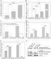
Figure 2. Activation of NF-κB signaling increases RANKL and PTHrP proteins expression in PCa cells.
(A) PCa cells capable of growth in the bone microenvironment exhibit increased NF-κB activity. NF-κB activity in LNCaP, C4-2B and PC3 cells were measured by using a NF-κB responsive NGL reporter. (B) Expression of PTHrP is correlated with the activity of NF-κB signaling in PCa cells. Expression of PTHrP in LNCaP, NF-κB activated LNCaP-EE, C4-2B and PC3 cells was measured by qRT-PCR. (C) Generating NF-κB inactivated PCa cell lines. NF-κB signaling was inactivated in C4-2B and PC3 cells by infecting with IKK2-KD retroviral vector (C4-2B-KD and PC3-KD), while PCa cells infected with the empty vector were used as the controls (C4-2B and PC3-EV). NF-κB activity was measured by using a NF-κB responsive NGL reporter. (D and E) Expression of RANKL (D) and PTHrP (E) proteins in NF-κB activated LNCaP-EE and NF-κB inactivated PC3-KD cells were measured by ELISA and RIA assays, respectively. The cells infected with empty vector (LNCaP-EV and PC3-EV) were used as control. (F) Expression of RANKL and PTHrP are correlated with the activity of NF-κB signaling in PCa cells. RANKL and PTHrP expression were evaluated by Western blot analysis. The values plotted represent the mean of at least three individual samples ± SEM. Statistical significance was determined by two-sample t-test. **, P<0.01.
Figure 3
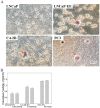
Figure 3. Secretions from NF-κB activated PCa cells induce osteoclast-like cell formation.
(A) Conditioned medium from NF-κB activated PCa cells induces osteoclast-like cell formation in vitro. Bone marrow-derived macrophages were treated with conditioned media from LNCaP, NF-κB activated LNCaP-EE, C4-2B and PC3 cells. TRAP staining was performed after 10 days of additional culture with conditioned media. Arrows indicate the osteoclast-like cells. (B) Activation of NF-κB signaling in PCa cells has no significant effect on the osteoblasts proliferation. Primary cultured osteoblasts were treated with conditioned media from NF-κB activated (LNCaP-EE, C4-2B-EV and PC3-EV) or inactivated (LNCaP-EV, C4-2B-KD and PC3-KD) PCa cells. Proliferation assay (MTT assay) was performed at 48 hours after treatment.
Figure 4
Figure 4. Activation of NF-κB signaling in PCa cells has no significant effect on the cell proliferation rate in vitro.
NF-κB activated/inactivated PCa cell lines were generated by stably infecting with IKK2-EE/IKK2-KD vectors. IKK2-EV was used as control vector. Proliferation rate was determined by MTT assay.
Figure 5
Figure 5. Inactivation of NF-κB signaling in PCa cells inhibits tumor establishment and growth in the bone microenvironment.
NF-κB inactivated PC3-KD and C4-2B-KD cells were grafted into the mouse bones by intratibial injection. PC3 and C4-2B cells infected with empty vector (PC3-EV and C4-2B-EV) were used as controls. (A) Tumor formation of PC3-EV and PC3-KD was determined by small animal X-ray radiograph imaging and histological analysis at 3 weeks after the grafting. (B) Tumor formation of C4-2B-EV and C4-2B-KD was determined by small animal X-ray radiograph imaging and histological analysis at 10 weeks after the grafting. Arrows indicate the sites of tumor formation in the bone.
Figure 6
Figure 6. Activation of NF-κB signaling in PCa cells contributes to tumor establishment and growth in the bone microenvironment.
NF-κB activated LNCaP-EE cells were grafted into the mouse bones by intratibial injection. LNCaP cells infected with empty vector (LNCaP-EV) were used as controls. (A) NF-κB activated LNCaP-EE cells growth in the bone was detected by small animal X-ray radiograph imaging and histological analysis (H&E staining) at 10 weeks after the grafting. (B) Immunohistochemical staining of AR, PSA and FoxA1 was performed to confirm that the cells growing in the bone are human PCa cells. Arrow indicates tumor formation in the bone. (C) Trichrome blue and TRAP staining were performed to confirm osteoblastic lesions and osteoclasts differentiation surrounding the new formed bone. Micro CT scanning showed osteoblastic lesions in the bone when injected with LNCaP-EE cells.
Figure 7
Figure 7. Schematic representation of the role of NF-κB signaling in the establishment of PCa bone metastases.
(A) The activation of NF-κB signaling in PCa cells increases the expression of osteoclastogenesis-associated genes (such as RANKL, PTHrP etc.). (B) These elevated osteolytic factors act as paracrine growth factors to affect directly/indirectly the osteoclast precursor to promote osteoclastogenesis in the bone microenvironment. (C) Activated osteoclastogenesis will induce osteoclastic bone resorption. (D) Therefore, osteoclastic bone resorption induced by the osteolytic factors from PCa cells is sufficient for cancer cells attachment and the establishment of tumors in the skeleton.

Similar articles

Cited by

References

    1. Mundy GR (2002) Metastasis to bone: causes, consequences and therapeutic opportunities. Nat Rev Cancer 2: 584–593. - PubMed
    1. Cheville JC, Tindall D, Boelter C, Jenkins R, Lohse CM, et al. (2002) Metastatic prostate carcinoma to bone: clinical and pathologic features associated with cancer-specific survival. Cancer 95: 1028–1036. - PubMed
    1. Pound CR, Partin AW, Eisenberger MA, Chan DW, Pearson JD, et al. (1999) Natural history of progression after PSA elevation following radical prostatectomy. Jama 281: 1591–1597. - PubMed
    1. Roodman GD (2004) Mechanisms of bone metastasis. N Engl J Med 350: 1655–1664. - PubMed
    1. Garnero P, Buchs N, Zekri J, Rizzoli R, Coleman RE, et al. (2000) Markers of bone turnover for the management of patients with bone metastases from prostate cancer. Br J Cancer 82: 858–864. - PMC - PubMed

Publication types

MeSH terms