. 2013;8(4):e60987.
doi: 10.1371/journal.pone.0060987. Epub 2013 Apr 5.

Th1-Th17 cells contribute to the development of uropathogenic Escherichia coli-induced chronic pelvic pain

Affiliations

Th1-Th17 cells contribute to the development of uropathogenic Escherichia coli-induced chronic pelvic pain

Marsha L Quick et al. PLoS One. 2013.

Abstract

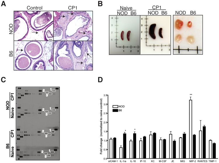
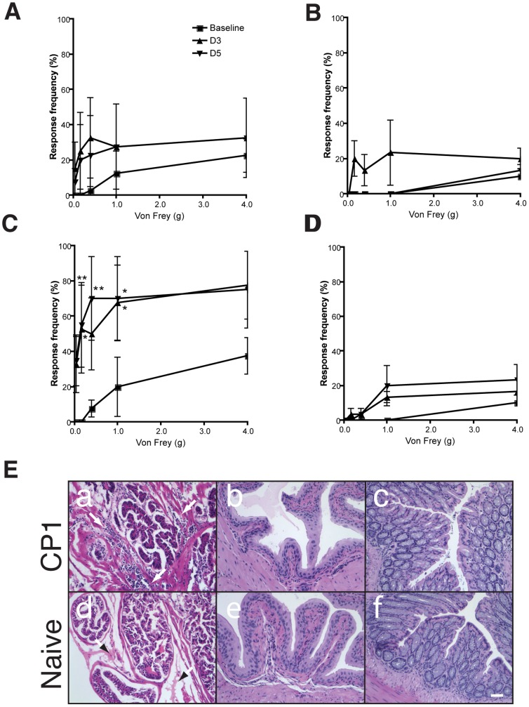
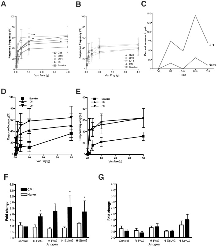
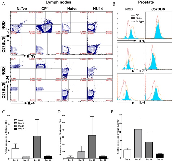
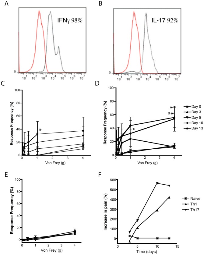
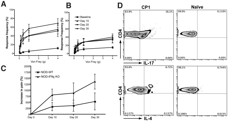
The etiology of chronic prostatitis/chronic pelvic pain syndrome in men is unknown but may involve microbes and autoimmune mechanisms. We developed an infection model of chronic pelvic pain in NOD/ShiLtJ (NOD) mice with a clinical Escherichia coli isolate (CP-1) from a patient with chronic pelvic pain. We investigated pain mechanisms in NOD mice and compared it to C57BL/6 (B6) mice, a strain resistant to CP-1-induced pain. Adoptive transfer of CD4+ T cells, but not serum, from CP-1-infected NOD mice was sufficient to induce chronic pelvic pain. CD4+ T cells in CP-1-infected NOD mice expressed IFN-γ and IL-17A but not IL-4, consistent with a Th1/Th17 immune signature. Adoptive transfer of ex-vivo expanded IFN-γ or IL-17A-expressing cells was sufficient to induce pelvic pain in naïve NOD recipients. Pelvic pain was not abolished in NOD-IFN-γ-KO mice but was associated with an enhanced IL-17A immune response to CP1 infection. These findings demonstrate a novel role for Th1 and Th17-mediated adaptive immune mechanisms in chronic pelvic pain.


Conflict of interest statement

Competing Interests: The authors have declared that no competing interests exist.

Figures

Figure 1
Figure 1. CP1 infection in NOD and B6 mice elicit differential immune responses.
(A) Representative histopathology of the prostate of NOD and B6 mice 28 days after CP1 infection or no infection (controls). Arrows represent areas of leukocytic infiltrate in the prostate. Bar, 50 µM. (B) Representative images of the spleen and lumbar lymph node in naïve or CP1 infected NOD and B6 mice 28 days following infection. Lymph nodes from infected mice are displayed in the upper row. (C and D) Profile of chemokines in prostates of NOD and B6 mice 28 days following CP1 infection. Chemokine levels were normalized to naïve NOD and B6 prostates (C) and chemokines differentially expressed between NOD and B6 mice were expressed as relative fold change (D). Representative image of the immunoblot (C) indicates elevation in IL-1ra and IL-16 (1 and 2) in B6 mice and MIP-2 (3) in NOD mice. The experiment was performed with pooled prostates (n = 5/group) and repeated twice. Statistical significance is indicated at * p<0.05.
Figure 2
Figure 2. Adoptive transfer of T cells mediate pelvic pain in NOD mice.
(A and B) Naive NOD mice (n = 5) were injected with 100 µl of serum from CP-1 infected (28 days) (A) or naïve (B) NOD mice and referred visceral hyperalgesia in NOD mice was measured as responses to mechanical stimulation of the pelvic region using von Frey filaments of 5 calibrated forces. Data is shown as the mean percentage of positive response ± SEM before instillation of serum or pan T Cells (baseline) and at days 3 and 5 following injection. (C and D) Naïve NOD mice (n = 5) were injected with pan T cells isolated from CP-1 infected (C) or naïve (D) NOD mouse spleen and lymph nodes and responses to mechanical stimulation of the pelvic region was measured as before. (E) All experiments were repeated at least two times and significant increase in response frequency is represented by * p<0.05 and **p<0.001. The symbol key shown in panel A applies to panels B, C and D. (E) Representative histopathology of corresponding prostate (panels a and d), bladder (b and e), and colon (c and f) of naïve or CP-1-exposed pan T cell recipients. Arrowheads in a and d indicate comparison of leukocytic infiltrates. Scale bar, 50 µM.
Figure 3
Figure 3. Adoptive transfer of CD4+ T cells mediate pelvic pain in NOD mice
(A–E) Naive NOD mice (n = 5) were injected with 4×10⁶ CD4+ or CD8+ T cells from CP-1 infected (A and D) or naïve (B and E) NOD mice and referred visceral hyperalgesia in NOD mice was measured as responses to mechanical stimulation of the pelvic region using von Frey filaments of 5 calibrated forces. Data is shown as the mean percentage of positive response (+/−) SEM before transfer of T cells (baseline) and at PID 9, 14, 19 and 28 (A and B) or PID 5 and 9 (D and E). (C) Percent response in NOD mice injected with CD4+ T cells from CP-1 infected and naïve NOD mice monitored at day 0, 9, 14, 19 and 28 (n = 5). F) and G) CD4+ T cells from CP1 infected NOD mice show proliferative response to prostate antigens. Irradiated splenocytes from naïve NOD mice or naïve B6 mice were used to present 2 µg/ml human, mouse and rat prostate antigen to CD4+ T cells isolated from naïve or CP1 infected NOD (F) and B6 (G) mice. The proliferative response was measured as absolute cell counts per 100 µl on day 3 and expressed as fold change in cell numbers compared to day 1. Experiments were repeated independently twice and statistical significance is shown at *p<0.05.
Figure 4
Figure 4. CD4+ T cell subsets in lymph nodes and prostates of CP-1-infected mice.
(A) Representative flow cytometry analysis of CD4+ T cells from lymph nodes of naïve and CP-1 infected NOD and B6 mice. Isolated lymph node cells were gated on CD4+ T cells followed by IFN-γ+, IL-17A+, IFN-γ+ IL-17A+, and IL-4+ expression. (B) Representative flow cytometry analysis of intraprostatic CD4+ T cells from naïve or CP-1 infected NOD and B6 mice. Histograms representing intracellular cytokine staining in CP-1 infected (solid line) NOD or B6 mice were overlaid with naïve T cells (tinted) and isotype controls (dotted line). Each experiment was independently repeated at least three times with 5 mice/group. In real-time PCR assays, prostates from NOD mice infected with CP1 were used to prepare cDNA and real-time PCR was performed for lineage-specific transcription factors Tbet, Foxp3, RORγt, as well as CD4. Expression levels are shown as Ct(TF)/Ct(CD4) and is the average of three independent experiments.
Figure 5
Figure 5. Adoptive transfer of IFNγ+ or IL-17A+ CD4+ T cells mediate pelvic pain.
(A and B) T cells from CP-1 infected NOD mice were cultured under Th1 (A) or Th17 (B) polarizing conditions followed by flow cytometry using anti-IFN-γ and anti-IL-17A (dotted line) or isotype control (solid line) antibodies. (C-F) Naive NOD mice were injected with 1×10⁶ IFN-γ+ (C), IL-17A+ (D), or naïve T cells (E) and referred visceral hyperalgesia in NOD mice was measured using von Frey filaments of 5 calibrated forces. Data is shown as the mean percentage of positive response (+/−) SEM before transfer of T cells (day 0) and at days 3, 5, 10 and 13 after transfer (C–E). (F) Percent increase in response frequency from baseline in NOD mice injected with CD4+ T cells. Experiments were repeated twice independently and statistical significance is shown at *p<0.05 or **p<0.01.
Figure 6
Figure 6. NOD-IFN-γ-KO mice exhibit enhanced pelvic pain and lymph node expression of IL-17A.
NOD-IFN-γ-KO (A, n = 5) or NOD mice (B, n = 5) or were infected with CP-1 bacteria and referred visceral hyperalgesia was measured as responses to mechanical stimulation of the pelvic region using von Frey filaments of 5 calibrated forces. Data is shown as the mean percentage of positive response ± SEM before infection (baseline) and at days 10, 20 and 30 days after infection. The symbol key shown in panel B applies to panels A. Percent increase in response in NOD and NOD-IFN-γ-KO mice were compared over the 30 days time-course (C). Representative flow cytometry analysis of CD4+ T cells from draining lymph nodes of naïve and CP-1 infected NOD-IFN-γ-KO mice. Isolated lymph node cells were gated on CD4+ T cells followed by intracellular cytokine staining for IL-17A+ and IL-4+ expression. Percent of CD4+ T cells representative of multiple animals are shown in individual quadrants. Experiments were repeated twice independently with 5 mice/group.

Comment in

References

    1. Collins MM, Stafford RS, O'Leary MP, Barry MJ (1998) How common is prostatitis? A national survey of physician visits. J Urol 159: 1224–1228. - PubMed
    1. Hua VN, Schaeffer AJ (2004) Acute and chronic prostatitis. Med Clin North Am 88: 483–494. - PubMed
    1. Nickel JC, Alexander RB, Schaeffer AJ, Landis JR, Knauss JS, et al. (2003) Leukocytes and bacteria in men with chronic prostatitis/chronic pelvic pain syndrome compared to asymptomatic controls. J Urol 170: 818–822. - PubMed
    1. Leskinen MJ, Rantakokko-Jalava K, Manninen R, Leppilahti M, Marttila T, et al. (2003) Negative bacterial polymerase chain reaction (PCR) findings in prostate tissue from patients with symptoms of chronic pelvic pain syndrome (CPPS) and localized prostate cancer. Prostate 55: 105–110. - PubMed
    1. Krieger JN, Riley DE, Roberts MC, Berger RE (1996) Prokaryotic DNA sequences in patients with chronic idiopathic prostatitis. J Clin Microbiol 34: 3120–3128. - PMC - PubMed

Publication types

MeSH terms