. 2013;8(4):e61158.
doi: 10.1371/journal.pone.0061158. Epub 2013 Apr 5.

Isolation of recombinant phage antibodies targeting the hemagglutinin cleavage site of highly pathogenic avian influenza virus

Affiliations

Isolation of recombinant phage antibodies targeting the hemagglutinin cleavage site of highly pathogenic avian influenza virus

Jinhua Dong et al. PLoS One. 2013.

Abstract

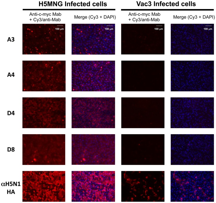
Highly pathogenic avian influenza (HPAI) H5N1 viruses, which have emerged in poultry and other wildlife worldwide, contain a characteristic multi-basic cleavage site (CS) in the hemagglutinin protein (HA). Because this arginine-rich CS is unique among influenza virus subtypes, antibodies against this site have the potential to specifically diagnose pathogenic H5N1. By immunizing mice with the CS peptide and screening a phage display library, we isolated four antibody Fab fragment clones that specifically bind the antigen peptide and several HPAI H5N1 HA proteins in different clades. The soluble Fab fragments expressed in Escherichia coli bound the CS peptide and the H5N1 HA protein with nanomolar affinity. In an immunofluorescence assay, these Fab fragments stained cells infected with HPAI H5N1 but not those infected with a less virulent strain. Lastly, all the Fab clones could detect the CS peptide and H5N1 HA protein by open sandwich ELISA. Thus, these recombinant Fab fragments will be useful novel reagents for the rapid and specific detection of HPAI H5N1 virus.


Conflict of interest statement

Competing Interests: The authors have declared that no competing interests exist.

Figures

Figure 1
Figure 1. Selection of anti-HPAI HA antibodies.
(A) The structure of BSA-MBS-HA331 used for mice immunization. (B) Flow chart for the development of monoclonal Fabs. The RNA extracted from the spleen cells of immunized mice was used for RT-PCR, which produced VH and VL cDNAs. These fragments were used to make the pDong1 phagemid library that was subsequently used for biopanning the phage libraries. (C) ELISA of the binding of positive Fab-phages to HA 331 peptide and HA proteins. HA331: cleavage site peptide; MBS: m-maleimidobenzoic acid N-hydroxysuccinimide ester; BSA: bovine serum albumin; H1N1-HA: recombinant A/California/04/2009 H1N1 HA; H5N1-HA: recombinant A/Vietnam/1194/2004 H5N1 HA.
Figure 2
Figure 2. Binding specificity of the clones.
(A) ELISA to determine binding to several other HA proteins. H5N1-HA(An): A/Anhui/1/05(H5N1) HA; H5N1-HA(Tu): A/Turkey/1/2005(H5N1) HA; H7N7-HA(Ne): A/Netherlands/219/03(H7N7) HA. (B) ELISA to determine epitope sequence. The inhibitory effect of 7-mer partial peptide sequences shown on the binding of Fab-phages to SAv-HA331 peptide was investigated. The peptide NSPQRER showed maximal inhibitory effect. (C) The cleavage site peptide sequence of HA proteins used. The presumptive peripheral and core epitope sequences are shown in brown and red, respectively. H6 is shown as a reference.
Figure 3
Figure 3. Characterization of soluble Fab fragments.
(A) SDS-PAGE of purified Fab fragments. Upper and lower bands correspond to Fd and L chains, respectively. M: Molecular weight standards. (B) ELISA for evaluating binding to H5N1 HA protein. H5N1-HA: recombinant HA from H5N1 A/Vietnam/1194/2004.
Figure 4
Figure 4. SPR sensorgrams obtained for the purified Fab fragments bound to HA331 peptide.
(A) A3, (B) A4, (C) D4, and (D) D8 at 200 (black line), 100 (hatched line), 50 (gray line), and 25 nM (gray hatched line) were applied.
Figure 5
Figure 5. Fab fragment-mediated immunofluorescent staining of Madin-Darby canine kidney (MDCK) cells infected with H5N1 Mongolia virus or Vac (low pathogenic) virus.
An anti-H5N1 antibody 9F2E3F3 was also used for staining.
Figure 6
Figure 6. Detection of (A) HA331 peptide and (B) recombinant H5N1 HA protein by open sandwich phage ELISAs.
The antigen-dependency of the VH/VL interaction was measured with the pDong system.

References

    1. Eccles R (2005) Understanding the symptoms of the common cold and influenza. Lancet Infect Dis 5: 718–725. - PMC - PubMed
    1. Wright F, Neumann G, Kawaoka Y (2007) Orthomyxoviruses. Fields Virology. Philadelphia: Wolters Kluwer, Lippincott Willams & Wilkins. pp. 1691–1740.
    1. Webster RG, Bean WJ, Gorman OT, Chambers TM, Kawaoka Y (1992) Evolution and ecology of influenza A viruses. Microbiol Rev 56: 152–179. - PMC - PubMed
    1. Fouchier RA, Munster V, Wallensten A, Bestebroer TM, Herfst S, et al. (2005) Characterization of a novel influenza A virus hemagglutinin subtype (H16) obtained from black-headed gulls. J Virol 79: 2814–2822. - PMC - PubMed
    1. Olsen B, Munster VJ, Wallensten A, Waldenstrom J, Osterhaus AD, et al. (2006) Global patterns of influenza a virus in wild birds. Science 312: 384–388. - PubMed

Publication types

MeSH terms