. 2013:2013:516051.
doi: 10.1155/2013/516051. Epub 2013 Mar 21.

Oxidative stress and mitogen-activated protein kinase pathways involved in cadmium-induced BRL 3A cell apoptosis

Affiliations

Oxidative stress and mitogen-activated protein kinase pathways involved in cadmium-induced BRL 3A cell apoptosis

Zhang Yiran et al. Oxid Med Cell Longev. 2013.

Abstract

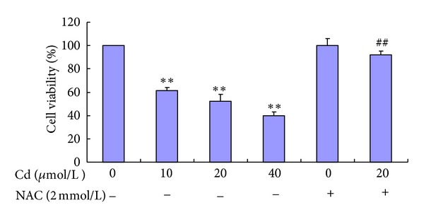
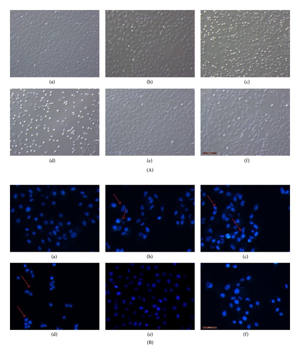
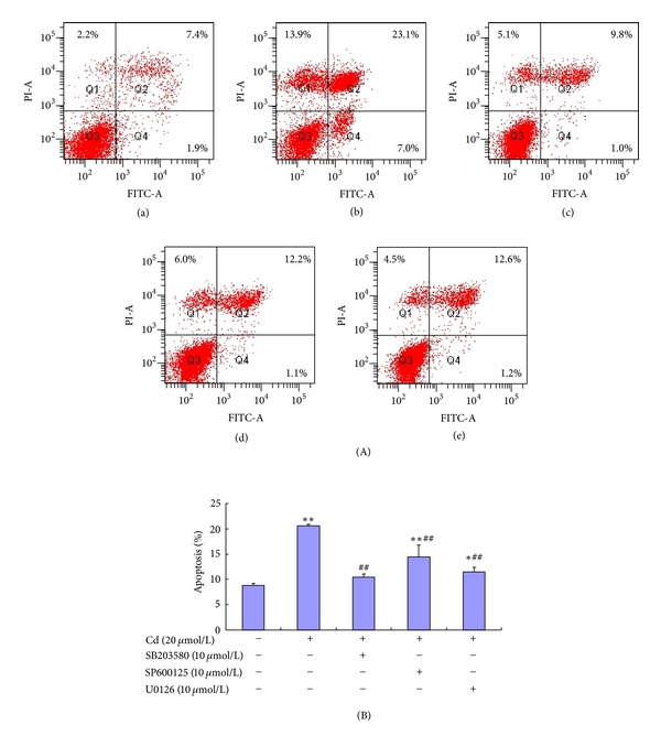
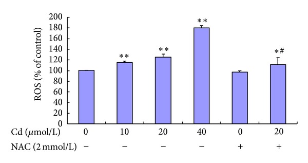
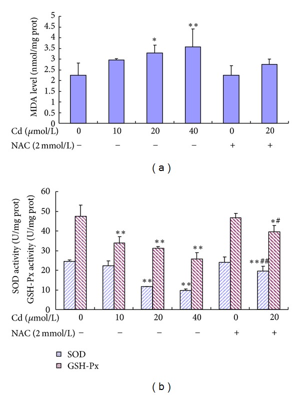
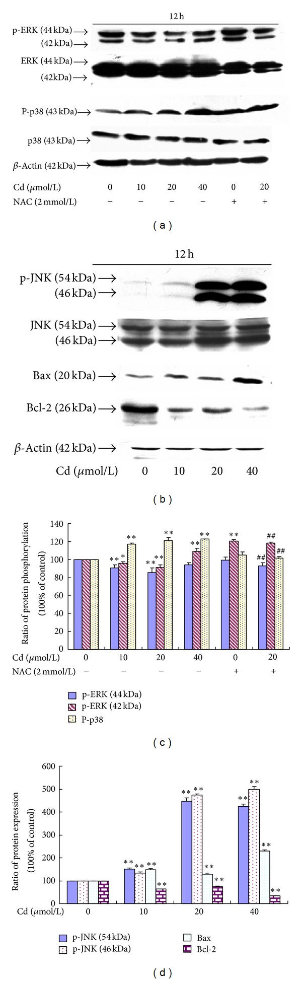
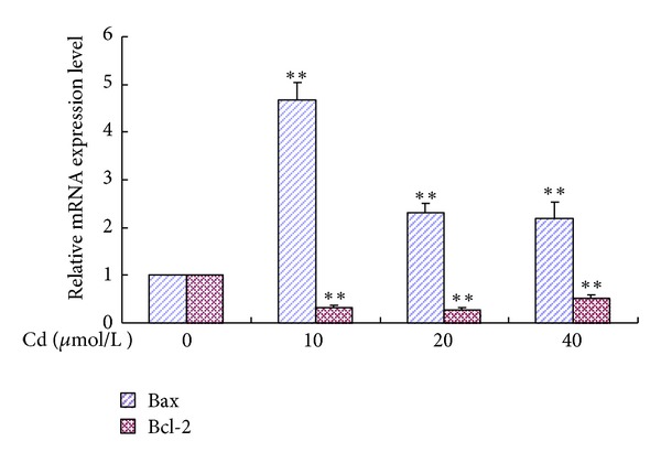
In this study, BRL 3A cells were treated with different Cd concentrations (0, 10, 20, and 40 μmol/L) for 12 h and preincubated with or without N-acetyl-L-cysteine (NAC) (2 mmol/L) for 30 min, and cells were treated with Cd (0 and 20 μmol/L), pretreated with p38 inhibitor (SB203580), JNK (c-Jun NH2-terminal kinases) inhibitor (SP600125), and extracellular signal-regulated kinase (ERK) inhibitor (U0126) for 30 min, and then treated with 20 μmol/L Cd for 12 h. Cd decreased cell viability, SOD, and GSH-Px activity in a concentration-dependent manner. Increased MDA level, ROS generation, nuclear condensation, shrinkage, and fragmentation in cell morphology were inhibited by NAC. Cd-induced apoptosis was attenuated by pretreatment with SB203580, SP600125, and U0126. The results of western blot showed that NAC preincubation affected Cd-activated MAPK pathways, p38 and ERK phosphorylation. Cd treatment elevated the mRNA levels of Bax and decreased the mRNA levels of Bcl-2, respectively. The same effect was found in their protein expression levels. These results suggest that oxidative stress and MAPK pathways participate in Cd-induced apoptosis and that the balance between pro- and antiapoptotic genes (Bax and Bcl-2) is important in Cd-induced apoptosis.


Figures

Figure 1
Figure 1
Cell viability of BRL 3A cells treated with Cd and NAC. Cells were treated with 0 μmol/L to 40 μmol/L Cd and pre-treated with or without 2 mmol/L NAC for 12 h. Data are presented as mean ± SD of three independent experiments performed in triplicate. Significant difference: *P < 0.05, **P < 0.01, compared with the control. # P < 0.05, ## P < 0.01, compared with the 20 μmol/L Cd group.
Figure 2
Figure 2
Cd and NAC induce morphological changes in BRL 3A cells. Cultured cells were exposed to 0, 10, 20, and 40 μmol/L Cd for 12 h (a, b, c, and d). In the other experiment, the cells were pre-incubated with 2 mmol/L NAC for 30 min and incubated with 20 μmol/L Cd or only incubated with 2 mmol/L NAC for 12 h (f and e). (A) After the treatment, images were taken with an Olympus inverted phase-contrast microscope. (B) Thereafter, the cells were fixed, stained with Hoechst 33258, and then observed under a fluorescence microscope. Arrows indicate morphological changes (blebbing cells, chromatin condensation, or fragmentation) in the nuclei of BRL 3A cells. Scale bar: 100 μm.
Figure 3
Figure 3
Flow cytometric analysis of BRL 3A cells after treatment with Cd and MAKP inhibitors. Cellular apoptosis was tested using an apoptosis detection kit. (A) BRL 3A cells were treated with 0 and 20 μmol/L Cd for 12 h (a and b). In the other three experiments, the cells were pre-incubated with 10 μmol/L SB203580, SP600125, and U0126 for 30 min, followed by incubation with 20 μmol/L Cd for 12 h (c, d, and e). (B) The apoptotic percentage shows that 10 μmol/L p38 inhibitor (SB203580), JNK inhibitor (SP600125), and ERK inhibitor (U0126) can reduce cell apoptosis significantly. Data are presented as mean ± SD of three independent experiments performed in triplicate. Significant difference: *P < 0.05, **P < 0.01, compared with the control. # P < 0.05, ## P < 0.01, compared with the 20 μmol/L Cd group.
Figure 4
Figure 4
Effect of Cd and NAC on ROS generation of BRL 3A cells. Cultured cells were exposed to 0 μmol/L to 40 μmol/L Cd and pre-treated with or without 2 mmol/L NAC for 12 h. Data are presented as mean ± SD of three independent experiments performed in triplicate. Significant difference: *P < 0.05, **P < 0.01, compared with the control. # P < 0.05, ## P < 0.01, compared with the 20 μmol/L Cd group.
Figure 5
Figure 5
Effect of Cd and NAC on MDA level (A), SOD activity, and GSH-Px activity (B) in BRL 3A cells. Cells were treated with 0 μmol/L to 40 μmol/L Cd and pre-treated with or without 2 mmol/L NAC for 12 h. Data are presented as mean ± SD of three independent experiments performed in triplicate. Significant difference: *P < 0.05, **P < 0.01, compared with the control. # P < 0.05, ## P < 0.01, compared with the 20 μmol/L Cd group.
Figure 6
Figure 6
(a) Effect of Cd and NAC on ERK and p38 phosphorylation of BRL 3A cells. (b) Effect of Cd on the JNK phosphorylation and Bax and Bcl-2 protein expression levels of BRL 3A cells. (c) Quantitative analysis of the immunoreactive phosphorylated ERK and p38 proteins in treated BRL 3A cells. (d) Quantitative analysis of the immunoreactive phosphorylated JNK, Bax, and Bcl-2 proteins in Cd-treated BRL 3A cells. Each value is expressed as the phospho/total MAPK percentage of phosphorylation and the ratio of OD in Bax and Bcl-2 with respect to β-actin. Significant difference: *P < 0.05, **P < 0.01, compared with the control. # P < 0.05, ## P < 0.01, compared with the 20 μmol/L Cd group.
Figure 7
Figure 7
Relative quantification of Bax and Bcl-2 gene expression levels by real-time PCR in relation to β-actin. Cells were treated with 0 μmol/L to 40 μmol/L for 12 h. Data are presented as mean ± SD of three independent experiments performed in triplicate. Significant difference: *P < 0.05, **P < 0.01, compared with the control; # P < 0.05, ## P < 0.01, compared with the 20 μmol/L Cd group.

References

    1. Méndez-Armenta M, Ríos C. Cadmium neurotoxicity. Environmental Toxicology and Pharmacology. 2007;23(3):350–358. - PubMed
    1. Bertin G, Averbeck D. Cadmium: cellular effects, modifications of biomolecules, modulation of DNA repair and genotoxic consequences (a review) Biochimie. 2006;88(11):1549–1559. - PubMed
    1. Thompson J, Doi T, Power E, Balasubramanian I, Puri P, Bannigan J. Evidence against a direct role for oxidative stress in cadmium-induced axial malformation in the chick embryo. Toxicology and Applied Pharmacology. 2010;243(3):390–398. - PubMed
    1. Bharavi K, Reddy AG, Rao GS, Reddy AR, Rao SVR. Reversal of cadmium-induced oxidative stress in chicken by herbal adaptogens Withania somnifera and Ocimum sanctum . Toxicology International. 2010;17(2):59–63. - PMC - PubMed
    1. Brzóska MM, Rogalska J, Kupraszewicz E. The involvement of oxidative stress in the mechanisms of damaging cadmium action in bone tissue: a study in a rat model of moderate and relatively high human exposure. Toxicology and Applied Pharmacology. 2011;250(3):327–335. - PubMed

Publication types

MeSH terms

LinkOut - more resources