. 2013:2013:370169.
doi: 10.1155/2013/370169. Epub 2013 Mar 17.

Radiographically occult and subtle fractures: a pictorial review

Affiliations

Radiographically occult and subtle fractures: a pictorial review

Mohamed Jarraya et al. Radiol Res Pract. 2013.

Abstract

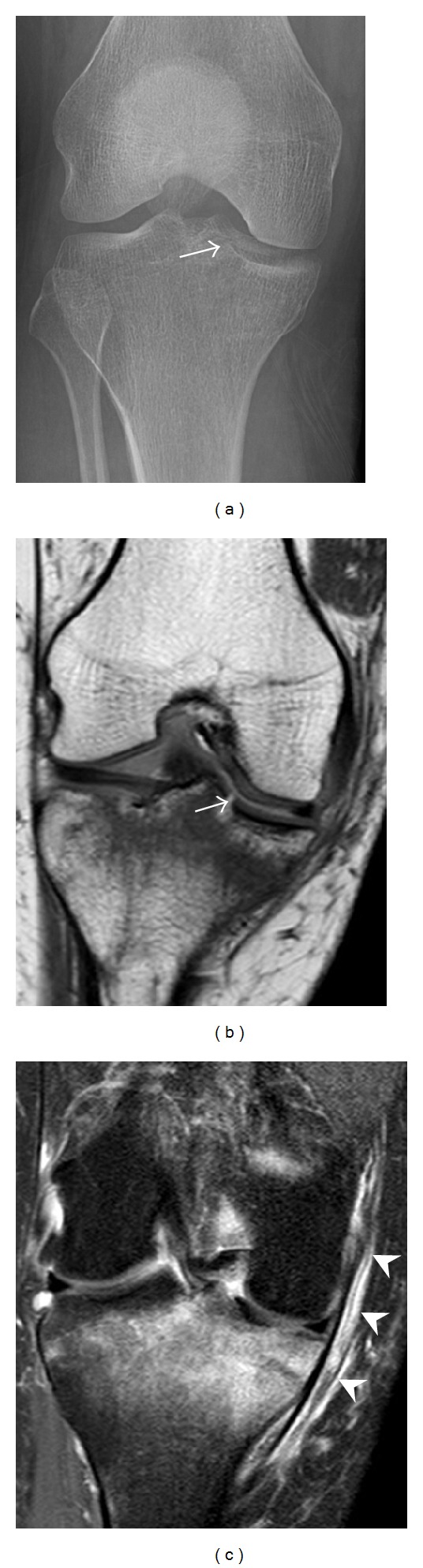
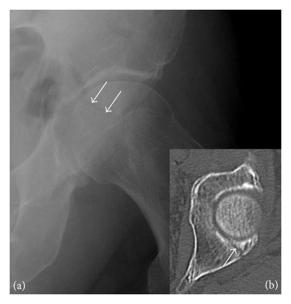
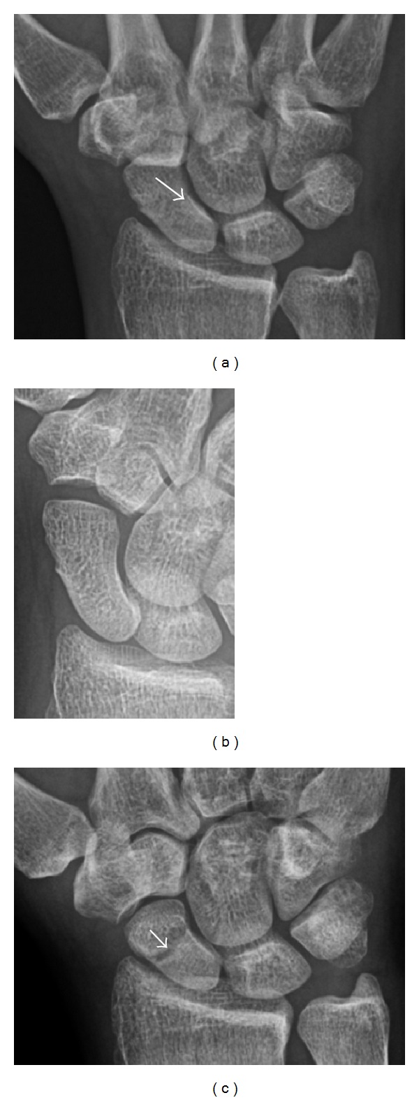
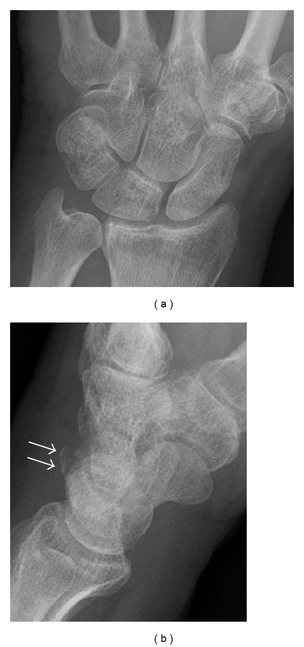
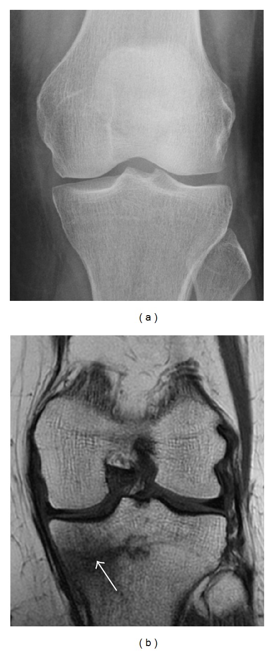
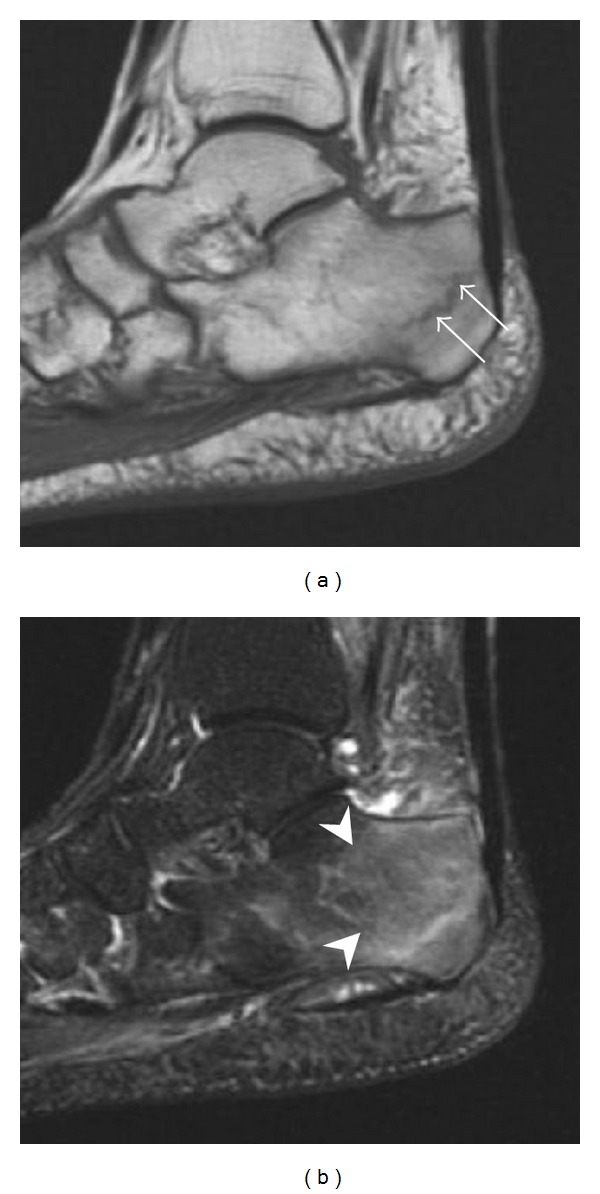
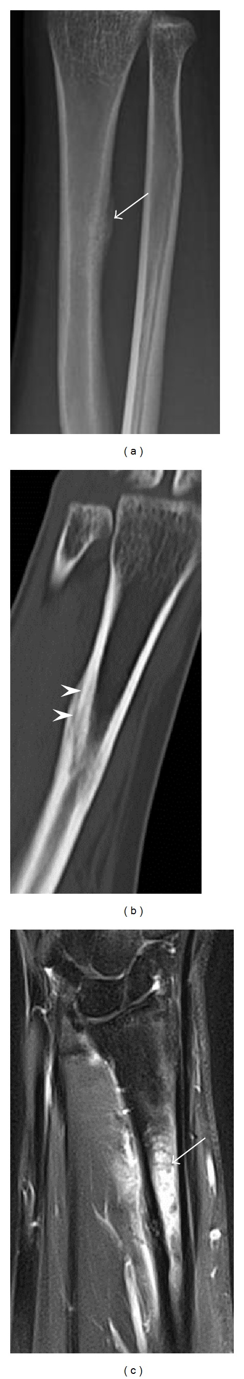
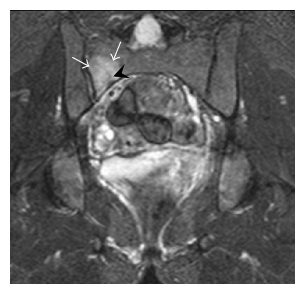
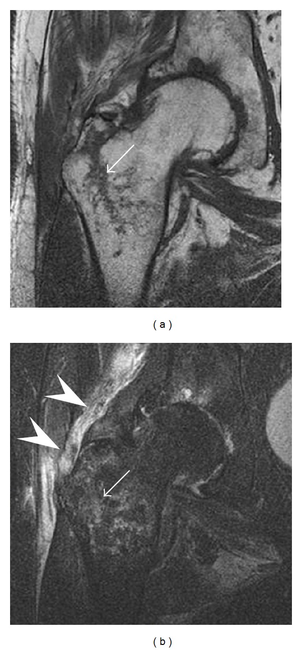
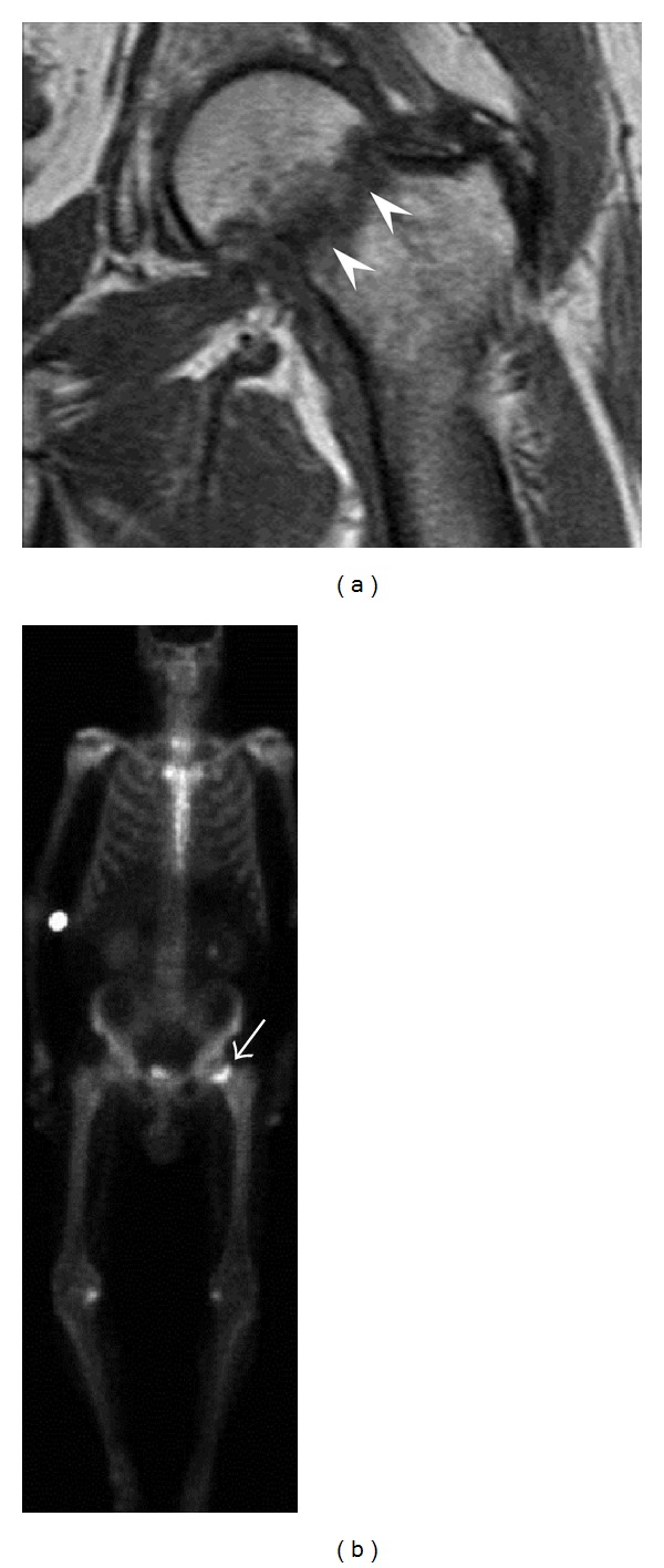
Radiographically occult and subtle fractures are a diagnostic challenge. They may be divided into (1) "high energy trauma fracture," (2) "fatigue fracture" from cyclical and sustained mechanical stress, and (3) "insufficiency fracture" occurring in weakened bone (e.g., in osteoporosis and postradiotherapy). Independently of the cause, the initial radiographic examination can be negative either because the findings seem normal or are too subtle. Early detection of these fractures is crucial to explain the patient's symptoms and prevent further complications. Advanced imaging tools such as computed tomography, magnetic resonance imaging, and scintigraphy are highly valuable in this context. Our aim is to raise the awareness of radiologists and clinicians in these cases by presenting illustrative cases and a discussion of the relevant literature.


Figures

Figure 1
Figure 1
A 56-year-old woman presenting with left knee pain after a fall. (a) Initial anteroposterior radiograph was considered normal, however, subtle cortical disruption of the anterior rim of the medial tibial plateau, medial to the tibial spine, is noted (arrow). (b) Coronal T1-weighted MRI confirms the cortical disruption (arrow) and shows extensive fracture through the proximal tibia. (c) Coronal proton density-weighted image with fat saturation shows extensive edema in the subchondral bone. Note also hypersignal adjacent to the medial collateral ligament corresponding to a grade I sprain (arrowheads).
Figure 2
Figure 2
Posterior acetabular fracture in a 49-year-old woman presenting with hip pain after a fall. (a) Anteroposterior radiograph of the left hip shows a radiolucent line through the posterior acetabular wall (arrows). (b) Axial CT confirms the acetabular fracture (arrow).
Figure 3
Figure 3
A 26-year-old man presenting with wrist pain after being assaulted. (a) Initial anteroposterior radiograph shows a subtle linear lucency within the scaphoid extending to the scaphocapitate articular surface that was overlooked (arrow). (b) Initial “scaphoid” view was negative. (c) Followup anteroposterior radiographs, 12 days later, shows obvious scaphoid fracture (arrows).
Figure 4
Figure 4
Dorsal triquetral fracture of the left wrist in a 30-year-old man after a trauma. Anteroposterior radiograph shows a normal appearance. (b) Lateral radiograph of the same wrist demonstrates a chip fracture off the dorsal aspect of the triquetrum (arrow).
Figure 5
Figure 5
Traumatic fracture of the greater tuberosity in a 51-year-old man presenting with left shoulder pain after a fall on ice. Initial radiographs were normal. Coronal inversion recovery MRI shows a fracture line (arrow) through the greater tuberosity surrounded by a bone marrow edema pattern.
Figure 6
Figure 6
Subtle anterior talar fracture in a 39-year-old man presenting with ankle pain after a fall. (a) Anteroposterior radiograph shows a subtle oblique radiolucent line through the talus (white arrows). (b) Sagittal CT reformation confirms the presence of an anterior talar fracture with cortical offset (black arrow).
Figure 7
Figure 7
Fatigue fracture of the talus in a 25-year-old male basketball player with right hind foot and ankle pain, without history of trauma, and a normal initial radiograph (not shown). (a) One-month followup lateral radiograph shows normal appearance. (b) Sagittal T1-weighted MRI shows an irregular fracture line (arrow) within an ill-defined area of hypointensity corresponding to bone marrow edema.
Figure 8
Figure 8
Proximal diaphyseal fatigue fracture of the tibia in a 20-year-old man with a history of regular jogging. (a) Lateral radiograph shows no obvious fracture lines but a subtle localized medial tibial cortex periosteal reaction (arrows). (b) Sagittal reformatted CT image acquired 1-month after the radiograph shows a linear hypoattenuation in the tibial cortex (arrowhead), as well as obvious periosteal thickening (arrows). (c) Sagittal T2-weighted fat-saturated image acquired the same day shows an area of hyperintensity spreading over the proximal tibia (arrows), which is consistent with the presence of proximal tibial fracture.
Figure 9
Figure 9
Proximal metaphyseal fatigue fracture of the tibia in a 27-year-old recent male military recruit. (a) Anteroposterior radiograph is within normal limits. (b) Coronal T1-weighted MR image shows a marked linear hypoattenuation along the medial tibial metaphysis (arrow) surrounded by diffuse hypointensity in keeping with posttraumatic edema.
Figure 10
Figure 10
Calcaneal fatigue fracture in a 30-year-old male runner. Radiographs were normal (not shown). (a) Sagittal T1-weighted and (b) short tau inversion recovery images show a linear hypointensity (arrows) of calcaneal tuberosity within diffuse bone marrow edema, which appears as an ill-defined area of hyperintensity on a fluid sensitive pulse sequence (arrowheads).
Figure 11
Figure 11
Stress fracture of the right radius in a 40-year-old man, a semiprofessional billiard player, with no history of trauma and complaining of pain of the right forearm for one month. (a) Anteroposterior radiograph shows medial radial cortex periosteal reaction (arrow) but no fracture line is seen. (b) Coronal reformatted CT depicts monocortical fracture line through the periosteal thickening (arrowheads). (c) Coronal T2-weighted fat-suppressed MRI shows intramedullary hyperintensity within the bone marrow (arrow) corresponding to bone marrow edema.
Figure 12
Figure 12
Right sacral alar insufficiency fracture in a 29-year-old woman with a 9-year history of corticosteroid therapy for systemic lupus erythematous. Conventional radiographs showed normal appearance (not shown). Coronal inversion recovery MRI shows an area of hyperintensity in the right sacral ala (white arrows), centered on a linear hypointensity corresponding to the fracture line (black arrowhead).
Figure 13
Figure 13
Partial osseous avulsion of the gluteal muscles at the greater trochanter in a 59-year-old man who presented with the right hip pain without a history of trauma. Lauenstein view and anteroposterior and radiographs (not shown) did not show an obvious fracture line or disruption of bony contours in the acetabulum or the right femoral neck. (a) Coronal T1-weighted MRI displays an incomplete fracture line extending partially from the greater trochanter (arrow). (b) Coronal short tau inversion recovery MRI shows heterogeneous hyperintensity in the same region (arrow) as well as hyperintensity within the gluteus medius and minimus muscles (arrowheads) consistent with tissue edema and hematoma.
Figure 14
Figure 14
Subcapital insufficiency fracture in a 55-year-old man with a left hip pain without a history of trauma. Anteroposterior and Lauenstein view radiographs centered on the left hip do not show an obvious fracture line, but mild acetabular osteophytosis was noted consistent with hip osteoarthritis (not shown). (a) Coronal T1-weighted MRI shows a linear low-signal band through the femoral neck corresponding to a fracture line (arrowheads). (b) Bone scintigraphy shows focal uptake (arrow) corresponding to the fracture.

References

    1. Guly HR. Diagnostic errors in an accident and emergency department. Emergency Medicine Journal. 2001;18(4):263–269. - PMC - PubMed
    1. Sshwartz DT. ACEP Scientific Assembly. Boston Convention and Exhibition Center; 2009. Ten most coommonly missed radiographic findings in the ED.
    1. Goldfarb CA, Yin Y, Gilula LA, Fisher AJ, Boyer MI. Wrist fractures: what the clinician wants to know. Radiology. 2001;219(1):11–28. - PubMed
    1. Pentecost RL, Murray RA, Brindley HH. Fatigue, insufficiency, and pathologic fractures. The Journal of the American Medical Association. 1964;187:1001–1004. - PubMed
    1. Fayad LM, Kamel IR, Kawamoto S, Bluemke DA, Frassica FJ, Fishman EK. Distinguishing stress fractures from pathologic fractures: a multimodality approach. Skeletal Radiology. 2005;34(5):245–259. - PubMed

LinkOut - more resources