

Modeling Tumor-Associated Edema in Gliomas during Anti-Angiogenic Therapy and Its Impact on Imageable Tumor
- PMID: 23577324
- PMCID: PMC3616256
- DOI: 10.3389/fonc.2013.00066
Modeling Tumor-Associated Edema in Gliomas during Anti-Angiogenic Therapy and Its Impact on Imageable Tumor
Abstract
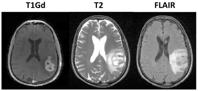
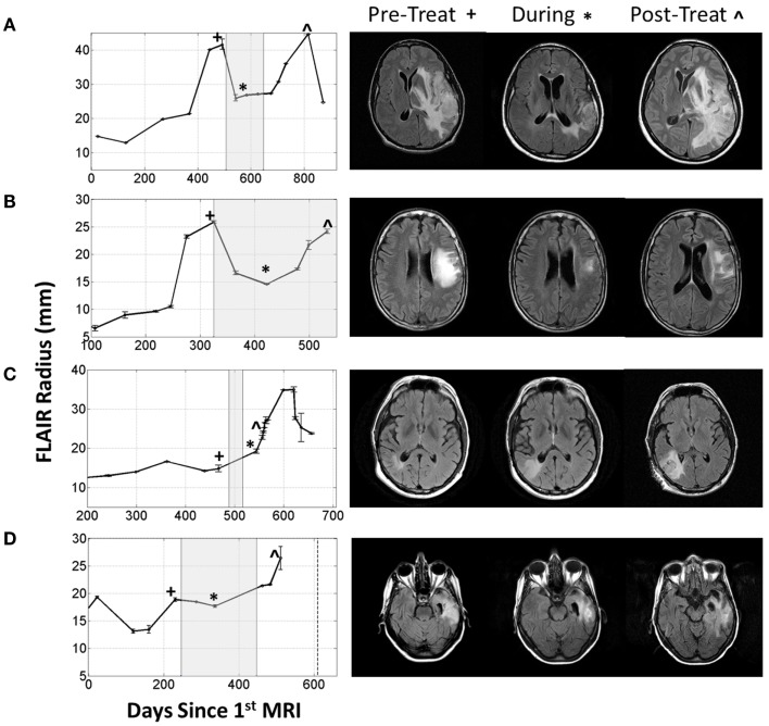
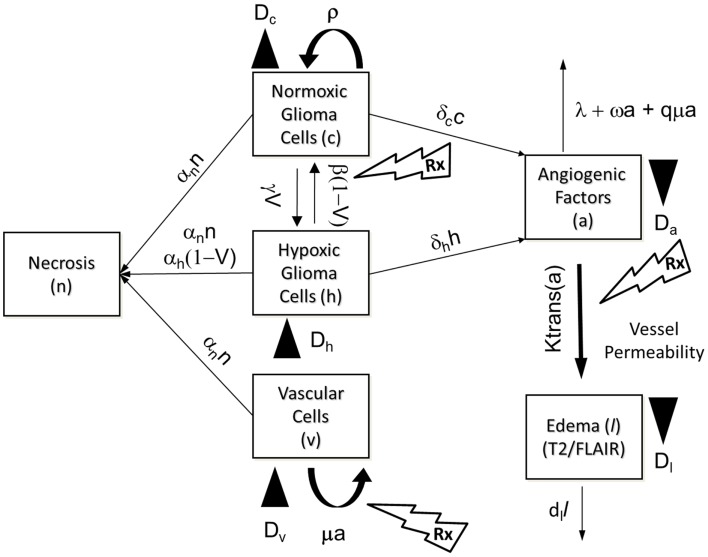
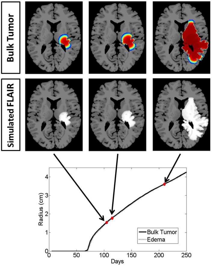
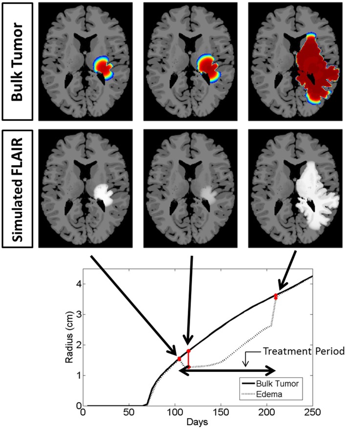
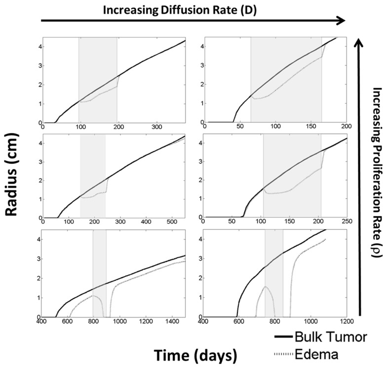
Glioblastoma, the most aggressive form of primary brain tumor, is predominantly assessed with gadolinium-enhanced T1-weighted (T1Gd) and T2-weighted magnetic resonance imaging (MRI). Pixel intensity enhancement on the T1Gd image is understood to correspond to the gadolinium contrast agent leaking from the tumor-induced neovasculature, while hyperintensity on the T2/FLAIR images corresponds with edema and infiltrated tumor cells. None of these modalities directly show tumor cells; rather, they capture abnormalities in the microenvironment caused by the presence of tumor cells. Thus, assessing disease response after treatments impacting the microenvironment remains challenging through the obscuring lens of MR imaging. Anti-angiogenic therapies have been used in the treatment of gliomas with spurious results ranging from no apparent response to significant imaging improvement with the potential for extremely diffuse patterns of tumor recurrence on imaging and autopsy. Anti-angiogenic treatment normalizes the vasculature, effectively decreasing vessel permeability and thus reducing tumor-induced edema, drastically altering T2-weighted MRI. We extend a previously developed mathematical model of glioma growth to explicitly incorporate edema formation allowing us to directly characterize and potentially predict the effects of anti-angiogenics on imageable tumor growth. A comparison of simulated glioma growth and imaging enhancement with and without bevacizumab supports the current understanding that anti-angiogenic treatment can serve as a surrogate for steroids and the clinically driven hypothesis that anti-angiogenic treatment may not have any significant effect on the growth dynamics of the overall tumor cell populations. However, the simulations do illustrate a potentially large impact on the level of edematous extracellular fluid, and thus on what would be imageable on T2/FLAIR MR. Additionally, by evaluating virtual tumors with varying growth kinetics, we see tumors with lower proliferation rates will have the most reduction in swelling from such treatments.
Keywords: anti-angiogenic therapy; edema; glioma; mathematical model.
Figures
Similar articles
-
Complementary but distinct roles for MRI and 18F-fluoromisonidazole PET in the assessment of human glioblastomas.J Nucl Med. 2009 Jan;50(1):36-44. doi: 10.2967/jnumed.108.055467. Epub 2008 Dec 17. J Nucl Med. 2009. PMID: 19091885 Free PMC article.
-
Relationship between contrast enhancement on fluid-attenuated inversion recovery MR sequences and signal intensity on T2-weighted MR images: visual evaluation of brain tumors.J Magn Reson Imaging. 2005 Jun;21(6):694-700. doi: 10.1002/jmri.20331. J Magn Reson Imaging. 2005. PMID: 15906343
-
Tumor invasion after treatment of glioblastoma with bevacizumab: radiographic and pathologic correlation in humans and mice.Neuro Oncol. 2010 Mar;12(3):233-42. doi: 10.1093/neuonc/nop027. Epub 2010 Jan 6. Neuro Oncol. 2010. PMID: 20167811 Free PMC article.
-
Advances in MRI assessment of gliomas and response to anti-VEGF therapy.Curr Neurol Neurosci Rep. 2011 Jun;11(3):336-44. doi: 10.1007/s11910-011-0179-x. Curr Neurol Neurosci Rep. 2011. PMID: 21234719 Free PMC article. Review.
-
Imaging biomarkers guided anti-angiogenic therapy for malignant gliomas.Neuroimage Clin. 2018 Jul 5;20:51-60. doi: 10.1016/j.nicl.2018.07.001. eCollection 2018. Neuroimage Clin. 2018. PMID: 30069427 Free PMC article. Review.
Cited by
-
A spatial model predicts that dispersal and cell turnover limit intratumour heterogeneity.Nature. 2015 Sep 10;525(7568):261-4. doi: 10.1038/nature14971. Epub 2015 Aug 26. Nature. 2015. PMID: 26308893 Free PMC article.
-
Recombinant interleukin-1 receptor antagonist conjugated to superparamagnetic iron oxide nanoparticles for theranostic targeting of experimental glioblastoma.Neoplasia. 2015 Jan;17(1):32-42. doi: 10.1016/j.neo.2014.11.001. Neoplasia. 2015. PMID: 25622897 Free PMC article.
-
Simulating cancer: computational models in oncology.Front Oncol. 2013 Sep 13;3:233. doi: 10.3389/fonc.2013.00233. eCollection 2013. Front Oncol. 2013. PMID: 24062986 Free PMC article. No abstract available.
-
ENvironmental Dynamics Underlying Responsive Extreme Survivors (ENDURES) of Glioblastoma: A Multidisciplinary Team-based, Multifactorial Analytical Approach.Am J Clin Oncol. 2019 Aug;42(8):655-661. doi: 10.1097/COC.0000000000000564. Am J Clin Oncol. 2019. PMID: 31343422 Free PMC article. Review.
-
Advanced magnetic resonance imaging of the physical processes in human glioblastoma.Cancer Res. 2014 Sep 1;74(17):4622-4637. doi: 10.1158/0008-5472.CAN-14-0383. Cancer Res. 2014. PMID: 25183787 Free PMC article. Review.
References
-
- Blinkov S. M., Glezer I. I. (1968). The Human Brain in Figures and Tables: A Quantitative Handbook. New York: Basic Books
Grants and funding
LinkOut - more resources
Full Text Sources
Other Literature Sources