

Mitochondrial myopathy: a rare cause of early-onset vocal fold atrophy
- PMID: 23577570
- PMCID: PMC3951151
- DOI: 10.1177/000348941312200306
Mitochondrial myopathy: a rare cause of early-onset vocal fold atrophy
Abstract
Objectives: We present the second published case of laryngeal involvement in mitochondrial myopathy.
Methods: A patient with laryngeal involvement of mitochondrial myopathy is presented, together with a literature review.
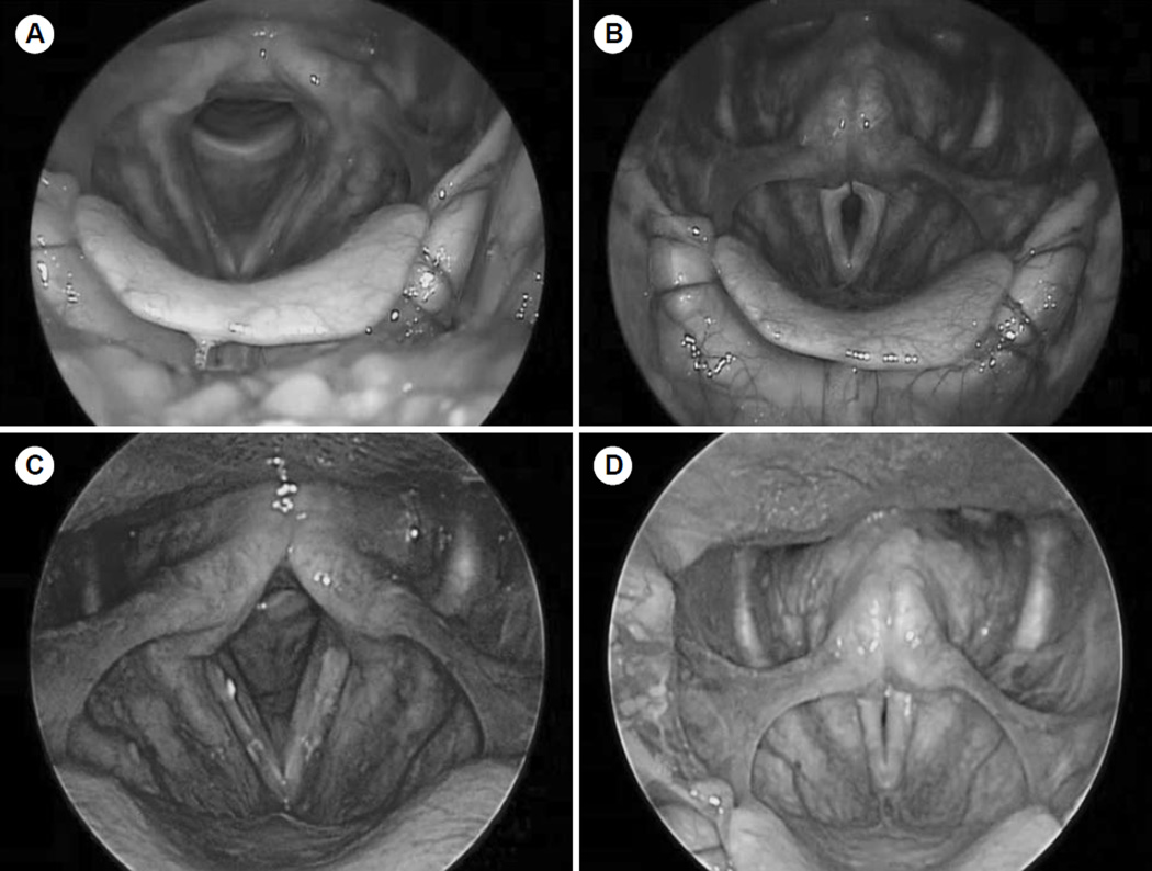
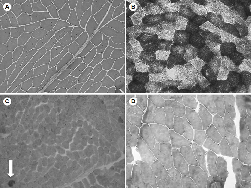
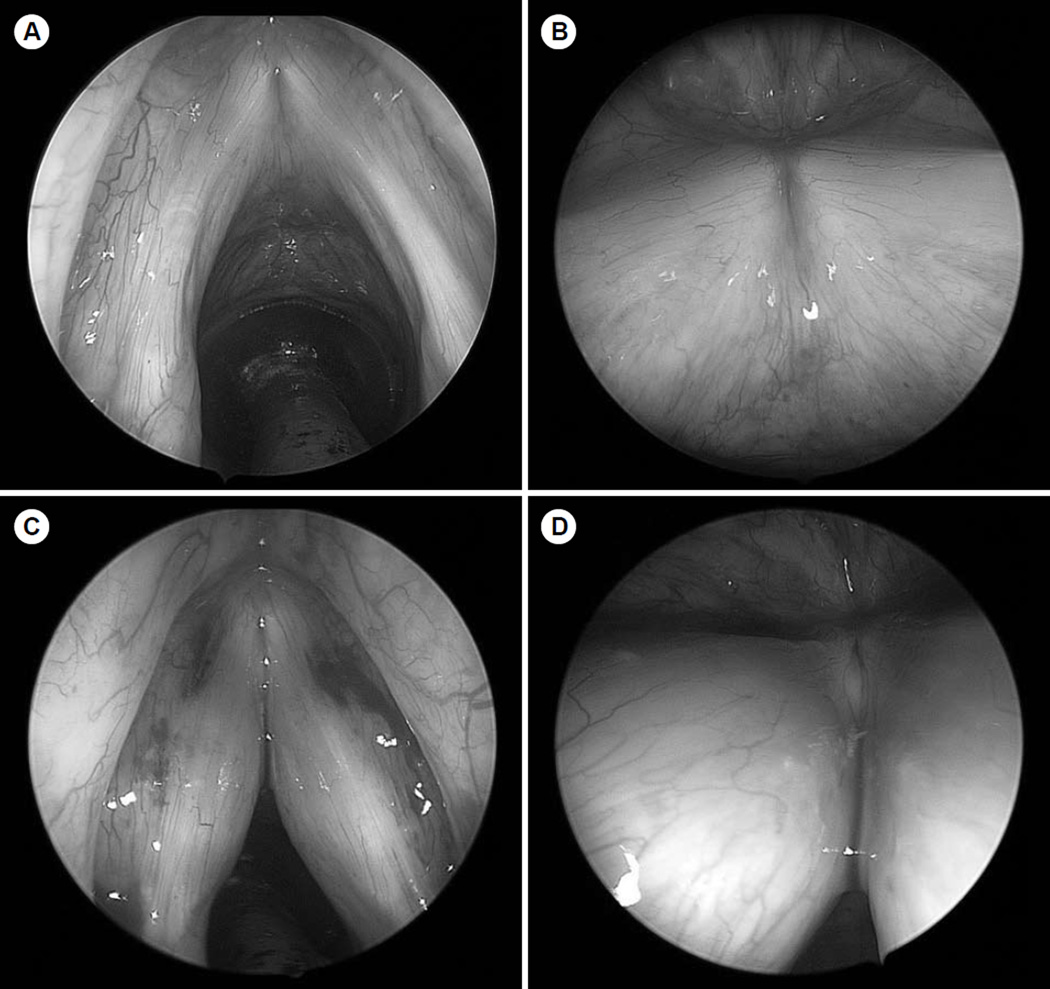
Results: A 41-year-old man presented with progressive breathy dysphonia. His brother had mitochondrial myopathy. Biopsy of the biceps muscle demonstrated cytochrome C oxidase-negative ragged blue fibers confirming mitochondrial myopathy. Videostroboscopy showed marked vocal fold atrophy, but subsequent injection laryngoplasty did not significantly improve the patient's voice, despite improved postoperative glottic closure.
Conclusions: Mitochondrial myopathy should be considered in the differential diagnosis of severe early-onset vocal fold atrophy.
Figures
References
-
- Woodson G. Management of neurologic disorders of the larynx. Ann Otol Rhinol Laryngol. 2008;117:317–326. - PubMed
-
- Hartley C, Ascott F. Laryngeal involvement in mitochondrial myopathy. J Laryngol Otol. 1994;108:685–687. - PubMed
-
- Cros D, Palliyath S, DiMauro S, Ramirez C, Shamsnia M, Wizer B. Respiratory failure revealing mitochondrial myopathy in adults. Chest. 1992;101:824–828. - PubMed
-
- Taylor RW, Schaefer AM, Barron MJ, McFarland R, Turnbull DM. The diagnosis of mitochondrial muscle disease. Neuromuscul Disord. 2004;14:237–245. - PubMed
Publication types
MeSH terms
Grants and funding
LinkOut - more resources
Full Text Sources
Other Literature Sources
Medical
