Review
. 2013:6:305-28.
doi: 10.1146/annurev-anchem-062012-092631. Epub 2013 Apr 1.

Structure determination of membrane proteins by nuclear magnetic resonance spectroscopy

Affiliations
Review

Structure determination of membrane proteins by nuclear magnetic resonance spectroscopy

Stanley J Opella. Annu Rev Anal Chem (Palo Alto Calif). 2013.

Abstract

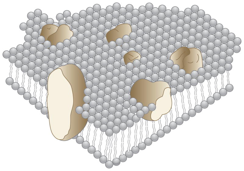
Many biological membranes consist of 50% or more (by weight) membrane proteins, which constitute approximately one-third of all proteins expressed in biological organisms. Helical membrane proteins function as receptors, enzymes, and transporters, among other unique cellular roles. Additionally, most drugs have membrane proteins as their receptors, notably the superfamily of G protein-coupled receptors with seven transmembrane helices. Determining the structures of membrane proteins is a daunting task because of the effects of the membrane environment; specifically, it has been difficult to combine biologically compatible environments with the requirements for the established methods of structure determination. There is strong motivation to determine the structures in their native phospholipid bilayer environment so that perturbations from nonnatural lipids and phases do not have to be taken into account. At present, the only method that can work with proteins in liquid crystalline phospholipid bilayers is solid-state NMR spectroscopy.


Figures

Figure 1
Figure 1
Schematic three-dimensional and cross-sectional views of membrane proteins embedded in a lipid matrix, illustrating the fluid mosaic model of biological membranes. The proteins appear to be randomly distributed but may form specific aggregates in order to function. Modified from Reference .
Figure 2
Figure 2
A model of a single-protein molecule in the membrane, viewed roughly parallel to the plane of the membrane. The top and bottom of the model correspond to the parts of the protein in contact with the solvent; the rest is in contact with the lipid. The most strongly tilted helices are in the foreground. Modified from Reference .
Figure 3
Figure 3
Comparison between α-carbon resonances in the 13C NMR spectra of two different proteins. (a) The membrane-bound form of fd coat protein solubilized in SDS (sodium dodecyl sulfate). (b) The enzyme lysozyme in an aqueous solution. Modified from Reference .
Figure 4
Figure 4
Experimental three-dimensional 1H chemical shift/15N–1H dipolar coupling/15N chemical shift correlation spectra of a model peptide. (a) Polycrystalline sample. (b) Single-crystal sample. (c) Diagram of tensors in a peptide plane. Modified from Reference .
Figure 5
Figure 5
Example of spectroscopic data for residue L31 obtained from magic angle spinning solid-state NMR spectra of uniformly 13C/15N-labeled MerFt in DMPC (dimyristoyl phosphatidylcholine) proteoliposomes at 25°C. (a) Two-dimensional 1H–15N dipolar coupling (DC)/13C shift separated local field (SLF) spectrum. (b) Two-dimensional 1H–15N DC/13C shift SLF spectra plane selected from a three-dimensional spectrum at an isotropic 15N chemical shift frequency of 118.6 ppm. The 1H–15N DC motionally averaged powder pattern for residue L31 has a perpendicular edge frequency of 4.7 kHz, corresponding to a splitting of 9.4 kHz, and a DC value of 18.8 kHz. (c) Two-dimensional 1H–13CαDC/13C shift SLF spectral plane selected from a three-dimensional spectrum at an isotropic 15N chemical shift frequency of 54.6 ppm. All three spectral planes are associated with residue L31. The dashed line traces the correlation among the frequencies, which were obtained from three separate experiments. The DC frequencies in the spectra correspond to the perpendicular edge frequencies of the corresponding powder patterns. Modified from Reference .
Figure 6
Figure 6
Three-dimensional structure of CXCR1. The backbone representation of CXCR1 shows helices (TM1–TM7 and H8) in aqua; extracellular loops (ECLs) in gray; and intracellular loops (ICLs) in blue (ICL1), green (ICL2), and red (ICL3). Disulfide-bonded cysteine (Cys) pairs (C30–C277 and C110–C187) are shown as sticks. (a) Side view. (b) View from the extracellular side. (c) View from the intracellular side. Modified from Reference .

References

LITERATURE CITED

    1. Sanders CR, Myers JK. Disease-related misassembly of membrane proteins. Annu Rev Biophys Biomol Struct. 2004;33:25–51. - PubMed
    1. Gilman AG. G proteins: transducers of receptor-generated signals. Annu Rev Biochem. 1987;56:615–49. - PubMed
    1. Page RC, Moore JD, Nguyen HB, Sharma M, Chase R, et al. Comprehensive evaluation of solution nuclear magnetic resonance spectroscopy sample preparation for helical integral membrane proteins. J Struct Funct Genomics. 2006;7:51–64. - PubMed
    1. McDermott AE. Structural and dynamic studies of proteins by solid-state NMR spectroscopy: rapid movement forward. Curr Opin Struct Biol. 2004;14:554–61. Reviews MAS methods for studying proteins. Although it is an early review, many of the methods it describes are still in use. - PubMed
    1. Opella SJ, Marassi FM. Structure determination of membrane proteins by NMR spectroscopy. Chem Rev. 2004;104:3587–606. - PMC - PubMed

RELATED RESOURCES

    1. Drorlist. http://www.drorlist.com. Lists membrane proteins with structures determined by NMR spectroscopy.

    1. Membrane Proteins of Known 3D Structure. http://blanco.biomol.uci.edu/mpstruc/listAll/list. Lists all membrane proteins with structures determined by all methods.

    1. Protein Data Bank. http://www.rcsb.org. Lists all protein structures.

    1. Biological Magnetic Resonance Data Bank. http://www.bmrb.wisc.edu. Contains NMR data for spectroscopic and structural analysis.

Publication types

MeSH terms

LinkOut - more resources