Review
. 2013;13(7):843-56.
doi: 10.2174/1568026611313070007.

Molecular modeling and ligand docking for solute carrier (SLC) transporters

Affiliations
Review

Molecular modeling and ligand docking for solute carrier (SLC) transporters

Avner Schlessinger et al. Curr Top Med Chem. 2013.

Abstract

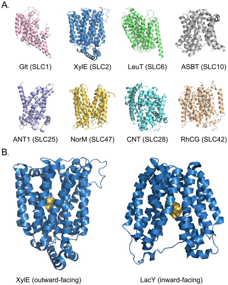
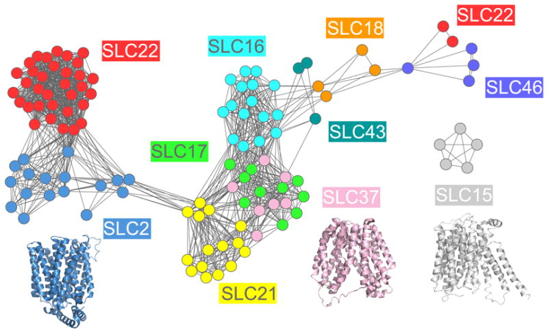
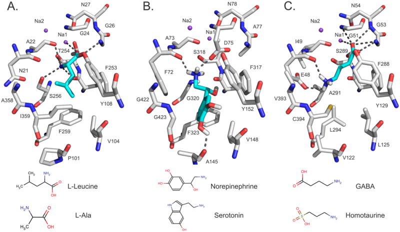
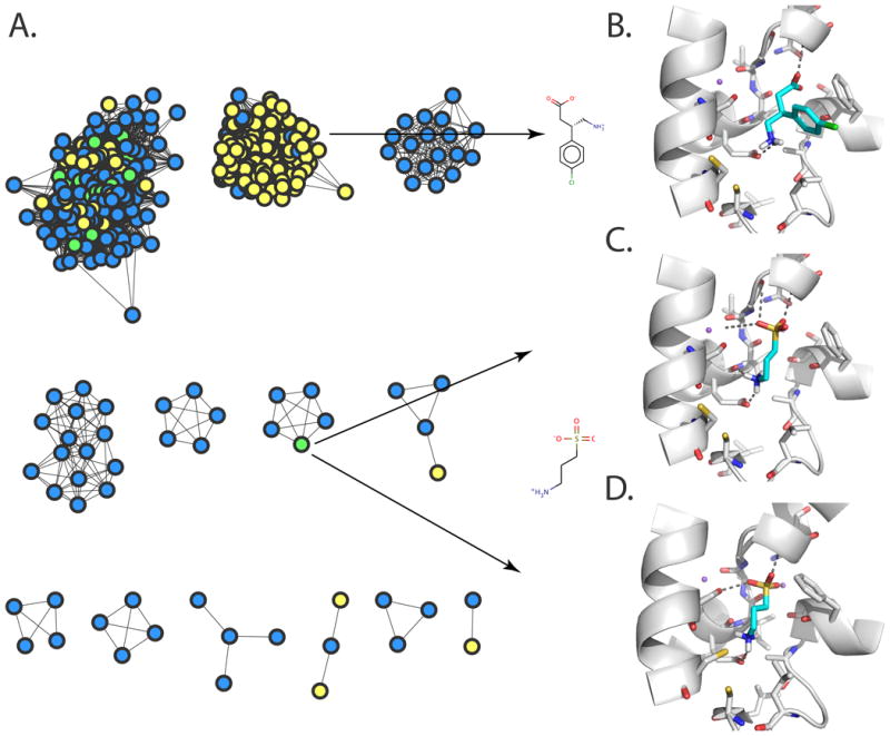
Solute Carrier (SLC) transporters are membrane proteins that transport solutes, such as ions, metabolites, peptides, and drugs, across biological membranes, using diverse energy coupling mechanisms. In human, there are 386 SLC transporters, many of which contribute to the absorption, distribution, metabolism, and excretion of drugs and/or can be targeted directly by therapeutics. Recent atomic structures of SLC transporters determined by X-ray crystallography and NMR spectroscopy have significantly expanded the applicability of structure-based prediction of SLC transporter ligands, by enabling both comparative modeling of additional SLC transporters and virtual screening of small molecules libraries against experimental structures as well as comparative models. In this review, we begin by describing computational tools, including sequence analysis, comparative modeling, and virtual screening, that are used to predict the structures and functions of membrane proteins such as SLC transporters. We then illustrate the applications of these tools to predicting ligand specificities of select SLC transporters, followed by experimental validation using uptake kinetic measurements and other assays. We conclude by discussing future directions in the discovery of the SLC transporter ligands.


Figures

Fig. 1
Fig. 1. Structures of SLC transporters
(A) Structures of SLC transporters representing select known structural classes and related human families. The structures are of the aspartate transporter Glt [153], the xylose transporter XylE [53], the leucine transporter LeuT [154], the sodium / bile acid symporter ASBT [155], the ADP/ATP translocase 1 ANT1 [156], the drug and toxin transporter NorM [142], the concentrative nucleoside transporter CNT [157], and the human rhesus glycoprotein RhCG ammonium transporter [23]. (B) The alternating access mechanism for structures with the MFS fold. The structures are of XylE in a ligand-bound outward-facing conformation (left) and the lactose permease LacY in a ligand-bound inward-facing conformation [54] (right). All structures are visualized with PyMol [158].
Fig. 2
Fig. 2. Sequence similarity network of human SLC families predicted to have the MFS fold
(A) The relationships between SLC sequences are visualized using Cytoscape 2.6.1 [51, 159]. (A) The nodes represent SLC sequences, including splice variants, that are similar to each other or to sequences of proteins with known MFS structures; the colors of the nodes indicate the SLC family [1]. The edges between the nodes correspond to a pairwise alignment with sequence identity of at least 10% and an E-value of less than 1 [51, 160]. The structures of XylE (blue) [53], LacY (pink) [54], and PEPTSO (grey) [55] are visualized using PyMol [158] and their colors are based on the color of the most similar human family in the similarity network.
Fig. 3
Fig. 3. Binding sites and modes of ligand binding in SLC6 members
(A) The X-ray structure of LeuT as well as the comparative models of (B) NET and (C) GAT-2, in the ligand-bound occluded conformation, are visualized with PyMol [158]. Atoms are displayed as sticks, with oxygen, nitrogen, and hydrogen atoms in red, blue, and white, respectively. The sodium ions Na1 and Na2 are visualized with purple spheres. The ligands L-Leucine, norepinephrine, and GABA are illustrated in cyan sticks and their hydrogen bonds with the binding-site residues of LeuT (Ala22, Gly26, Thr254, Ser256, and Na1), GAT-2 (Glu48, Gly51, Gly53, Asn54, and Na1), and NET (Ala145, Phe72, and Asp75) are displayed as dotted gray lines. Sketches of two representative ligands of each transporter are shown at the bottom.
Fig. 4
Fig. 4. Structure-based ligand prediction for GAT-2
(A) A network view of predicted GAT-2 ligand drugs and their similarities [91]. The nodes represent top small molecule hits predicted to bind GAT-2, using the occluded model (blue), the outward-facing model (yellow), or both models (green). Predicted structures of GAT-2 in the outward-facing (B and C) and occluded (D) conformations, in complex with the representative experimentally confirmed hits baclofen (B) and homotaurine (C and D). Small molecules ligands are colored in cyan, with oxygen, nitrogen, sulfur, and hydrogen atoms in red, blue, yellow, and white, respectively. The sodium ions Na1 and Na2 are visualized as purple spheres. The GAT-2 TMH regions are illustrated in white ribbons. Important residues for binding are depicted as sticks; predicted hydrogen bonds between ligands and GAT-2 are displayed as dotted gray lines.
Fig. 5
Fig. 5. Ligand discovery for MATE-1
(A) An interaction network for prescription drugs and SLC transporters in kidney [140]. SLC transporters are depicted as yellow diamonds. Positively charged drugs are shown as solid blue circles, negatively charged drugs as solid red circles, and uncharged drugs as gray circles. Edges between transporters and drugs correspond to known transporter-drug interactions. (B) A preliminary human MATE-1 comparative model based on the structure of its prokaryotic homolog NorM. The model is visualized using the Coulombic Surface Coloring in UCSF Chimera [161]. Negative and positive electrostatic potentials are illustrated in red and blue, respectively.

References

    1. Hediger MA, Romero MF, Peng JB, Rolfs A, Takanaga H, Bruford EA. The ABCs of solute carriers: physiological, pathological and therapeutic implications of human membrane transport proteinsIntroduction. Pflugers Arch. 2004;447(5):465–468. - PubMed
    1. Povey S, Lovering R, Bruford E, Wright M, Lush M, Wain H. The HUGO Gene Nomenclature Committee (HGNC) Hum Genet. 2001;109(6):678–680. - PubMed
    1. Saier MH., Jr A functional-phylogenetic classification system for transmembrane solute transporters. Microbiol Mol Biol Rev. 2000;64(2):354–411. - PMC - PubMed
    1. Saier MH, Jr, Yen MR, Noto K, Tamang DG, Elkan C. The Transporter Classification Database: recent advances. Nucleic Acids Res. 2009;37(Database issue):D274–278. - PMC - PubMed
    1. Forrest LR, Rudnick G. The rocking bundle: a mechanism for ion-coupled solute flux by symmetrical transporters. Physiology (Bethesda) 2009;24:377–386. - PMC - PubMed

Publication types