Review
. 2013 Sep;140(1):1-11.
doi: 10.1111/imm.12110.

Anti-Gal: an abundant human natural antibody of multiple pathogeneses and clinical benefits

Affiliations
Review

Anti-Gal: an abundant human natural antibody of multiple pathogeneses and clinical benefits

Uri Galili. Immunology. 2013 Sep.

Abstract

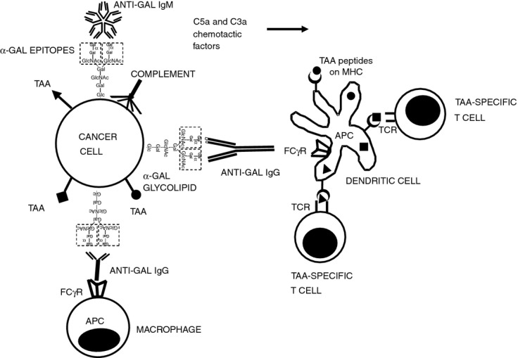
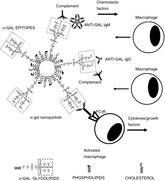
Anti-Gal is the most abundant natural antibody in humans, constituting ~ 1% of immunoglobulins. Anti-Gal is naturally produced also in apes and Old World monkeys. The ligand of anti-Gal is a carbohydrate antigen called the 'α-gal epitope' with the structure Galα1-3Galβ1-4GlcNAc-R. The α-gal epitope is present as a major carbohydrate antigen in non-primate mammals, prosimians and New World monkeys. Anti-Gal can contributes to several immunological pathogeneses. Anti-Gal IgE produced in some individuals causes allergies to meat and to the therapeutic monoclonal antibody cetuximab, all presenting α-gal epitopes. Aberrant expression of the α-gal epitope or of antigens mimicking it in humans may result in autoimmune processes, as in Graves' disease. α-Gal epitopes produced by Trypanosoma cruzi interact with anti-Gal and induce 'autoimmune like' inflammatory reactions in Chagas' disease. Anti-Gal IgM and IgG further mediate rejection of xenografts expressing α-gal epitopes. Because of its abundance, anti-Gal may be exploited for various clinical uses. It increases immunogenicity of microbial vaccines (e.g. influenza vaccine) presenting α-gal epitopes by targeting them for effective uptake by antigen-presenting cells. Tumour lesions are converted into vaccines against autologous tumour-associated antigens by intra-tumoral injection of α-gal glycolipids, which insert into tumour cell membranes. Anti-Gal binding to α-gal epitopes on tumour cells targets them for uptake by antigen-presenting cells. Accelerated wound healing is achieved by application of α-gal nanoparticles, which bind anti-Gal, activate complement, and recruit and activate macrophages that induce tissue regeneration. This therapy may be of further significance in regeneration of internally injured tissues such as ischaemic myocardium and injured nerves.

Keywords: anti-Gal; increased immunogenicity; tumour vaccine; wound healing; α-gal epitopes.


Figures

Figure 1
Figure 1
Insertion of α-gal glycolipids into cell membranes of injected tumours. α-Gal glycolipids are injected into solid tumours in the form of micelles in which the hydrophobic ceramide tails form the core of the micelle whereas the hydrophilic carbohydrate chains protrude into the surrounding aqueous environment. The glycolipids spontaneously insert into the outer phospholipid leaflet of tumour cell membranes because the hydrophobic tail of glycolipids is energetically much more stable when surrounded by the fatty acid tails of phospholipids in tumour cell membranes than within the micelle where they are surrounded by water molecules. This insertion results in presentation of multiple α-gal epitopes on the tumour cell membranes (described as phospholipid bilayer) and binding of the natural anti-Gal antibody molecules to these epitopes. The representative α-gal glycolipid has 10 carbohydrate units (ceramide decasaccharide) and two branches (antennae), each capped by an α-gal epitope marked by a dashed line rectangle.
Figure 2
Figure 2
Targeting of tumour cells to antigen-presenting cell (APC) by the natural anti-Gal antibody. Following insertion of injected α-gal glycolipids into tumour cell membranes both anti-Gal IgM and IgG antibodies (released from ruptured capillaries) bind to α-gal epitopes on the tumour cells. Binding of anti-Gal IgM results in effective complement activation and generation of the chemotactic complement cleavage peptides C5a and C3a, which induce rapid migration of APC (dendritic cells and macrophages) into the treated tumour. Following anti-Gal IgG binding to the α-gal epitopes on tumour cells (i.e. opsonization), its Fc portion interacts with Fcγ receptor (FcγR) on APC. This interaction stimulates APC to internalize intact or lysed tumour cells and the tumour-associated antigens (TAA) they carry. Internalized TAA are transported by the APC to regional lymph nodes, processed and various immunogenic TAA peptides (•, ▪, ▴) are presented by the APC in association with class I and class II MHC molecules. These immunogenic peptides interact with the corresponding T-cell receptors (TCR) and activate the multiple TAA-specific cytotoxic and helper T-cell clones that mediate a protective anti-tumour immune response.
Figure 3
Figure 3
Acceleration of wound healing by α-gal nanoparticles applied to wounds. Binding of the natural anti-Gal antibody to α-gal epitopes on α-gal nanoparticles activates the complement system within the treated wound. The resulting complement cleavage chemotactic factors induce rapid recruitment of macrophages into the wound. The recruited macrophages undergo activation as a result of interaction between the Fc ‘tails’ of anti-Gal immunocomplexed to the α-gal nanoparticles with Fcγ receptor (FcγR) on macrophages. The activated macrophages produce and secrete cytokines/growth factors that accelerate healing and may recruit stem cells. Symbols for α-gal glycolipids, phospholipids and cholesterol are illustrated at the bottom of the figure.

References

    1. Galili U, Rachmilewitz EA, Peleg A, Flechner I. A unique natural human IgG antibody with anti-α-galactosyl specificity. J Exp Med. 1984;160:1519–31. - PMC - PubMed
    1. Galili U, Mandrell RE, Hamadeh RM, Shohet SB, Griffis JM. Interaction between human natural anti-α-galactosyl immunoglobulin G and bacteria of the human flora. Infect Immun. 1988;56:1730–7. - PMC - PubMed
    1. Galili U, Anaraki F, Thall A, Hill-Black C, Radic M. One percent of circulating B lymphocytes are capable of producing the natural anti-Gal antibody. Blood. 1993;82:2485–93. - PubMed
    1. Wang L, Radic MZ, Galili U. Human anti-Gal heavy chain genes: preferential use of VH3 and the presence of somatic mutations. J Immunol. 1995;155:1276–85. - PubMed
    1. Kearns-Jonker M, Swensson J, Ghiuzeli C, et al. The human antibody response to porcine xenoantigens is encoded by IGHV3–11 and IGHV3–74 IgVH germline progenitors. J Immunol. 1999;163:4399–412. - PubMed