

Comparison of an all-inside suture technique with traditional pull-out suture and suture anchor repair techniques for flexor digitorum profundus attachment to bone
- PMID: 23578439
- PMCID: PMC4426886
- DOI: 10.1016/j.jhsa.2013.02.015
Comparison of an all-inside suture technique with traditional pull-out suture and suture anchor repair techniques for flexor digitorum profundus attachment to bone
Abstract
Purpose: One goal in repairing zone 1 flexor digitorum profundus (FDP) injuries is to create a tendon-bone construct strong enough to allow early rehabilitation while minimizing morbidity. This study compares an all-inside suture repair technique biomechanically with pull-out suture and double-suture anchor repairs.
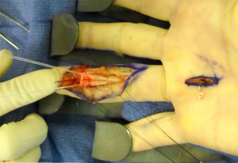
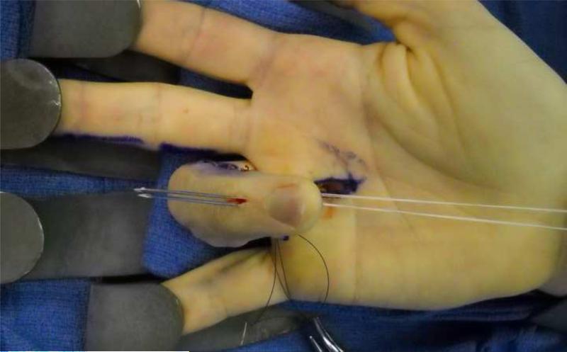
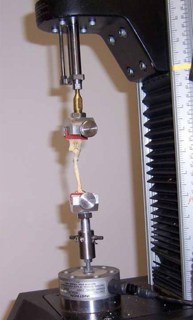
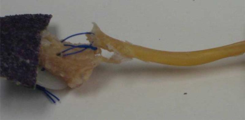
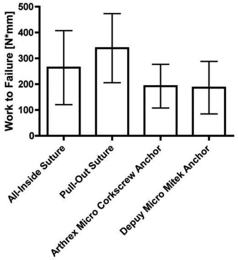
Methods: Repairs were performed on 30 cadaver fingers. In all-inside suture repairs (n = 8), the FDP tendon was attached to bone with two 3-0 Ethibond sutures and tied over the dorsal aspect of distal phalanx. Pull-out suture repairs (n = 8) were performed with 2-0 Prolene suture and tied over a dorsal button. There were 2 suture anchor repair groups: Arthrex Micro Corkscrew anchors preloaded with 2-0 FiberWire suture (n = 7) and Depuy Micro Mitek anchors preloaded with 3-0 Orthocord suture (n = 7). Repair constructs were tested using a servohydraulic materials testing system and loaded until the repair lost 75% of its strength.
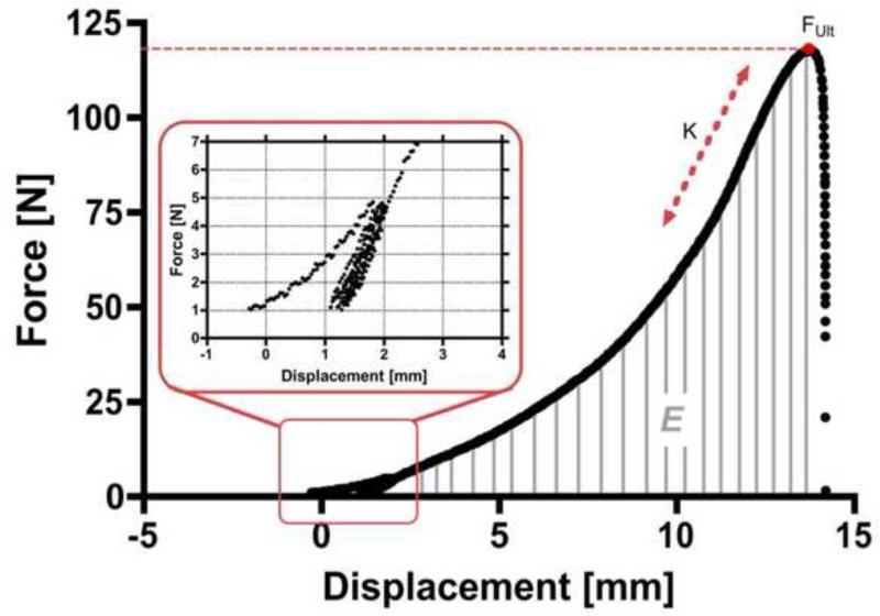
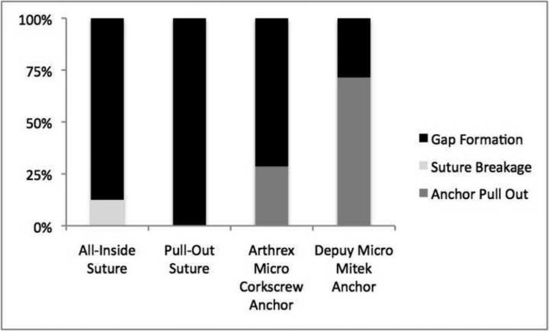
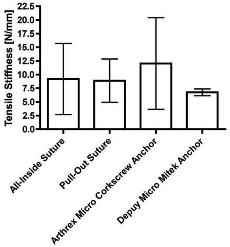
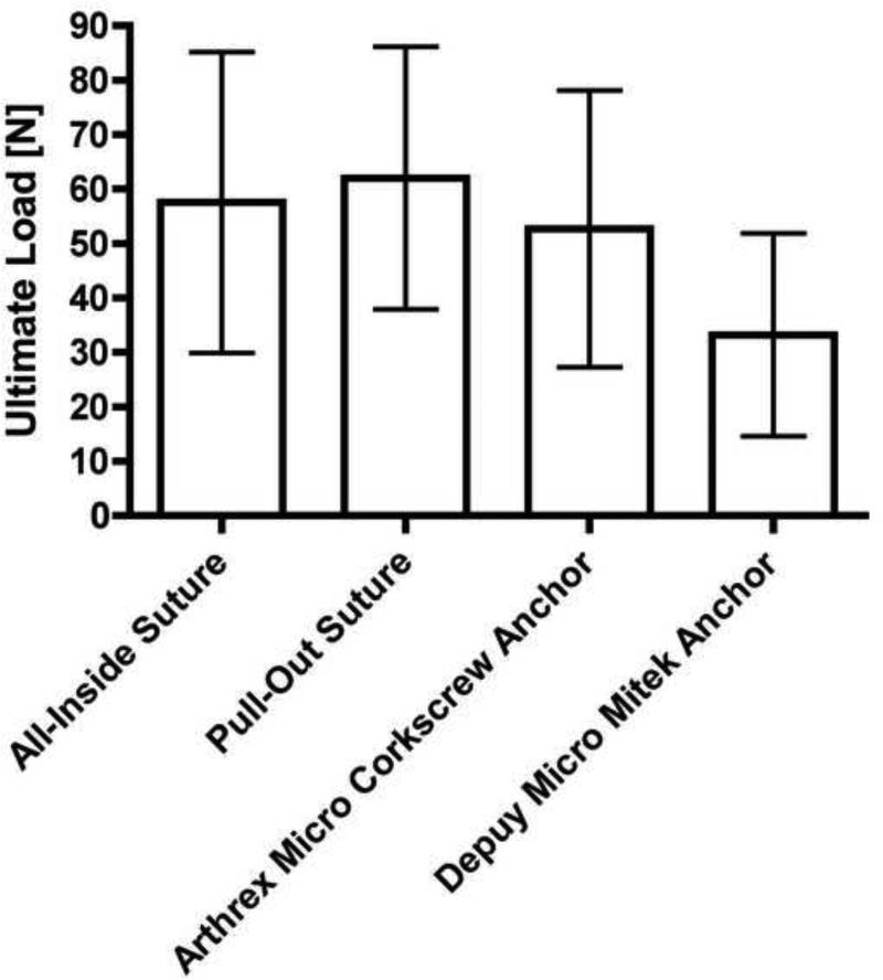
Results: There were no statistically significant differences in tensile stiffness, ultimate load, or work to failure between the repairs. Failure mode was suture stretch and gap formation greater than 2 mm at the repair site for all pull-out suture repairs and for 7 of 8 all-inside suture repairs. Two of the Arthrex Micro Corkscrew repairs and 5 of the Depuy Micro Mitek repairs failed by anchor pull-out.
Conclusions: This cadaveric biomechanical study showed no difference in tensile stiffness, ultimate load, and work to failures between an all-inside suture repair technique for zone 1 FDP repairs and previously described pull-out suture and suture anchor repair techniques. The all-inside suture technique also has the advantages of avoiding an external button and the cost of anchors. Therefore, it should be considered as an alternative to other techniques.
Clinical relevance: This study introduces a new FDP reattachment technique that avoids some of the shortcomings of current techniques.
Copyright © 2013 American Society for Surgery of the Hand. Published by Elsevier Inc. All rights reserved.
Figures
Similar articles
-
Effect of suture material and bone quality on the mechanical properties of zone I flexor tendon-bone reattachment with bone anchors.J Hand Surg Am. 2008 May-Jun;33(5):709-17. doi: 10.1016/j.jhsa.2008.01.025. J Hand Surg Am. 2008. PMID: 18590854 Free PMC article.
-
Biomechanical characteristics of suture anchor implants for flexor digitorum profundus repair.J Hand Surg Am. 2014 Feb;39(2):256-61. doi: 10.1016/j.jhsa.2013.11.023. J Hand Surg Am. 2014. PMID: 24480686
-
Flexor tendon repairs: the impact of fiberwire on grasping and locking core sutures.J Hand Surg Am. 2007 May-Jun;32(5):591-6. doi: 10.1016/j.jhsa.2007.03.003. J Hand Surg Am. 2007. PMID: 17481994
-
A Meta-Analysis of Biomechanical Studies for Suture Button Pullout Versus Suture Anchor Repair of Flexor Digitorum Profundus Avulsions.Hand (N Y). 2024 Jun;19(4):671-678. doi: 10.1177/15589447221126760. Epub 2022 Oct 5. Hand (N Y). 2024. PMID: 36196928 Free PMC article.
-
The Accommodation of Bone Anchors Within the Distal Phalanx for Repair of Flexor Digitorum Profundus Avulsions.J Hand Surg Am. 2019 Nov;44(11):986.e1-986.e6. doi: 10.1016/j.jhsa.2018.12.012. Epub 2019 Feb 15. J Hand Surg Am. 2019. PMID: 30777399 Review.
Cited by
-
Modified Pull-Out Technique for Zone One Flexor Digitorum Profundus Repair.Arch Bone Jt Surg. 2022 Nov;10(11):976-981. doi: 10.22038/ABJS.2022.65724.3146. Arch Bone Jt Surg. 2022. PMID: 36561223 Free PMC article.
-
Biomechanical Comparison of Flexor Digitorum Profundus Avulsion Repair.J Wrist Surg. 2019 Aug;8(4):312-316. doi: 10.1055/s-0039-1685470. Epub 2019 Apr 22. J Wrist Surg. 2019. PMID: 31402995 Free PMC article.
-
The Transosseous Internal Four Strand Technique: A New All-Inside Technique for Zone 1 Flexor Tendon Repairs.Hand (N Y). 2023 Jun;18(4):628-634. doi: 10.1177/15589447211060430. Epub 2021 Dec 28. Hand (N Y). 2023. PMID: 34963321 Free PMC article.
-
A review of mallet finger and jersey finger injuries in the athlete.Curr Rev Musculoskelet Med. 2017 Mar;10(1):1-9. doi: 10.1007/s12178-017-9395-6. Curr Rev Musculoskelet Med. 2017. PMID: 28188545 Free PMC article. Review.
-
Four Anchor Repair of Jersey Finger.Iowa Orthop J. 2021 Dec;41(2):95-100. Iowa Orthop J. 2021. PMID: 34924876 Free PMC article.
References
-
- McCallister WV, Ambrose HC, Katolik LI, Trumble TE. Comparison of pullout button versus suture anchor for zone I flexor tendon repair. J Hand Surg Am. 2006;31(2):246–251. - PubMed
-
- Bunnell S. Surgery of the Hand. Ed 2. Lippincott; Philadephia, PA: 1948. pp. 381–466.
-
- Guinard D, Montanier F, Thomas D, Corcella D, Moutet F. The Mantero flexor tendon repair in zone 1. J Hand Surg Br. 1999;24(2):148–151. - PubMed
-
- Brustein M, Pellegrini J, Choueka J, Heminger H, Mass D. Bone suture anchors versus the pullout button for repair of distal profundus tendon injuries: a comparison of strength in human cadaveric hands. J Hand Surg Am. 2001;26(3):489–496. - PubMed
-
- Silva MJ, Hollstien SB, Fayazi AH, Adler P, Gelberman RH, Boyer MI. The effects of multiple-strand suture techniques on the tensile properties of repair of the flexor digitorum profundus tendon to bone. J Bone Joint Surg Am. 1998;80(10):1507–1514. - PubMed
Publication types
MeSH terms
Substances
Grants and funding
LinkOut - more resources
Full Text Sources
Other Literature Sources
Medical
Miscellaneous
