. 2013 Jun:43:44-54.
doi: 10.1016/j.jaut.2013.03.002. Epub 2013 Apr 8.

Neuropsychiatric disease in murine lupus is dependent on the TWEAK/Fn14 pathway

Affiliations

Neuropsychiatric disease in murine lupus is dependent on the TWEAK/Fn14 pathway

Jing Wen et al. J Autoimmun. 2013 Jun.

Abstract

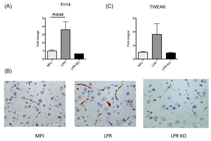
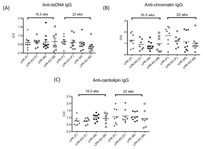
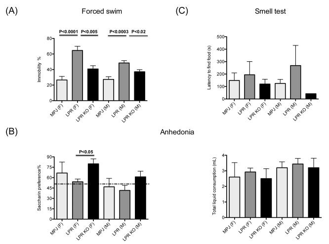
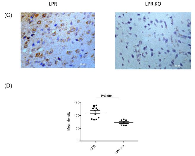
Given the early onset of neuropsychiatric disease and the potential response to immunosuppressive therapy, neuropsychiatric disease is considered a primary disease manifestation in systemic lupus erythematosus (SLE). However, the pathogenesis is not fully understood and optimal treatment has yet to be determined. TWEAK is a TNF family ligand that mediates pleotropic effects through its receptor Fn14, including the stimulation of inflammatory cytokine production by astrocytes, endothelial cells, and other non-hematopeotic cell types, and induction of neuronal death. Furthermore, TWEAK-inducible mediators are implicated in neuropsychiatric lupus. Thus, we hypothesized that the TWEAK/Fn14 pathway may be involved in the pathogenesis of neuropsychiatric SLE. We generated MRL-lpr/lpr (MRL/lpr) mice deficient for Fn14, the sole known signaling receptor for TWEAK. Neuropsychiatric disease was compared in age- and gender-matched MRL/lpr Fn14 wild type (WT) and knockout (KO) mice, using a comprehensive battery of neurobehavioral tests. We found that MRL/lpr Fn14WT mice displayed profound depression-like behavior as seen by increased immobility in a forced swim test and loss of preference for sweetened fluids, which were significantly ameliorated in Fn14KO mice. Similarly, MRL/lpr Fn14WT mice had impaired cognition, and this was significantly improved in Fn14KO mice. To determine the mechanism by which Fn14 deficiency ameliorates neuropsychiatric disease, we assessed the serum levels of autoantibodies and local expression of cytokines in the cortex and hippocampus of lupus mice. No significant differences were found in the serum levels of antibodies to nuclear antigens, or autoantibodies specifically associated with neuropsychiatric disease, between MRL/lpr Fn14WT and KO mice. However, MRL/lpr Fn14KO mice had significantly decreased brain expression of RANTES, C3, and other proinflammatory mediators. Furthermore, MRL/lpr Fn14KO mice displayed improved blood brain barrier integrity. In conclusion, several central manifestations of neuropsychiatric lupus, including depression-like behavior and altered cognition, are normalized in MRL/lpr mice lacking Fn14. Our results are the first to indicate a role for the TWEAK/Fn14 pathway in the pathogenesis of neuropsychiatric lupus, and suggest this ligand-receptor pair as a potential therapeutic target for a common and dangerous disease manifestation.


Figures

Figure 1
Figure 1. Fn14 expression is upregulated in brains of MRL/lpr mice
Fn14 (A, B) and TWEAK (C) expression were measured in cortical tissue of 14 week old female MRL/+ (“MPJ”) (n=4), MRL/lpr Fn14WT (“LPR”) (n=4), and MRL/lpr Fn14KO (“LPR KO”) (n=3) by real-time PCR (A,C) and Fn14 immunohistochemistry (B). For the PCR, fold changes were calculated in reference to the control MRL/+ mice, whose mean value was set at 1. In (B), representative images of Fn14 staining in frontal cortical tissue from MRL/+, MRL/lpr Fn14WT, and MRL/lpr Fn14KO (“LPR KO”) mice are shown. The arrows in the panels point to endothelial cells. Sections were examined using a Zeiss AxioObserver at 40X magnification.
Figure 2
Figure 2. Titers of serum autoantibodies are similar in MRL/lpr Fn14WT and KO mice
Titers of serum IgG anti-dsDNA (A), anti-chromatin (B), anti-cardiolipin (C), anti-ribosomal P (D), and anti-NMDA receptor (E) antibodies in MRL/lpr Fn14WT and KO mice at 16.5 and 22 weeks of age were determined by ELISA, as described in the Materials and methods.
Figure 3
Figure 3. Depression like behavior is ameliorated in MRL/lpr Fn14KO mice
(A) Forced swim test was performed on 10-20 week old MRL/+ (“MPJ”)(10 females (F), 10 males (M)), MRL/lpr Fn14WT (“LPR”) (10F/14M) and MRL/lpr Fn14KO (“LPR KO”)(8F/13M) mice. (B) Lack of preference for sweetened liquids (anhedonia) was measured by comparing the amount of fluid intake in individual 10-20 week old MRL/+ (“MPJ”) (5F/5M), MRL/lpr Fn14WT (“LPR”) (8F/9M), and MRL/lpr Fn14KO (“LPR KO”) (6F/6M) mice given a choice of drinking from two identical water bottles, one containing artificially sweetened water (left panel). Normal mice are expected to have >50% preference for sweetened fluid (above the dotted line). The right panel shows the total amount of fluid (sweetened and unsweetened) consumed by the mice in each group in this experiment. (C) Olfactory competency was assessed in 10-20 week old MRL/+ (“MPJ”)(5F/5M), MRL/lpr Fn14WT (“LPR”) (5F/2M) and MRL/lpr Fn14KO (“LPR KO”) (4F/1M) mice by measuring the time to uncover buried food. Subsequent figures utilize the same designations for each genotype, with the (F) and (M) label after the genotype in the figure legend denoting female and male mice, respectively.
Figure 4
Figure 4. Cognitive deficits in the object placement test in MRL/lpr mice are reversed by Fn14 deficiency
In social preference (A), the % of the time 10-20 week old MRL/+ (10F/10M), MRL/lpr Fn14WT (10F/14M) and MRL/lpr Fn14KO (8F/13M) mice spend interacting with another mouse relative to an inanimate object is measured. In the object placement test (B), the relative preference of MRL/+, MRL/lpr Fn14WT and KO mice to explore a familiar object in a novel location was measured. At 10-20 weeks of age, the number of mice in the MRL/+, MRL/lpr Fn14WT and MRL/lpr Fn14KO groups were 5F/5M, 3F/7M, and 2F/6M, respectively. In mice 21-40 weeks of age, the number of mice in the MRL/+, MRL/lpr Fn14WT, and MRL/lpr Fn14KO groups were 9F/10M, 7F/7M, and 5F/6M, respectively. Only for the 10-20 week old mice in the object placement test were the MRL/+ mice tested at a different time than the MRL/lpr mice, for technical reasons. Otherwise, the MRL/+, MRL/lpr Fn14WT, and MRL/lpr Fn14KO mice groups had their neurobehavioral assessments done concurrently. In object recognition (C), the relative preference of MRL/lpr Fn14WT and KO mice to explore a novel object was measured. At 10-20 weeks of age, the number of mice in the MRL/+, MRL/lpr Fn14WT and MRL/lpr Fn14KO groups was 5F/5M, 8F/9M, and 6F/6M, respectively. In mice 21-40 weeks of age, the number of mice in the MRL/+, MRL/lpr Fn14WT, and MRL/lpr Fn14KO groups were 10F/10M, 7F/7M, and 4F/7M, respectively. The dotted line in panels A-C denotes normal (>50% preference).
Figure 5
Figure 5. General locomotor activity and motor coordination are preserved and do not differ between MRL/lpr Fn14WT and KO mice
(A-C) General locomotor activity was assessed in individual 10-20 week old MRL/+ (10F/9M), MRL/lpr Fn14WT (10F/12M), and MRL/lpr Fn14KO (8F/10M) mice for 6 minutes in an 39 × ***39 cm open field, with the center 15 × ***15 cm defined as the center zone. Total (A) and center (C) track length and the time spent in the center zone (B) were analyzed by Viewer software. (D) Motor coordination was assessed by crossing a 100 cm long, 1.5 cm diameter balance beam, with the number of slips defined as at least one of the paws passing below the midline of the beam.
Figure 6
Figure 6. Fn14 deficiency decreases expression of RANTES, C3 and CXCL11 in the brain of MRL/lpr mice
(A) The gene expression of RANTES, C3 and CXCL11 in the cortex of 19 week old, female and male MRL/lpr Fn14WT and Fn14KO mice was measured by real-time PCR, and normalized to GAPDH. For the PCR, fold changes were calculated in reference to the MRL/lpr Fn14KO mice, whose mean value was set at 1. (B) The gene expression of RANTES, C3 and CXCL11 in the whole brain of 40 week old, female and male MRL/lpr Fn14WT and Fn14KO mice was measured by real-time PCR, and normalized to GAPDH. For the PCR, fold changes were calculated in reference to the MRL/lpr Fn14KO mice, whose mean value was set at 1. (C) Representative images of the immunohistochemical staining for RANTES in the frontal cortex of female MRL/lpr Fn14WT and Fn14KO mice at 40 weeks of age. Sections were examined using a Zeiss AxioObserver at 40X magnification. (D) RANTES staining quantitation was performed by Image J software. Staining intensity in 10 randomly selected areas was quantitated in each section, and the average of the density for each section was then calculated. Female mice, full dots; Male mice, open dots.
Figure 7
Figure 7. MRL/lpr Fn14KO mice display improved BBB permeability
The permeability of the blood brain barrier in individual 10-20 week old female MRL/lpr Fn14KO (n=6) and MRL/lpr Fn14WT (n=6) mice was assessed by calculating the albumin quotient, defined as cerebrospinal fluid (CSF) albumin/serum albumin. Intrathecal IgG synthesis was studied by calculating the IgG index, defined as IgG index = (CSF IgG/serum IgG)/albumin quotient). (A) Albumin quotient. (B) IgG ratio (CSF IgG/serum IgG). (C) IgG index. (D) Anti-dsDNA antibody titers in CSF obtained from 15-23 week old female MRL/lpr Fn14KO and WT mice were measured as described in the Materials and methods. Age and gender matched MRL/+ mice served as controls. Fold change was calculated relative to the OD values in MRL/+ control mice, which were normalized to a value of 1.

References

    1. The American College of Rheumatology nomenclature and case definitions for neuropsychiatric lupus syndromes. Arthritis Rheum. 1999;42:599–608. - PubMed
    1. Gulinello M, Putterman C. The MRL/lpr mouse strain as a model for neuropsychiatric systemic lupus erythematosus. J Biomed Biotechnol. 2011;2011:207504. - PMC - PubMed
    1. Petri M, Naqibuddin M, Carson KA, Wallace DJ, Weisman MH, Holliday SL, et al. Depression and cognitive impairment in newly diagnosed systemic lupus erythematosus. J Rheumatol. 2010;37:2032–38. - PubMed
    1. Sakic B, Denburg JA, Denburg SD, Szechtman H. Blunted sensitivity to sucrose in autoimmune MRL-lpr mice: a curve-shift study. Brain Res Bull. 1996;41:305–11. - PubMed
    1. Diamond B, Huerta PT, Mina-Osorio P, Kowal C, Volpe BT. Losing your nerves? Maybe it’s the antibodies. Nature Rev Immunol. 2009;9:449–56. - PMC - PubMed

Publication types

MeSH terms