. 2013 Apr 11;4(4):e583.
doi: 10.1038/cddis.2013.98.

Menin mediates epigenetic regulation via histone H3 lysine 9 methylation

Affiliations

Menin mediates epigenetic regulation via histone H3 lysine 9 methylation

Y-J Yang et al. Cell Death Dis. .

Abstract

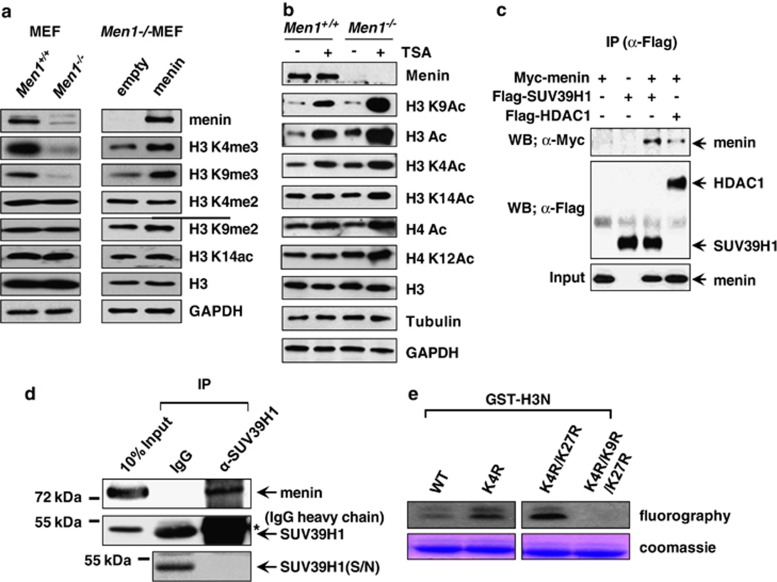
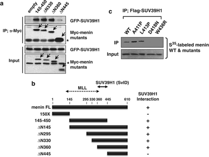
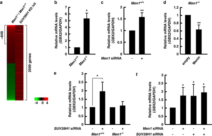
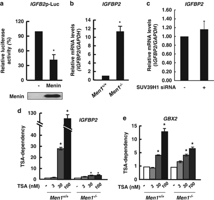
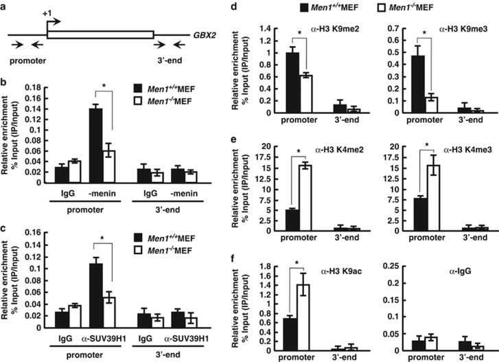
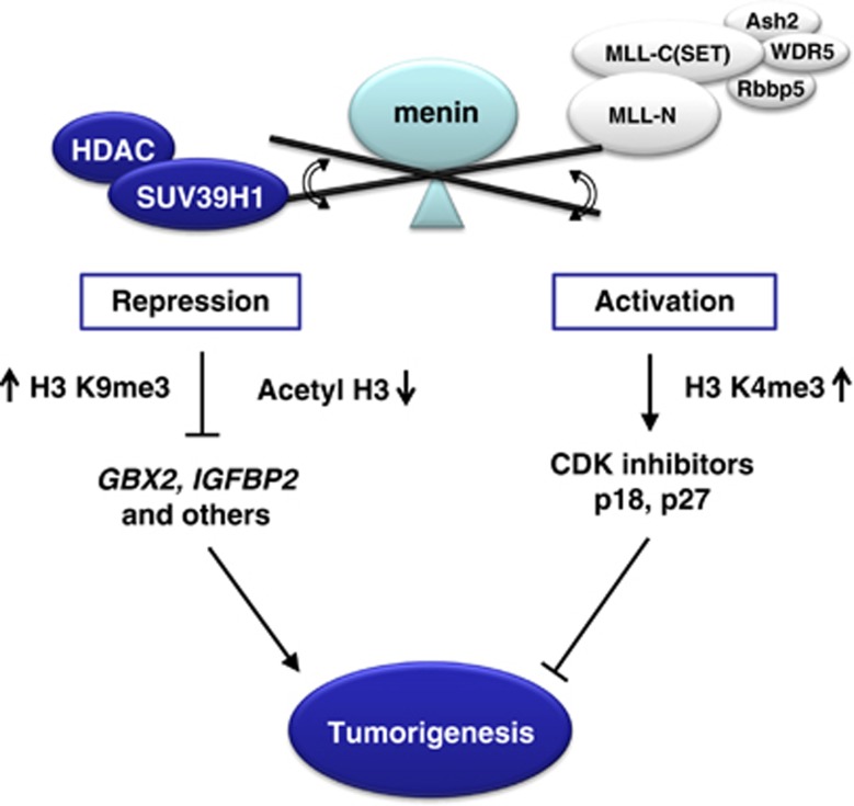
Menin, encoded by the multiple endocrine neoplasia type 1 (MEN1) gene, is a tumor suppressor that leads to multiple endocrine tumors upon loss of its function. Menin functions as a transcriptional activator by tethering MLL complex to mediate histone H3 K4 methylation. It also functions as a repressor. However, the molecular mechanism of how menin contributes to the opposite outcome in gene expression is largely unknown. Here, we investigated the role of menin in the epigenetic regulation of transcription mediated by histone covalent modification. We show that the global methylation level of histone H3 K9, as well as H3 K4, was decreased in Men1(-/-) MEF cells. Consistently, menin was able to interact with the suppressor of variegation 3-9 homolog family protein, SUV39H1, to mediate H3 K9 methylation. This interaction decreased when patient-derived MEN1 mutation was introduced into the SUV39H1-interaction domain. We show that menin mediated different chromatin changes depending on target genes. Chromatin immunoprecipitation studies showed that menin directly associated with the GBX2 promoter and menin-dependent recruitment of SUV39H1 was essential for chromatin remodeling and transcriptional regulation. These results provide a molecular basis of how menin functions as a transcriptional repressor and suggest that menin-dependent integration of H3 K9 methylation might play an important role in preventing tumors.


Figures

Figure 1
Figure 1
Menin specifically associates with SUV39H1. (a) Whole-cell lysates of Men1+/+and Men1/MEFs were used for western blot analysis with antibodies against menin, H3K4me2, H3K4me3, H3K9me2, H3K9me3, H3K14ac, H3, and GAPDH (left panel). H3 and GAPDH served as loading controls. Men1/ MEF cells were infected with control or menin-expressing retroviruses. Cell lysates from the infected cells were subjected to western blot analysis (right panel). (b) Menin's effect on histone covalent modification. MEF cells were treated with TSA (100 nℳ) for 24 h and whole-cell extract was prepared for western blot analysis. H3, Tubulin, and GAPDH are loading controls. (c) 293T cells were transfected with Flag-SUV39H1, Flag-HDAC1, and Myc-menin expression vectors as indicated. Whole-cell extracts were immunoprecipitated with anti-Flag antibody, and the immunoprecipitates were analyzed by western blotting using anti-Flag and anti-Myc antibodies. (d) Co-immunoprecipitation assay was performed to detect interaction between endogenous proteins in 293T cells. Anti-SUV39H1 antibody was used for immunoprecipitation. The asterisks * denote cross-reactive IgG heavy chain bands. As SUV39H1 co-migrates with and is masked by IgG heavy chain on SDS-PAGE, the supernatant fraction remained after IP is shown in parallel to show that SUV39H1 was efficiently precipitated by its antibody. (e) In vitro HMT activity associated with menin. HMT reaction was performed with menin immunoprecipitates and [3H]-SAM. Each reaction contains bacterially expressed GST-H3N (residues 1–57) wild-type (wt) or mutants (K4R, K4R/K27R and K4R/K9R/K27R) as indicated. Proteins were resolved on a SDS-polyacrylamide gel. The gel was stained with Coomassie (bottom) and exposed to film for fluorography (top)
Figure 2
Figure 2
Mapping of HMT-binding domains in menin. (a) 293T cells were transfected with Myc-menin- and GFP-SUV39H1-expressing vectors. Whole-cell extracts were immunoprecipitated with anti-Myc antibody, and the immunoprecipitates were analyzed by western blotting with antibodies against GFP, or Myc epitope. The asterisks * denote nonspecific cross-reactive bands. (b) Schematic diagram showing association of SUV39H1 with the indicated versions of menin. MLL-binding region is denoted. (c) Each35[S]-labeled menin mutant derived from MEN1 patients was mixed with Flag-SUV39H1-expressing cell lysates and subjected to IP with anti-Flag antibody. Proteins were resolved by SDS-PAGE, and their interaction was detected by autoradiography. (A, alanine; P, proline; D, aspartic acid; N, asparagine; L, leucine; W, tryptophan; R, arginine)
Figure 3
Figure 3
Menin and SUV39H1 contribute to GBX2 repression. (a) The cDNA microarray analysis of MEF cells reveals common targets of menin and SUV39H1. Commonly upregulated genes by depletion of menin (Men//Men+/+) and SUV39H1 (SUV39H1 siRNA/control) were indicated. Expression of GBX2 was measured using qRT-PCR in Men1+/+ or Men1/ MEFs (b) as well as in MEFs that had been treated with Men1 siRNA (c). (d) The level of GBX2 mRNA was analyzed in Men1/ MEFs infected with retroviral vectors expressing empty (control) or menin. (e) Knockdown of SUV39H1 increased the mRNA level of GBX2 in Men1+/+MEF cells, but not in Men1/ MEF cells. Men1+/+ and Men1/ MEF cells were treated for 1 day with siRNA specifically targeting SUV39H1 and total RNA was isolated to detect the steady state level of GBX2 by qRT-PCR. (f) Double knockdown of both menin and SUV39H1 did not have an additive effect on GBX2 mRNA level. The PCR values were normalized for GAPDH and presented as relative values by considering GBX2/GAPDH of control as 1. Error bars represent S.D., n=3. Significance of differences was evaluated (*P-value<0.05, **P-value<0.005)
Figure 4
Figure 4
Epigenetic regulation of IGFBP2 is mediated by menin via HDAC. (a) Menin represses IGFBP2 promoter activity. The 293T cells were transfected with the IGFBP2 promoter-luciferase reporter and epitope-tagged menin or empty vector control. After incubation for 24 h, luciferase activity was measured. Each transfection was performed in duplicate and repeated three times. The means±S.D. of duplicate determinations from three separate experiments are shown. The expression level of transfected menin is shown at the bottom. (b) Increased level of genomic IGFBP2 expression in Men1/ MEFs was confirmed using qRT-PCR. (c) Men1+/+ MEFs cells were treated with siRNA targeting SUV39H1. The expression level of IGFBP2 was analyzed by qRT-PCR. Error bars represent S.D., n=3. Significance of differences was evaluated (*P-value<0.05). (d and e) Men1+/+ and Men1/ MEF cells were treated with TSA for 24 h with increasing dose as indicated. Total RNA was prepared to perform qRT-PCR. The expression level of IGFBP2 and GBX2 were normalized by GAPDH. TSA-dependency was shown as the fold difference between samples by considering expression levels obtained from each Men1+/+ and Men1/ MEF cells without TSA treatment as 1, respectively. Error bars represent S.D., n=3 (*P-value<0.05)
Figure 5
Figure 5
Menin recruits SUV39H1 to promote trimethylation of H3K9 around the GBX2 locus. (a) GBX2 locus showing regions subjected to ChIP analysis. (b and c) Recruitment of menin and SUV39H1 along the GBX2 locus was analyzed by ChIP assay. Chromatin solution was prepared from Men1+/+ and Men1/ cells and subjected to ChIP using control rabbit IgG and the antibodies against endogenous menin (anti-menin Ab) (b) or SUV39H1(anti-SUV39H1 Ab) (c). Immunoprecipitated DNA was analyzed in duplicates by quantitative real-time PCR with primers shown above (a) and the relative enrichment of proteins was shown. (df) ChIP assay with Men1+/+ and Men1/ MEFs using antibodies against indicated H3 modifications was performed to measure the relative levels of H3 K4 or H3 K9 di-, trimethylation, and acetylation along the GBX2. The data represent the percentage of ChIP (IP/input) and error bars indicate the S.D., n=3. Significance of differences was evaluated (*P-value<0.05)
Figure 6
Figure 6
A model for a dual role of menin in gene regulation during tumorigenic pathways. Menin may function as a transcriptional activator or repressor in a context-dependent manner, by selective mediation of chromatin remodeling, which provides an efficient mechanism for the regulation of gene expression and cell tumorigenesis

References

    1. Chandrasekharappa SC, Guru SC, Manickam P, Olufemi SE, Collins FS, EmmertBuck MR, et al. Positional cloning of the gene for multiple endocrine neoplasia-type 1. Science. 1997;276:404–407. - PubMed
    1. Chandrasekharappa SC, Teh BT. Functional studies of the MEN1 gene. J Intern Med. 2003;253:606–615. - PubMed
    1. Bertolino P, Radovanovic I, Casse H, Aguzzi A, Wang ZQ, Zhang CX. Genetic ablation of the tumor suppresor menin causes lethality at mid-gestation with defects in multiple organs. Mech Dev. 2003;120:549–560. - PubMed
    1. Crabtree JS, Scacheri PC, Ward JM, Garrett-Beal L, Emmert-Buck MR, Edgemon KA, et al. A mouse model of multiple endocrine neoplasia, type 1, develops multiple endocrine tumors. Proc Natl Acad Sci USA. 2001;98:1118–1123. - PMC - PubMed
    1. Yang Y, Hua X. In search of tumor suppressing functions of menin. Mol Cell Endocrinol. 2007;265:34–41. - PMC - PubMed

Publication types

MeSH terms

LinkOut - more resources