

doi: 10.1126/science.1233936.
Epub 2013 Apr 11.
Self-assembling cages from coiled-coil peptide modules
Affiliations
- PMID: 23579496
- PMCID: PMC6485442
- DOI: 10.1126/science.1233936
Item in Clipboard
Self-assembling cages from coiled-coil peptide modules
Science.
.
Abstract
An ability to mimic the boundaries of biological compartments would improve our understanding of self-assembly and provide routes to new materials for the delivery of drugs and biologicals and the development of protocells. We show that short designed peptides can be combined to form unilamellar spheres approximately 100 nanometers in diameter. The design comprises two, noncovalent, heterodimeric and homotrimeric coiled-coil bundles. These are joined back to back to render two complementary hubs, which when mixed form hexagonal networks that close to form cages. This design strategy offers control over chemistry, self-assembly, reversibility, and size of such particles.
Figures
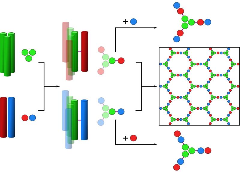
Left to right: Homotrimeric coiled coil (CC-Tri3, green) and heterodimeric coiled coils (CC-Di-AB); the latter comprises CC-Di-A and CC-Di-B, colored red and blue, respectively. CC-Tri and CC-Di-AB are linked via asymmetric disulfide bonds (purple lines) to render hub A (green-red) and hub B (green-blue). Mixing hub A with CC-Di-B, or hub B with CC-Di-A produces discrete 9-helix assemblies; whereas, mixing the hubs directly produces a hexagonal network, which should close.
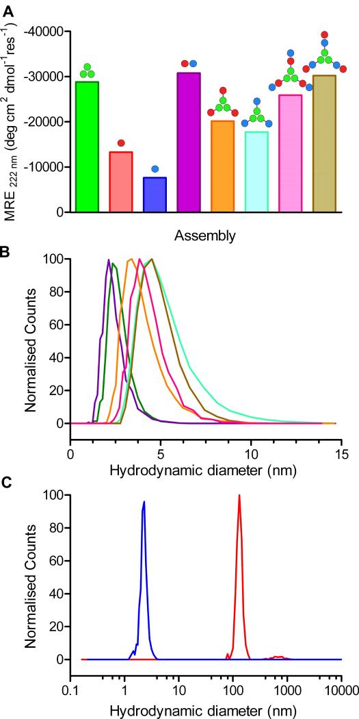
(A) Mean Residue Ellipticity (MRE) at 222 nm (MRE222) from CD spectroscopy in phosphate-buffered saline (PBS) at 20 °C for: CC-Tri3 (50 μM, green); CC-Di-A (50 μM, red); CC-Di-B (50 μM, blue); the mixture of CC-Di-A and CC-Di-B, i.e., CC-Di-AB, (50 μM + 50 μM, purple); hub A (CC-Tri3—CC-Di-A, 50 μM, orange); hub B (CC-Tri3—CC-Di-B, 50 μM, cyan); hub A plus CC-Di-B (50 μM + 50 μM, pink); hub B plus CC-Di-A (50 μM + 50 μM, brown). Full CD spectra and thermal denaturation curves are provided in Fig. S3 (19). The leucine-zipper peptide, GCN4-p1, gives a benchmark MRE222 for 100% α-helix of ≈ -36,000 deg cm2 dmol−1 res−1 (24). (B) Hydrodynamic diameters of the coiled-coil modules, hubs and 9-chain terminated assemblies as determined by dynamic light scattering; colors as per panel A. Expanded DLS data are given in Fig. S5. (C) DLS data for the assembled SAGE particles before (red) and after (blue) treatment with TCEP (tris(2-carboxyethyl)phosphine).
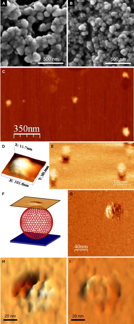
(A) Images recorded after mixing hub A and hub B in PBS, pH 7.4 (to final concentrations of 50 μM in each of the component peptides, CC-Tri3—CC-Di-A and CC-Tri3—CC-Di-B, i.e. 16.7 μM of each hub). After 1 h, resuspended material was transferred to a carbon-coated stub and sputter-coated with gold/palladium before imaging. (B) Smaller SAGE particles formed by mixing CC-Tri3—CC-Di-Ai and CC-Tri3—CC-Di-Bi . Full images are provided in Fig. S11. (C – I) Scanning probe microscopy of the SAGEs. (C) Tapping-mode AFM (TM-AFM) scan of 4 individual collapsed SAGEs dried onto a mica substrate. (D) 3D representation of the topography measured over a single collapsed SAGE via TM-AFM. (E) LMFM scan recorded in liquid in a noncontact regime with a constant separation of the tip from the glass substrate. (F) Schematic representation of the LMFM scanning regime. An optical feedback maintained the vertically oriented cantilever at a constant separation from the substrate. Mapping the shear-force interaction nanometers above the SAGE allowed an image to be collected in a non-contact mode. (G – I) LMFM images of the hexagonal ultra-structure on the surfaces of hydrated SAGEs.
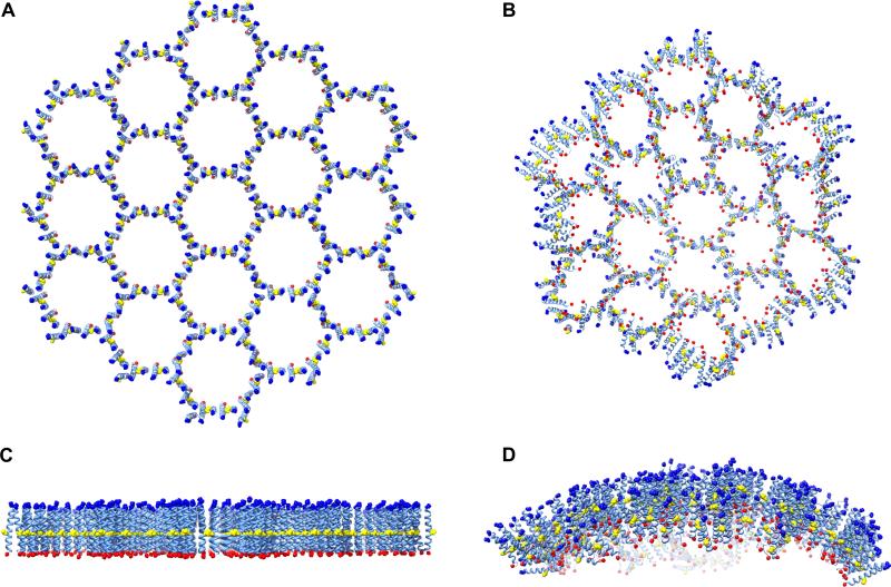
(A and C) Atomistic molecular model of 19 tessellated hexagons comprising hub A and hub B across three perspectives. These were generated from models for CC-Tri3 and CC-Di-AB aligned “back-to-back” along the 3- and 2-fold symmetry axes and linked through disulfide bonds. As for the synthesized peptides, the termini of the modeled peptides were capped and are represented in blue (acylated N-termini) and red (amidated C-termini), with the Cys-Cys disulfide linkers colored yellow. (B and D) Curvature observed following 5 ns molecular dynamics simulations. These were performed under periodic boundary conditions using explicit water (TIP3P) at pH 7 and with 150 mM NaCl present (full details provided as supplementary material (19), along with a movie S1).
Comment in
-
Materials science. Obey the peptide assembly rules.Science. 2013 May 3;340(6132):561-2. doi: 10.1126/science.1237708. Science. 2013. PMID: 23641105 No abstract available.
-
Cages from coils.Nat Biotechnol. 2013 Sep;31(9):809-10. doi: 10.1038/nbt.2670. Nat Biotechnol. 2013. PMID: 24022157 No abstract available.
References
Publication types
MeSH terms
Substances
Grants and funding
LinkOut - more resources
Full Text Sources
Other Literature Sources
