Review
. 2013 Nov;70(21):4085-99.
doi: 10.1007/s00018-013-1327-z. Epub 2013 Apr 12.

Life in the Fas lane: differential outcomes of Fas signaling

Affiliations
Review

Life in the Fas lane: differential outcomes of Fas signaling

Elizabeth Brint et al. Cell Mol Life Sci. 2013 Nov.

Abstract

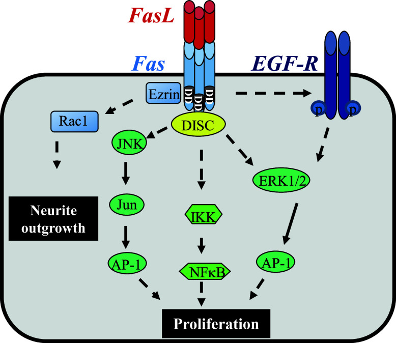
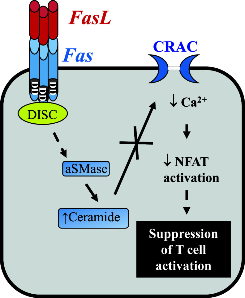
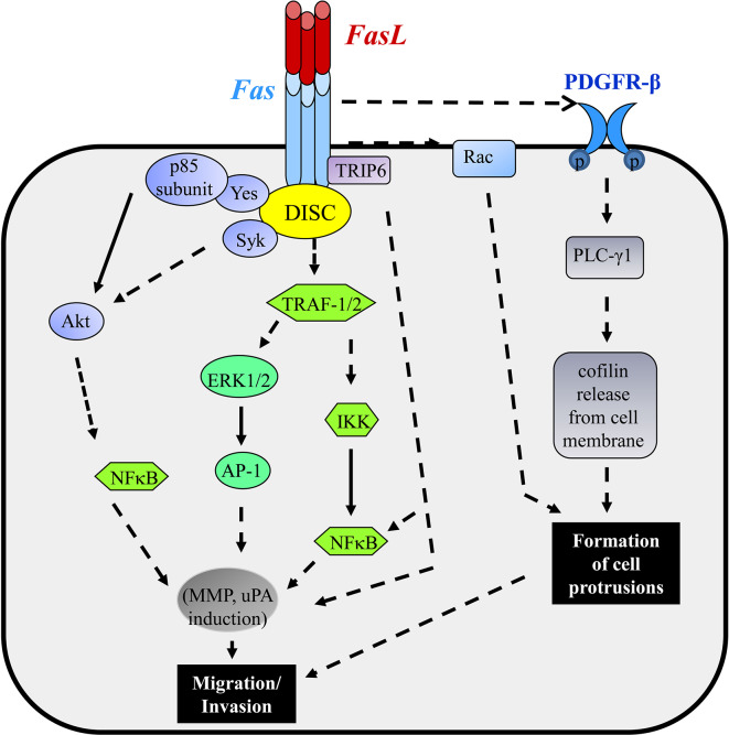
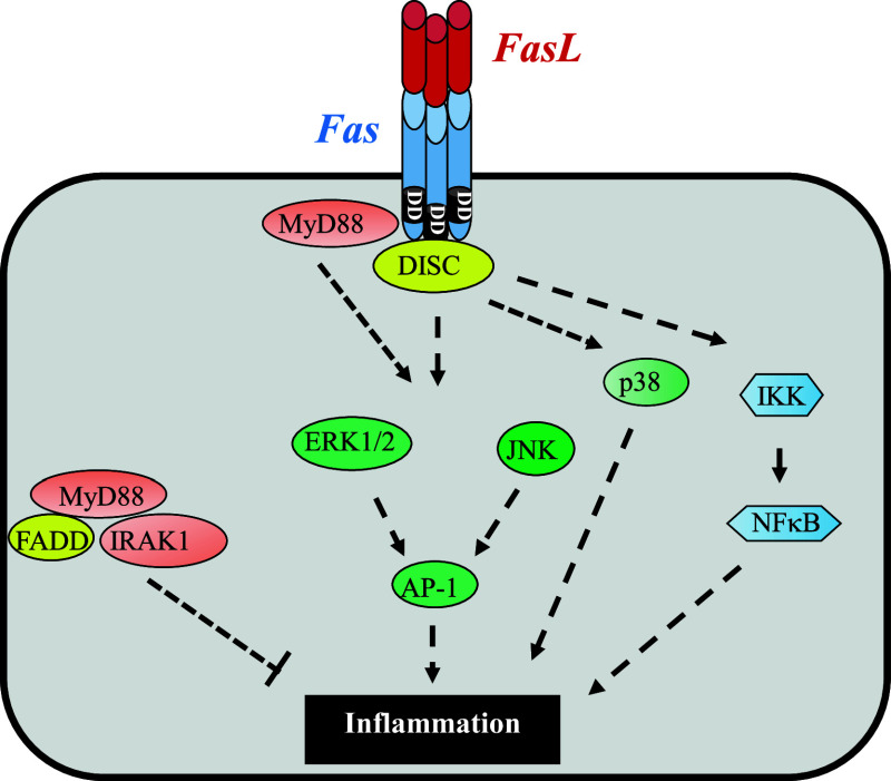
Fas, also known as CD95 or APO-1, is a member of the tumor necrosis factor/nerve growth factor superfamily. Although best characterized in terms of its apoptotic function, recent studies have identified several other cellular responses emanating from Fas. These responses include migration, invasion, inflammation, and proliferation. In this review, we focus on the diverse cellular outcomes of Fas signaling and the molecular switches identified to date that regulate its pro- and anti-apoptotic functions. Such switches occur at different levels of signal transduction, ranging from the receptor through to cross-talk with other signaling pathways. Factors identified to date including other extracellular signals, proteins recruited to the death-inducing signaling complex, and the availability of different intracellular components of signal transduction pathways. The success of therapeutically targeting Fas will require a better understanding of these pathways, as well as the regulatory mechanisms that determine cellular outcome following receptor activation.


Figures

Fig. 1
Fig. 1
Mitogenic signaling pathways implicated in Fas-induced proliferation. Following Fas stimulation, formation of the DISC occurs. Fas-mediated activation of the NF-κB transcription factor and the MAPK signaling pathways downstream of the DISC can promote cell proliferation. FasL can also induce ligand-dependent EGFR (epidermal growth factor receptor) activation, and phosphorylation, which triggers mitogenic signaling through ERK. Neurite growth requires recruitment of ezrin to Fas and activation of the small GTPase Rac1. Dashed lines represent signaling pathways/intermediaries that are not fully elucidated
Fig. 2
Fig. 2
Fas-mediated suppression of T cell activation. In T cells, activation of acidic sphingomyelinase (aSMase) following Fas ligation can increase the level of ceramide in the cells, which in turn inhibits Ca2+ release-activated Ca2+ channels (CRAC) in the plasma membrane. Impairment of CRAC opening prevents sustained Ca2+ influx, leading to suppressed NFAT activation and impaired T cell activation. Dashed lines represent signaling pathways/intermediaries that are not fully elucidated
Fig. 3
Fig. 3
Signaling pathways implicated in Fas-mediated migration. Multiple signaling pathways have been shown to play a role in the induction of cell migration and invasion by Fas. Ligation of Fas stimulates platelet-derived growth factor receptor-β (PDGFR-β) tyrosine phosphorylation by an unknown mechanism, leading to phosphorylation of phospholipase C-γ1 (PLC-γ1), with the subsequent release of cofilin from the cell membrane. Together with Fas-activated Rac, this leads to the formation of cell protrusions. Fas activation also results in the recruitment of the p85 subunit of PI3K and the tyrosine kinases Yes and Syk to Fas, activating Akt and NF-κB. Fas-induced NF-κB and ERK MAPK activation may also lead to cell migration in a TRAF2-mediated fashion. Association of TRIP6 with Fas following Fas activation can inhibit apoptosis, and promote NF-κB activation and cell migration. MMPs and uPA are among the genes most prominently upregulated in response to Fas ligation, and together facilitate migration and invasion. Dashed lines represent signaling pathways/intermediaries that not fully elucidated
Fig. 4
Fig. 4
Signaling pathways involved in Fas-mediated inflammation. Activation of Fas may promote inflammation via activation of the p38, ERK, and JNK MAPK signaling pathways and the NF-κB transcription factor downstream of the DISC. Interaction of MyD88 directly with Fas may also activate the JNK and ERK MAPK signaling pathways, independently of FADD, and mediate Fas-induced inflammation. Alternatively, in the absence of Fas ligation, the Fas adaptor molecule, FADD may interact with MyD88 in the cytoplasm, potentially blocking/limiting MyD88 signaling. Dashed lines represent signaling pathways/intermediaries that are not fully elucidated
Fig. 5
Fig. 5
Reverse signaling through FasL. In addition to initiating signaling through Fas, membrane-bound FasL can also transmit a co-stimulatory signal in the reverse direction upon ligation of Fas, a process known as reverse or retrograde signaling. Binding of Src homology 3 domain-containing proteins such as Fyn, Grb2, and PI3K to FasL leads to the subsequent activation of the MAPK ERK1/2 signaling pathway. Interaction of FasL with the hepatocyte growth factor receptor, Met, can also activate the Met-Stat3 signaling pathway. Dashed lines represent signaling pathways/intermediaries that are not fully elucidated
Fig. 6
Fig. 6
Multilevel regulation of the Fas signaling pathway. The outcome of Fas ligation is regulated at several points along the signaling pathway. a FasL may exist as either sFasL or mFasL, and they vary in their ability to induce apoptosis and/or non-apoptotic signaling. b At the receptor level, defective internalization of the Fas signaling complex or the presence of mutations in one Fas allele may inhibit apoptosis and promote non-apoptotic signaling. c The balance between the ratio of cFLIP and pro-caspase-8 at the DISC may determine the outcome of Fas ligation. d Recruitment of proteins such as TRIP6 and Toso to the DISC can inhibit cell death, and promote the activation of non-apoptotic signaling pathways by Fas. e In addition to inhibiting apoptosis, oncogenic K-Ras and mutated PI3K catalytic subunit α (PIK3CA) promote Fas-mediated invasion through effects on receptor internalization and the actin cytoskeleton. f Cell fate following Fas ligation was also shown to be regulated by growth factors and cytokines present in the cell microenvironment

References

    1. Trauth BC, Klas C, Peters AM, Matzku S, Moller P, Falk W, Debatin KM, Krammer PH. Monoclonal antibody-mediated tumor regression by induction of apoptosis. Science. 1989;245:301–305. - PubMed
    1. Yonehara S, Ishii A, Yonehara M. A cell-killing monoclonal antibody (anti-Fas) to a cell surface antigen co-downregulated with the receptor of tumor necrosis factor. J Exp Med. 1989;169:1747–1756. - PMC - PubMed
    1. Locksley RM, Killeen N, Lenardo MJ. The TNF and TNF receptor superfamilies: integrating mammalian biology. Cell. 2001;104:487–501. - PubMed
    1. Siegel RM, Frederiksen JK, Zacharias DA, Chan FK, Johnson M, Lynch D, Tsien RY, Lenardo MJ. Fas preassociation required for apoptosis signaling and dominant inhibition by pathogenic mutations. Science. 2000;288:2354–2357. - PubMed
    1. Leithauser F, Dhein J, Mechtersheimer G, Koretz K, Bruderlein S, Henne C, Schmidt A, Debatin KM, Krammer PH, Moller P. Constitutive and induced expression of APO-1, a new member of the nerve growth factor/tumor necrosis factor receptor superfamily, in normal and neoplastic cells. Lab Invest. 1993;69:415–429. - PubMed

Publication types