

Pulsatile tinnitus caused by a dilated mastoid emissary vein
- PMID: 23580003
- PMCID: PMC3617320
- DOI: 10.3346/jkms.2013.28.4.628
Pulsatile tinnitus caused by a dilated mastoid emissary vein
Abstract
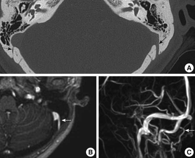
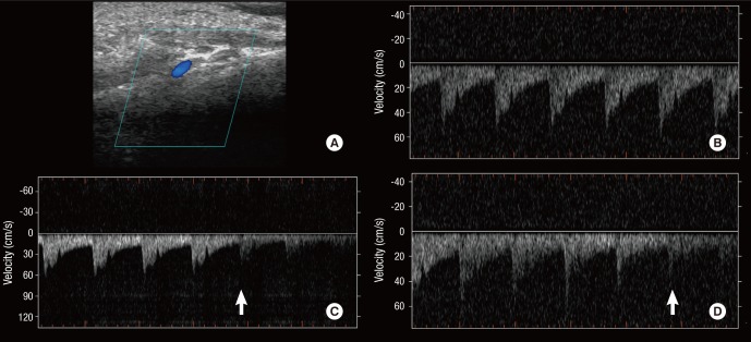
Although pulsatile tinnitus can be audible, objective demonstration of this heartbeat-synchronous sound has rarely been successful. We report a rare case of pulsatile tinnitus in a 44-yr-old female patient, which was induced by a large mastoid emissary vein (MEV) and objectively documented by Doppler sonography of the left posterior auricular region. The tinnitus was intermittent and the patient could adapt to the tinnitus without intervention on the mastoid emissary vein. These findings suggest that a single large MEV can cause pulsatile tinnitus in the absence of other vascular abnormalities, and imaging studies of the posterior fossa and Doppler ultrasonography can aid the diagnosis in such cases.
Keywords: Mastoid Vein; Pulsatile Tinnitus.
Figures
Similar articles
-
Bilateral Pulsatile Tinnitus Caused by Bilateral Dilated Mastoid Emissary Vein.Ear Nose Throat J. 2024 Oct;103(10):NP581-NP583. doi: 10.1177/01455613221077597. Epub 2022 Feb 16. Ear Nose Throat J. 2024. PMID: 35171065
-
Endovascular coiling of large mastoid emissary vein causing pulsatile tinnitus.Interv Neuroradiol. 2020 Dec;26(6):821-825. doi: 10.1177/1591019920926333. Epub 2020 May 14. Interv Neuroradiol. 2020. PMID: 32408784 Free PMC article.
-
Objective pulsatile tinnitus caused by an enlarged mastoid emissary vein in a child: A case report.Asian J Surg. 2023 Dec;46(12):5643-5645. doi: 10.1016/j.asjsur.2023.08.062. Epub 2023 Aug 23. Asian J Surg. 2023. PMID: 37625957 No abstract available.
-
State of the Art: Venous Causes of Pulsatile Tinnitus and Diagnostic Considerations Guiding Endovascular Therapy.Radiology. 2021 Jul;300(1):2-16. doi: 10.1148/radiol.2021202584. Epub 2021 May 25. Radiology. 2021. PMID: 34032509 Review.
-
Surgical management of pulsatile tinnitus secondary to jugular bulb or sigmoid sinus diverticulum with review of literature.Am J Otolaryngol. 2018 Mar-Apr;39(2):247-252. doi: 10.1016/j.amjoto.2017.12.019. Epub 2017 Dec 29. Am J Otolaryngol. 2018. PMID: 29336902 Review.
Cited by
-
[Clinical analysis of the vascular pulsatile tinnitus associated with sigmoid sinus-mastoid].Lin Chuang Er Bi Yan Hou Tou Jing Wai Ke Za Zhi. 2021 May;35(5):410-415. doi: 10.13201/j.issn.2096-7993.2021.05.006. Lin Chuang Er Bi Yan Hou Tou Jing Wai Ke Za Zhi. 2021. PMID: 34304464 Free PMC article. Chinese.
-
Prevalence, morphology, morphometry and associated clinical implications of mastoid emissary veins: narrative review.J Vasc Bras. 2023 Jul 17;22:e20230036. doi: 10.1590/1677-5449.202300362. eCollection 2023. J Vasc Bras. 2023. PMID: 37576721 Free PMC article. Review.
-
Pulsatile Tinnitus Due to a Large Mastoid Emissary Vein: Successfully Managed with Percutaneous Embolization in a Novel Approach.Indian J Otolaryngol Head Neck Surg. 2024 Oct;76(5):4858-4861. doi: 10.1007/s12070-024-04906-2. Epub 2024 Jul 19. Indian J Otolaryngol Head Neck Surg. 2024. PMID: 39376321
-
Incidence of vascular anomalies and variants associated with unilateral venous pulsatile tinnitus in 242 patients based on dual-phase contrast-enhanced computed tomography.Chin Med J (Engl). 2015 Mar 5;128(5):581-5. doi: 10.4103/0366-6999.151648. Chin Med J (Engl). 2015. PMID: 25698187 Free PMC article.
-
Radiological and Morphometric Study of the Emissary Foramens and Canal in the Posterior Cranial Fossa of the Human Skull with Its Neurosurgical Significance.Asian J Neurosurg. 2022 Oct 28;17(4):588-594. doi: 10.1055/s-0042-1757429. eCollection 2022 Dec. Asian J Neurosurg. 2022. PMID: 36570755 Free PMC article.
References
-
- Lo WW, Solti-Bohman LG, McElveen JT., Jr Aberrant carotid artery: radiologic diagnosis with emphasis on high-resolution computed tomography. Radiographics. 1985;5:985–993. - PubMed
-
- Levine SB, Snow JB., Jr Pulsatile tinnitus. Laryngoscope. 1987;97:401–406. - PubMed
-
- Forte V, Turner A, Liu P. Objective tinnitus associated with abnormal mastoid emissary vein. J Otolaryngol. 1989;18:232–235. - PubMed
-
- Louis RG, Jr, Loukas M, Wartmann CT, Tubbs RS, Apaydin N, Gupta AA, Spentzouris G, Ysique JR. Clinical anatomy of the mastoid and occipital emissary veins in a large series. Surg Radiol Anat. 2009;31:139–144. - PubMed
Publication types
MeSH terms
LinkOut - more resources
Full Text Sources
Other Literature Sources
Medical
