Three-dimensional LASIK flap thickness variability: topographic central, paracentral and peripheral assessment, in flaps created by a mechanical microkeratome (M2) and two different femtosecond lasers (FS60 and FS200)
- PMID: 23580024
- PMCID: PMC3621724
- DOI: 10.2147/OPTH.S40762
Three-dimensional LASIK flap thickness variability: topographic central, paracentral and peripheral assessment, in flaps created by a mechanical microkeratome (M2) and two different femtosecond lasers (FS60 and FS200)
Abstract
Purpose: To evaluate programmed versus achieved laser-assisted in situ keratomileusis (LASIK) flap central thickness and investigate topographic flap thickness variability, as well as the effect of potential epithelial remodeling interference on flap thickness variability.
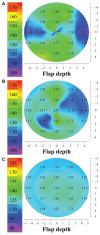
Patients and methods: Flap thickness was investigated in 110 eyes that had had bilateral myopic LASIK several years ago (average 4.5 ± 2.7 years; range 2-7 years). Three age-matched study groups were formed, based on the method of primary flap creation: Group A (flaps made by the Moria Surgical M2 microkeratome [Antony, France]), Group B (flaps made by the Abbott Medical Optics IntraLase™ FS60 femtosecond laser [Santa Ana, CA, USA]), and Group C (flaps made by the Alcon WaveLight(®) FS200 femtosecond laser [Fort Worth, TX, USA]). Whole-cornea topographic maps of flap and epithelial thickness were obtained by scanning high-frequency ultrasound biomicroscopy. On each eye, topographic flap and epithelial thickness variability was computed by the standard deviation of thickness corresponding to 21 equally spaced points over the entire corneal area imaged.
Results: The average central flap thickness for each group was 138.33 ± 12.38 μm (mean ± standard deviation) in Group A, 128.46 ± 5.72 μm in Group B, and 122.00 ± 5.64 μm in Group C. Topographic flap thickness variability was 9.73 ± 4.93 μm for Group A, 8.48 ± 4.23 μm for Group B, and 4.84 ± 1.88 μm for Group C. The smaller topographic flap thickness variability of Group C (FS200) was statistically significant compared with that of Group A (M2) (P = 0.004), indicating improved topographic flap thickness consistency - that is, improved precision - over the entire flap area affected.
Conclusions: The two femtosecond lasers produced a smaller flap thickness and reduced variability than the mechanical microkeratome. In addition, our study suggests that there may be a significant difference in topographic flap thickness variability between the results achieved by the two femtosecond lasers examined.
Keywords: 400 Hz excimer; IntraLase FS60; Moria M2; WaveLight® FS200, Allegretto Wave® Eye-Q; ultrasound biomicroscopy.
Figures


References
-
- Kanellopoulos AJ, Pe LH, Kleiman L. Moria M2 single use microkeratome head in 100 consecutive LASIK procedures. J Refract Surg. 2005;21(5):476–479. - PubMed
-
- Kanellopoulos AJ, Conway J, Pe LH. LASIK for hyperopia with the WaveLight excimer laser. J Refract Surg. 2006;22(1):43–47. - PubMed
-
- Reggiani-Mello G, Krueger RR. Comparison of commercially available femtosecond lasers in refractive surgery. Expert Rev Opthalmol. 2011;6(1):55–56.
-
- Juhasz T, Kastis GA, Suárez C, Bor Z, Bron WE. Time-resolved observations of shock waves and cavitation bubbles generated by femtosecond laser pulses in corneal tissue and water. Lasers Surg Med. 1996;19(1):23–31. - PubMed
LinkOut - more resources
Full Text Sources
Other Literature Sources

