. 2013 Apr 8:4:33.
doi: 10.3389/fneur.2013.00033. eCollection 2013.

Molecular events underlying Parkinson's disease - an interwoven tapestry

Affiliations

Molecular events underlying Parkinson's disease - an interwoven tapestry

Kah-Leong Lim et al. Front Neurol. .

Abstract

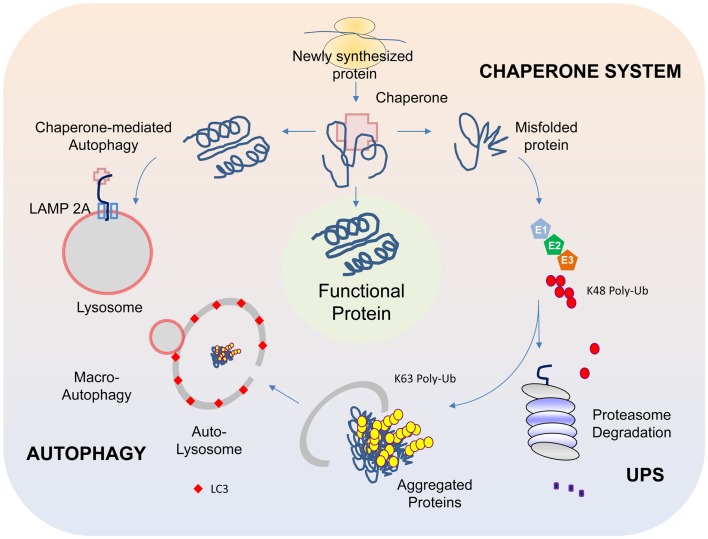
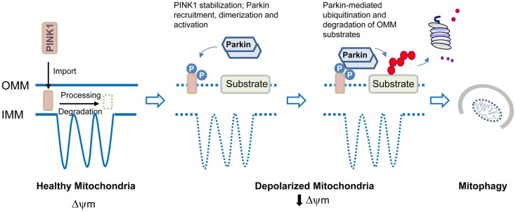
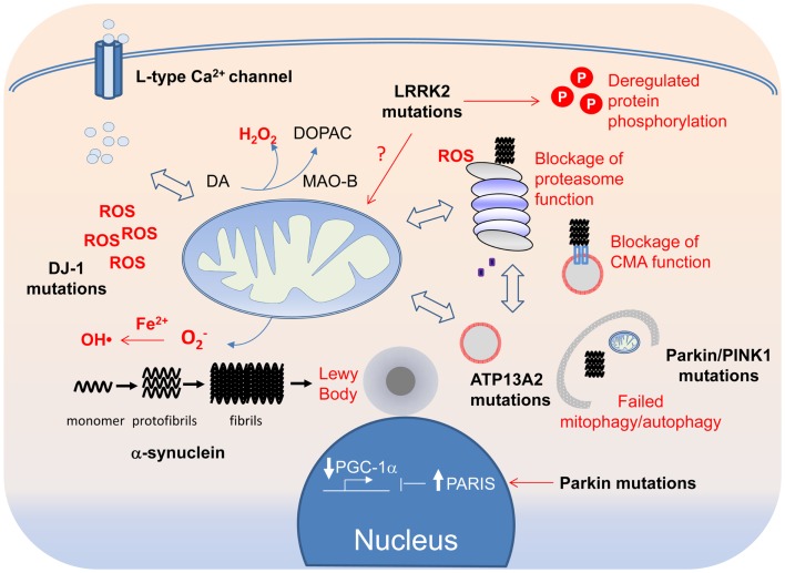
Although a subject of intense research, the mechanisms underlying dopaminergic neurodegeneration in Parkinson's disease (PD) remains poorly understood. However, a broad range of studies conducted over the past few decades, including epidemiological, genetic, and post-mortem analysis, as well as in vitro and in vivo modeling, have contributed significantly to our understanding of the pathogenesis of the disease. In particular, the recent identification and functional characterization of several genes, including α-synuclein, parkin, DJ-1, PINK1, and LRRK2, whose mutations are causative of rare familial forms of PD have provided tremendous insights into the molecular pathways underlying dopaminergic neurodegeneration. Collectively, these studies implicate aberrant mitochondrial and protein homeostasis as key contributors to the development of PD, with oxidative stress likely acting as an important nexus between the two pathogenic events. Aberrations in homeostatic processes leading to protein aggregation and mitochondrial dysfunction may arise intrinsically in substantia nigra pars compacta dopaminergic neurons as a result of impairments in the ubiquitin-proteasome system, failure in autophagy-mediated clearance, alterations of mitochondrial dynamics, redox imbalance, iron mishandling, dopamine dysregulation, or simply from the chronic pace-making activity of nigra-localized L-type calcium channels, or extrinsically from non-autonomous sources of stress. Given the myriad of culprits implicated, the pathogenesis of PD necessarily involves an intricate network of interwoven pathways rather than a linear sequence of events. Obviously, understanding how the various disease-associated pathways interact with and influence each other is of mechanistic and therapeutic importance. Here, we shall discuss some key PD-related pathways and how they are interwoven together into a tapestry of events.

Keywords: Parkinson disease; autophagy; mitophagy; oxidative stress; proteasome; protein aggregation.


Figures

Figure 1
Figure 1
Protein QC systems. The chaperone, ubiquitin-proteasome, and autophagy systems function co-ordinately to maintain intracellular protein homeostasis. The chaperones, comprising of members of the heat-shock proteins, represent the first line of defense in ensuring the correct folding and refolding of proteins. When a native folding state cannot be attained, the chaperones will direct the misfolded protein for degradation by the proteasome. Proteins that are destined for proteasome-mediated degradation usually have a chain of ubiquitin added via a reaction cascade that involves the ubiquitin-activating (E1), -conjugating (E2), and -ligating (E3) enzymes, whereby successive iso-peptide linkages are formed between the terminal residue (G76) of one ubiquitin molecule and a lysine (K) residue (most commonly K48) within another. The (G76-K48) polyubiquitinated substrate is then recognized by the 26S proteasome as a target for degradation. In some cases, proteins may be modified by K63-linked polyubiquitination, which can promote their aggregation into inclusion bodies and their subsequent removal by autophagy. The autophagy process involves the sequestration of substrates by a phagophore that expands into a double-membrane structure called autophagosome that engulfs the substrate. The autophagosome then fuses with a lysosome to form autolysosomes, within which the inner membrane of the autophagosome is broken down and the cargo degraded by acidic lysosomal hydrolases. Another form of autophagy is chaperone-mediated autophagy (CMA), which involves the direct translocation of unfolded substrate proteins across the lysosomal membrane through the actions of a cytosolic chaperone hsc70, and an integral lysosomal membrane receptor LACMP2A (lysosome-associated membrane protein type 2A).
Figure 2
Figure 2
Model of parkin/PINK1-mediated mitophagy. In healthy mitochondria, PINK1 imported through the outer mitochondrial membrane (OMM) is rapidly processed and degraded. Upon mitochondrial depolarization, PINK1 stabilization on the OMM leads to its dualautophosphorylation on Ser228 and Ser402. This event somehow triggers parkin recruitment, self-association, and catalytic activation. Parkin ubiquitinates several proteins on the OMM that results in their degradation by the proteasome. Mitophagy induction then occurs.
Figure 3
Figure 3
A tapestry of molecular events in PD pathogenesis. Disruption of mitochondrial and protein QCs can arise from overt PD-linked genetic mutations or through oxidative modifications of their components by ROS, the levels of which can be elevated by DJ-1 mutations, Fe2+-mediated Fenton reaction or increased Ca2+ influx through the L-type Ca2+ channel. Because of the crosstalk that exists between the QC systems, each can in turn affect the other in a reciprocal fashion. Aberrant mitochondrial and protein QCs and redox imbalance all can promote the formation of α-synuclein protofibrils and fibrils, which in turn can block the function of the proteasome and CMA. Aggregated α-synuclein species, if not cleared in a timely fashion, can also seed the formation of LBs, which can be physically obstructive to neuronal function if allowed to grow.

References

    1. Ahmed I., Liang Y., Schools S., Dawson V. L., Dawson T. M., Savitt J. M. (2012). Development and characterization of a new Parkinson’s disease model resulting from impaired autophagy. J. Neurosci. 32, 16503–16509 10.1523/JNEUROSCI.5110-11.2012 - DOI - PMC - PubMed
    1. Alam Z. I., Daniel S. E., Lees A. J., Marsden D. C., Jenner P., Halliwell B. (1997a). A generalised increase in protein carbonyls in the brain in Parkinson’s but not incidental Lewy body disease. J. Neurochem. 69, 1326–1329 10.1046/j.1471-4159.1997.69031326.x - DOI - PubMed
    1. Alam Z. I., Jenner A., Daniel S. E., Lees A. J., Cairns N., Marsden C. D., et al. (1997b). Oxidative DNA damage in the parkinsonian brain: an apparent selective increase in 8-hydroxyguanine levels in substantia nigra. J. Neurochem. 69, 1196–1203 10.1046/j.1471-4159.1997.69031196.x - DOI - PubMed
    1. Alexander C., Votruba M., Pesch U. E., Thiselton D. L., Mayer S., Moore A., et al. (2000). OPA1, encoding a dynamin-related GTPase, is mutated in autosomal dominant optic atrophy linked to chromosome 3q28. Nat. Genet. 26, 211–215 10.1038/79944 - DOI - PubMed
    1. Andres-Mateos E., Perier C., Zhang L., Blanchard-Fillion B., Greco T. M., Thomas B., et al. (2007). DJ-1 gene deletion reveals that DJ-1 is an atypical peroxiredoxin-like peroxidase. Proc. Natl. Acad. Sci. U.S.A. 104, 14807–14812 10.1073/pnas.0703219104 - DOI - PMC - PubMed

LinkOut - more resources