. 2013 Sep;31(9):1382-9.
doi: 10.1002/jor.22367. Epub 2013 Apr 11.

Electrospun fibers as a scaffolding platform for bone tissue repair

Affiliations

Electrospun fibers as a scaffolding platform for bone tissue repair

Seungyoun Lyu et al. J Orthop Res. 2013 Sep.

Abstract

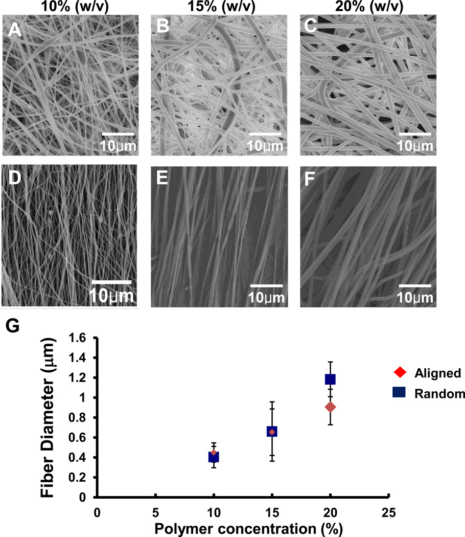
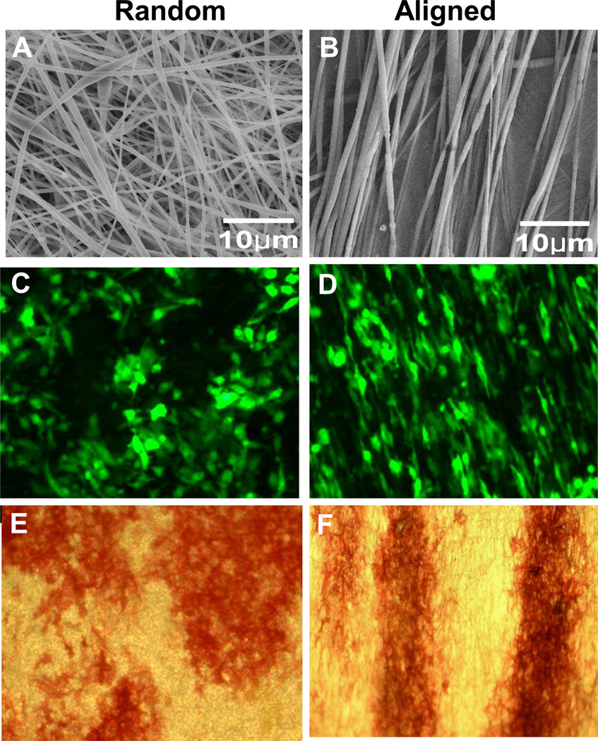
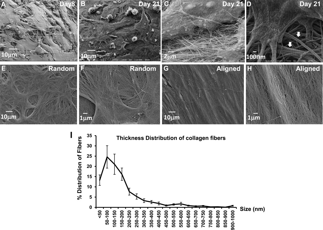
The purpose of the study is to investigate the effects of electrospun fiber diameter and orientation on differentiation and ECM organization of bone marrow stromal cells (BMSCs), in attempt to provide rationale for fabrication of a periosteum mimetic for bone defect repair. Cellular growth, differentiation, and ECM organization were analyzed on PLGA-based random and aligned fibers using fluorescent microscopy, gene analyses, electron scanning microscopy (SEM), and multiphoton laser scanning microscopy (MPLSM). BMSCs on aligned fibers had a reduced number of ALP+ colony at Day 10 as compared to the random fibers of the same size. However, the ALP+ area in the aligned fibers increased to a similar level as the random fibers at Day 21 following stimulation with osteogenic media. Compared with the random fibers, BMSCs on the aligned fibers showed a higher expression of OSX and RUNX2. Analyses of ECM on decellularized spun fibers showed highly organized ECM arranged according to the orientation of the spun fibers, with a broad size distribution of collagen fibers in a range of 40-2.4 μm. Taken together, our data support the use of submicron-sized electrospun fibers for engineering of oriented fibrous tissue mimetic, such as periosteum, for guided bone repair and reconstruction.

Keywords: electrospinning; extracellular matrix (ECM); periosteum.


Figures

Fig. 1
Fig. 1
A–G Representative SEM images showing randomly oriented (A–C) and aligned (D–F) electrospun fibers fabricated at a concentration of 10% (A, D), 15% (B, E), and 20% (C, F) 75:25 PLGA. The means of the fiber diameter are directly correlated with the indicated concentrations of polymer (w/v) for both aligned and random fibers (G).
Fig. 2
Fig. 2
A–F SEM analyses show distinctive topology of random (A, 1000×) or aligned (B, 1000×) PLGA fibers. Fluorescence images of GFP-tagged murine bone marrow stromal cells cultured on both types of fibers show distinctive morphology. Cells on randomly fibers adopted a rounder morphology (C, 25×) whereas cells on aligned fibers were elongated (D, 25×). ALP-positive colonies on scaffolds at day 10 displayed a similar morphology on random (E, 4×) and aligned fibers (F, 4×).
Fig. 3
Fig. 3
A–E Alkaline phosphatase staining were performed on random and aligned fibers on days 10, 15 and 21 as indicated (A). Photographs of the colony formation on fibrous meshes were taken at a magnification of 1× via a dissection microscope. Numbers of the ALP+ colony were quantified at day 10 (B) and areas of the ALP+ region were quantified at day 21 (C). Real time PCR analyses show osteogenic marker gene OSX (D) and RUNX2 (E) expressions at day 10 and day 21. * indicate p<0.05.
Fig. 4
Fig. 4
A–H Representative SEM images (×1000) of bone marrow stromal cells cultured on randomly oriented electrospun fibers on Day 8 (A) and Day 21 (B). Higher magnification image (×10,000) shows detailed morphology of and nano and micron-sized collagen matrix with polygonal cells (C). Characteristic collagen fibrils and collagen bundles (arrows) are shown at ×30,000 (D). Following removal of the cells (decellularization), the native collagen matrix produced by BMSCs cultured on randomly oriented (E, F) or an aligned fibers (G, H) is shown at ×1000 and ×5000.
Fig. 5
Fig. 5
A–D Fluorescent excitation and SHG of live GFP-positive cells cultured on random or aligned fibers were imaged via MPLSM at day 21. Cells are shown as green and collagen matrix produced by these cells shown as cyan. Distinctive cellular morphology and collagen matrix organization are shown in randomly oriented (A, C, E, G) and aligned (B, D, F, H) fibers. Multiphoton microscopy further allows optical sectioning of the live specimens at a depths of 5um (A–D, surface layer) and 50 um (E and F, deeper layer). G and H show collagen matrix in random or aligned decellularized electrospun fibers.

References

    1. Ma Z, Kotaki M, Inai R, Ramakrishna S. Potential of nanofiber matrix as tissue-engineering scaffolds. Tissue Eng. 2005;11:101–109. - PubMed
    1. Kumbar SG, James R, Nukavarapu SP, Laurencin CT. Electrospun nanofiber scaffolds: engineering soft tissues. Biomed Mater. 2008;3:034002. - PubMed
    1. Li D, Xia Y. Electrospinning of Nanofibers: Reinventing the Wheel? Advanced Materials. 2004;16:1151.
    1. Dahlin RL, Kasper FK, Mikos AG. Polymeric nanofibers in tissue engineering. Tissue Eng Part B Rev. 2011;17:349–364. - PMC - PubMed
    1. Holzwarth JM, Ma PX. Biomimetic nanofibrous scaffolds for bone tissue engineering. Biomaterials. 2011;32:9622–9629. - PMC - PubMed

Publication types

MeSH terms