. 2013 May 23;121(21):4295-302.
doi: 10.1182/blood-2012-07-442251. Epub 2013 Apr 11.

The large ectodomains of CD45 and CD148 regulate their segregation from and inhibition of ligated T-cell receptor

Affiliations

The large ectodomains of CD45 and CD148 regulate their segregation from and inhibition of ligated T-cell receptor

Shaun-Paul Cordoba et al. Blood. .

Abstract

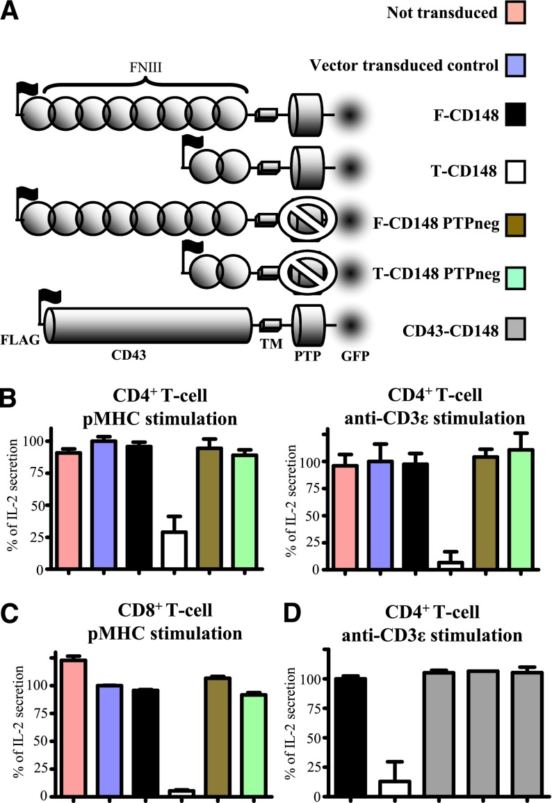
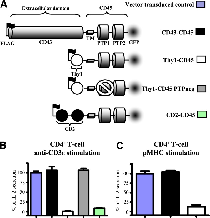
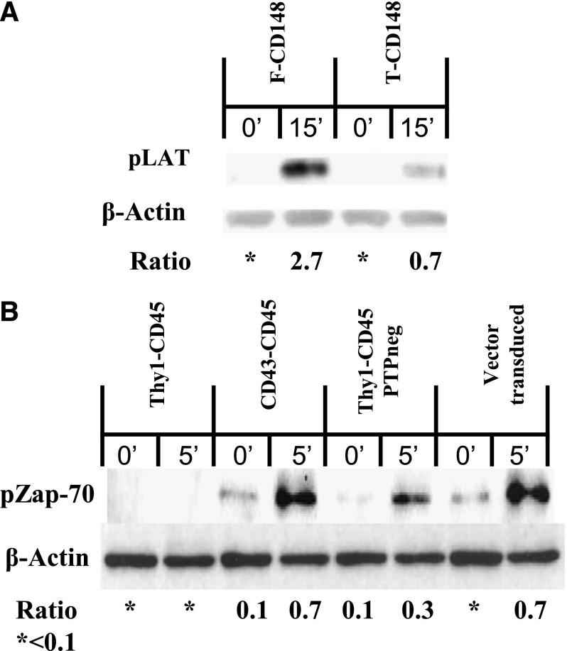
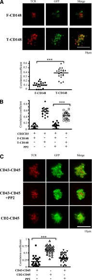
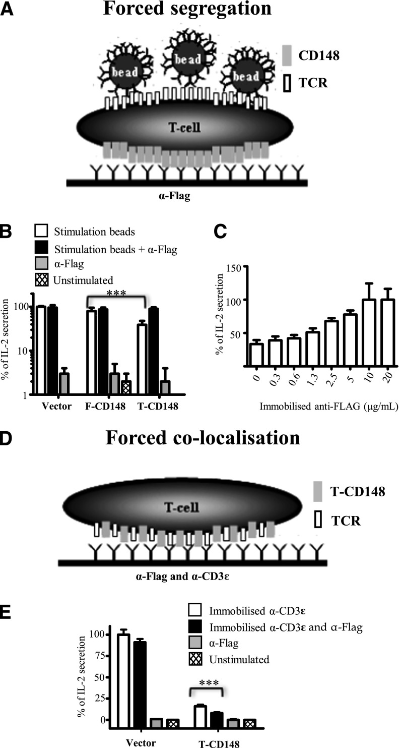
T-cell receptor (TCR) triggering results in a cascade of intracellular tyrosine phosphorylation events that ultimately leads to T-cell activation. It is dependent on changes in the relative activities of membrane-associated tyrosine kinases and phosphatases near the engaged TCR. CD45 and CD148 are transmembrane tyrosine phosphatases with large ectodomains that have activatory and inhibitory effects on TCR triggering. This study investigates whether and how the ectodomains of CD45 and CD148 modulate their inhibitory effect on TCR signaling. Expression in T cells of forms of these phosphatases with truncated ectodomains inhibited TCR triggering. In contrast, when these phosphatases were expressed with large ectodomains, they had no inhibitory effect. Imaging studies revealed that truncation of the ectodomains enhanced colocalization of these phosphatases with ligated TCR at the immunological synapse. Our results suggest that the large ectodomains of CD45 and CD148 modulate their inhibitory effect by enabling their passive, size-based segregation from ligated TCR, supporting the kinetic-segregation model of TCR triggering.


Figures

Figure 1
Figure 1
The size of CD148 ectodomain modulates its inhibitory effect on T-cell activation. (A) Schematic depiction of the CD148 constructs used in this study. The ectodomains comprised either the full-length form of CD148 or the truncated form of CD148 or the ectodomain from CD43 (CD43-CD148). Catalytically inactive (PTPneg) forms of full-length and truncated CD148 had a cysteine to serine substitution (C1140S) in the phosphatase domain. All constructs contained an N-terminal FLAG tag to stain and match surface expression levels by FACS and a C-terminal GFP tag for imaging. (B) Control (untransduced or vector-transduced) 2B4 T cells or 2B4 T cells expressing comparable levels of the indicated CD148 construct (supplemental Figure 1) were stimulated with either CHO cells expressing I-Ek, presenting the cognate MCC peptide (left) or plate-immobilized anti-mouse CD3ε (right) and IL-2 secretion analyzed after 14 to 18 hours. (C) Control or transduced B3Z T cells expressing comparable levels of the indicated CD148 construct (supplemental Figure 4A) were stimulated with CHO cells expressing cognate pMHC as a single-chain trimer and IL-2 secretion analyzed after 14 to 18 hours. (D) 2B4 T-cells transduced with the indicated CD148 constructs were stimulated with plate-immobilized anti-mouse CD3ε and IL-2 secretion examined after 14 to 18 hours. Three different CD43-CD148 transduced clones were tested. Error bars represent the SD of the mean from at least 3 replicates. For (B) and (C), IL-2 secretion data were normalized with 0% and 100%, representing unstimulated and stimulated vector-transduced controls, respectively. In (D), IL-2 secretion was normalized to full-length CD148 transduced cells.
Figure 2
Figure 2
The size of CD45 ectodomain modulates its inhibitory effect on T-cell activation. (A) Schematic depiction of the CD45 constructs used in this study. They comprised the mouse CD45 transmembrane and cytosolic domains and the ectodomains of rat Thy1 (1 Ig-like domain), rat CD2 (2 Ig-like domains), or rat CD43. A catalytically inactive form of Thy1-CD45 (Thy1-CD45 PTPneg) had a cysteine-to-serine substitution (C840S) in the membrane-proximal phosphatase domain. All constructs contained an N-terminal FLAG tag to stain and match surface expression levels by FACS and a C-terminal GFP tag for imaging. 2B4 T-cells expressing comparable levels of the indicated CD45 construct (supplemental Figure 5) were stimulated with either (B) plate-immobilized anti-mouse CD3ε or (C) CHO cells expressing I-Ek presenting the cognate MCC peptide (left) and IL-2 secretion analyzed after 14 to 18 hours. Error bars represent the SD of the mean from at least 3 replicates. Data normalized to vector-transduced controls as in Figure 2.
Figure 3
Figure 3
Forms of CD148 and CD45 with short ectodomains abrogate TCR proximal signaling. (A) Immunoblot of phosphorylated LAT (pLAT) and β-actin in B3Z T-cells transduced with either full-length CD148 or truncated CD148 and stimulated with anti-mouse CD3ε coated beads for 15 minutes. (B) Immunoblot of phosphorylated Zap-70 (pZap-70) and β-actin in 2B4 T cells transduced with the indicated CD45 constructs (or vector as control) and stimulated with anti-mouse CD3ε coated beads for 5 minutes. All immunoblots are representative of 3 replicates. The densitometry ratio between either pLAT or pZAP-70 and β-actin was calculated in ImageJ (National Institutes of Health).
Figure 4
Figure 4
Truncation of CD148 and CD45 ectodomains enhances colocalization with TCR microclusters. (A) B3Z T cells expressing the indicated CD148-GFP chimera (green) were imaged by TIRF microscopy after initial contact with a planar bilayer that contained glycosylphosphatidylinositol-ICAM-1 and Cy5-labeled (red) monobiotinylated anti-mouse CD3ε. Representative images are shown (upper). Pearson correlation coefficients between the GFP and Cy5 fluorescence were analyzed in 20 cell contacts (lower). (B) B3Z T cells expressing CD2-TCRζ-mCherry and the indicated CD148-GFP chimera were preincubated with or without PP2 and then imaged in contact with a planar bilayer that contained glycosylphosphatidylinositol-ICAM-1 and anti-rat CD2. Pearson’s correlation coefficients between GFP and mCherry fluorescence were analyzed in 20 cell contacts. (C) 2B4 cells expressing the indicated CD45-GFP chimera (green) were preincubated with or without PP2 and imaged by TIRF microscopy after initial contact with a planar bilayer that contained ICAM-1 and monobiotinylated anti-mouse CD3ε. CD45 protein was imaged through its C-terminal GFP tag, and TCRβ was detected using a specific antibody (H57) Fab′ fragment labeled with AF568 (upper). Pearson’s correlation coefficients between the GFP and AF568 were analyzed on more than 25 cell contacts (lower). ***P < .01, using Student t test.
Figure 5
Figure 5
Antibody-induced segregation of truncated CD148 rescues TCR signaling. (A) Schematic depiction of forced segregation experiment. B3Z T cells expressing FLAG-tagged CD48 are plated onto surfaces presenting immobilized anti-FLAG antibody before stimulation with anti-mouse CD3ε beads. By accumulating FLAG-tagged CD148 on the basal side of the cell, this forces segregation from apical areas where the beads engage TCR. (B) B3Z cells expressing the indicated form of CD148 and plated onto anti-FLAG or control surfaces were exposed to anti-mouse CD3ε (stimulation) or control beads IL-2 secretion determined after 16 hours after stimulation. Data are normalized with IL-2 secretion from unstimulated and stimulated vector transduced cells set to 0% and 100%, respectively. ***P < .01, using paired Student t test. (C) B3Z cells expressing the truncated form of CD148 were plated onto surfaces coated with the indicated concentration of immobilized anti-FLAG, stimulation with anti-mouse CD3ε beads, and IL-2 secretion determined after 16 hours. Data normalized to IL-2 secretion observed with highest levels of anti-FLAG (100%) and IL-2 secretion from unstimulated cells was set to 0%. (D) Schematic depiction of forced colocalization experiment. B3Z cells expressing FLAG-tagged truncated CD148 was plated onto surfaces containing both immobilized anti-FLAG and anti-mouse CD3ε. (E) Control B3Z cells or B3Z cells expressing the truncated form of CD148 were plated onto surfaces coated with the indicated combination of anti-FLAG and anti-mouse CD3ε antibodies and IL-2 secretion determined after 16 hours of stimulation. Data are normalized with IL-2 secretion from unstimulated and stimulated vector-transduced cells set to 0% and 100%, respectively. Error bars represent the SD of the mean from at least 3 replicates.

References

    1. Weiss A, Samelson LE. T-lymphocyte activation. In: Paul WE, ed. Fundamental Immunology. Philadelphia: Lippincott Williams & Wilkins; 2003: 321-364.
    1. van der Merwe PA, Dushek O. Mechanisms for T cell receptor triggering. Nat Rev Immunol. 2011;11(1):47–55. - PubMed
    1. Hermiston ML, Zikherman J, Zhu JW. CD45, CD148, and Lyp/Pep: critical phosphatases regulating Src family kinase signaling networks in immune cells. Immunol Rev. 2009;228(1):288–311. - PMC - PubMed
    1. O’Shea JJ, McVicar DW, Bailey TL, Burns C, Smyth MJ. Activation of human peripheral blood T lymphocytes by pharmacological induction of protein-tyrosine phosphorylation. Proc Natl Acad Sci USA. 1992;89(21):10306–10310. - PMC - PubMed
    1. Imbert V, Peyron JF, Farahi Far D, Mari B, Auberger P, Rossi B. Induction of tyrosine phosphorylation and T-cell activation by vanadate peroxide, an inhibitor of protein tyrosine phosphatases. Biochem J. 1994;297(Pt 1):163–173. - PMC - PubMed

Publication types

MeSH terms

Substances