. 2013 Apr 11;153(2):376-88.
doi: 10.1016/j.cell.2013.02.053.

Type 2 innate signals stimulate fibro/adipogenic progenitors to facilitate muscle regeneration

Affiliations

Type 2 innate signals stimulate fibro/adipogenic progenitors to facilitate muscle regeneration

Jose E Heredia et al. Cell. .

Abstract

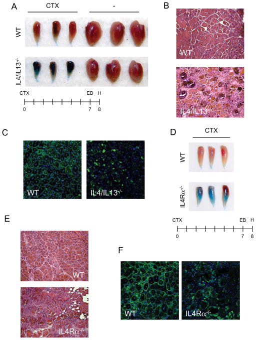
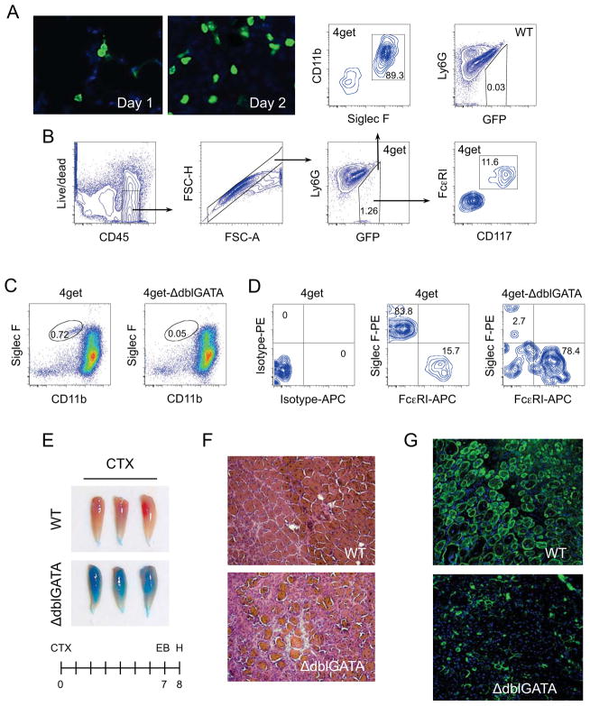
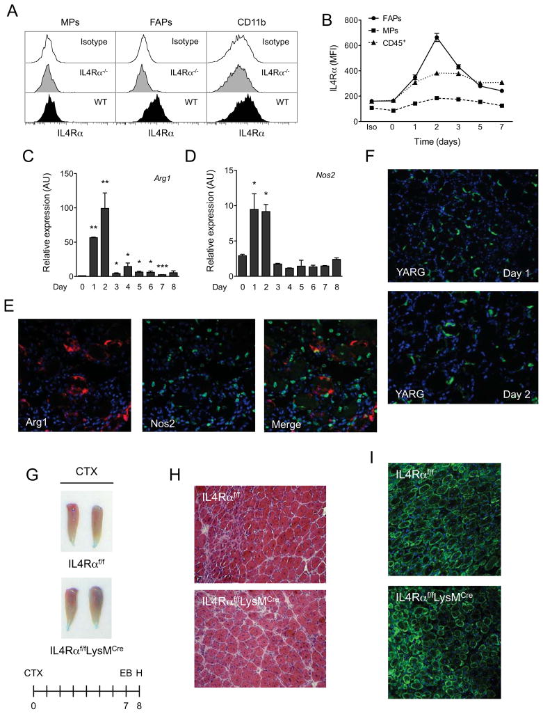
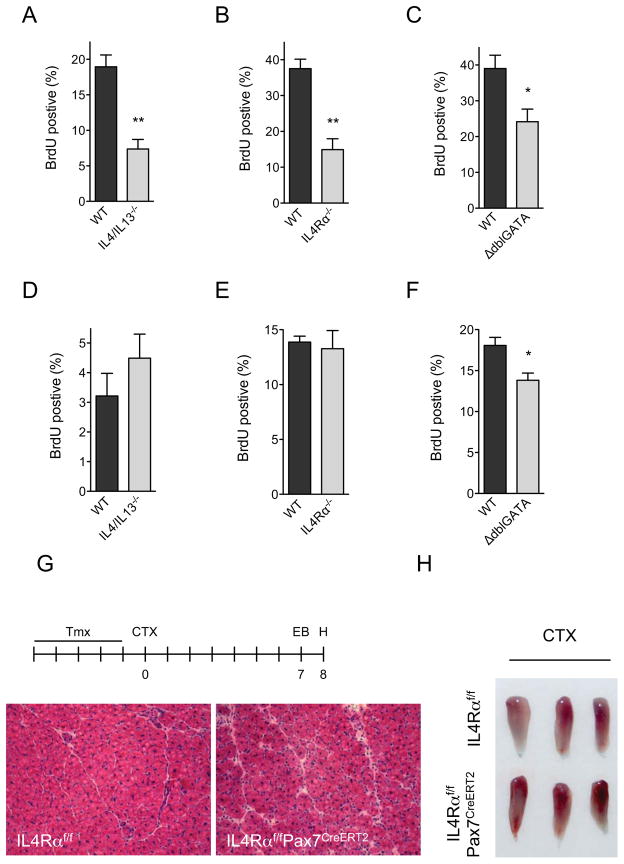
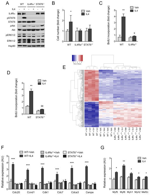
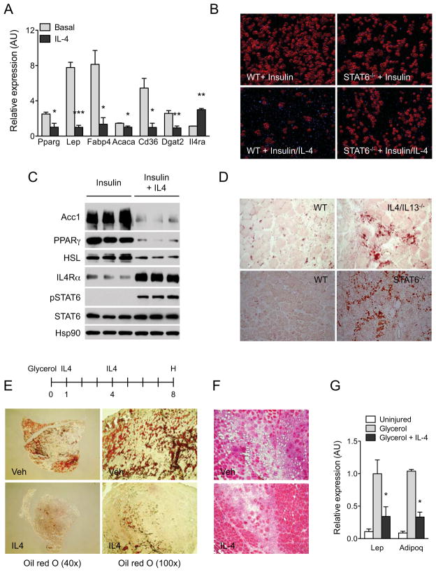
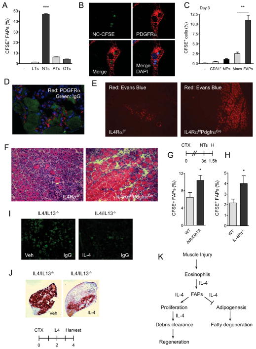
In vertebrates, activation of innate immunity is an early response to injury, implicating it in the regenerative process. However, the mechanisms by which innate signals might regulate stem cell functionality are unknown. Here, we demonstrate that type 2 innate immunity is required for regeneration of skeletal muscle after injury. Muscle damage results in rapid recruitment of eosinophils, which secrete IL-4 to activate the regenerative actions of muscle resident fibro/adipocyte progenitors (FAPs). In FAPs, IL-4/IL-13 signaling serves as a key switch to control their fate and functions. Activation of IL-4/IL-13 signaling promotes proliferation of FAPs to support myogenesis while inhibiting their differentiation into adipocytes. Surprisingly, type 2 cytokine signaling is also required in FAPs, but not in myeloid cells, for rapid clearance of necrotic debris, a process that is necessary for timely and complete regeneration of tissues.


Figures

Figure 1
Figure 1. IL-4/IL-13 signaling is required for muscle regeneration
(A, D) Tibialis anterior (TA) muscles of WT, IL-4/IL-13−/− (A) and IL-4Rα−/− (D) mice 8 days after injury by cardiotoxin (CTX). (B, D) Representative TA muscle sections of WT, IL-4/IL-13−/− (B) and IL-4Rα−/− (D) were stained with hematoxylin and eosin 8 days after injury, n=6 per genotype (magnification, X200). (C, F) Fluorescent microscopy of WT, IL-4/IL-13−/− (C) and IL-4Rα−/− (F) TA muscles 8 days after injury (magnification, X200). Desmin (green), and DAPI (blue). See also Table S1.
Figure 2
Figure 2. IL-4 expressing eosinophils are required for muscle regeneration after injury
(A) IL-4 expressing cells infiltrate injured muscles. TA muscles from 4get mice were stained for GFP 1 or 2 days after injury with CTX (magnification, X400). (B) Flow cytometric gating strategy for identification of IL-4 expressing cells in injured muscles. Eosinophils were defined as being CD11b+ and Siglec F+, whereas mast cells were CD117+ and FcεRI+. (C) IL-4 expressing cells in 4get and 4get-ΔdblGATA mice 36 hours after injury with CTX. (D) Eosinophils in 4get and 4get-ΔdblGATA mice 36 hours after muscle injury. (E) TA muscles from WT and ΔdblGATA mice 8 days after injury. (F) Hematoxylin and eosin staining of representative muscle sections taken from WT and 4get-ΔdblGATA mice 8 days after injury, n=6 per genotype (magnification, X200). (G) Immunostaining of TA muscle sections of WT and ΔdblGATA mice 8 days after CTX injury (magnification, X200). Desmin (green) and DAPI (blue). See also Figure S1.
Figure 3
Figure 3. IL-4/IL-13 signaling in myeloid cells is dispensable for muscle regeneration
(A) Expression of IL-4Rα in muscle progenitors (MPs), fibro/adipogenic progenitors (FAPs) and myeloid cells (CD11b+). (B) Time course of IL-4Rα expression in FAPs, MPs and CD45+ hematopoietic cells. (C, D) Expression of arginase 1 (Arg1) and iNOS (Nos2) mRNAs in regenerating TA muscles of wild type mice, n=4–6 per time point. (E) Fluorescent microscopy for Arg1 (red), Nos2 (green) and DAPI (blue) 2 days after muscle injury of WT mice (magnification, X400). (F) GFP staining of TA muscles of YARG mice after injury (magnification, X200). (G) TA muscles 8 days after injury in IL-4Rαf/f and IL-4Rαf/fLysMCre mice. (H) Representative day 8 muscle sections from IL-4Rαf/f and IL-4Rαf/fLysMCre mice stained with hematoxylin and eosin, n=6 per genotype. (I) Fluorescent microscopy of IL-4Rαf/f and IL-4Rαf/fLysMCre TA muscles 8 days after injury. Desmin in green and DAPI in blue. Error bars represent SEM. See also Figure S2.
Figure 4
Figure 4. IL-4/IL-13 signaling controls proliferation of FAPs but not satellite cells during muscle regeneration
(A–C) Quantification of BrdU incorporation in FAPs of WT, IL-4/IL-13−/− and ΔdblGATA mice 24 hours after muscle injury, n=4 per genotype. (D–F) Quantification of BrdU incorporation in MPs of WT, IL-4/IL-13−/− and ΔdblGATA mice 24 hours after muscle injury, n=4 per genotype. (G, H) Signaling via IL-4Rα in satellite cells is dispensable for muscle regrowth after injury. (G) Representative day 8 muscle sections from IL-4Rαf/f and IL-4Rαf/fPax7CreERT2 mice stained with hematoxylin and eosin, n=6 per genotype (magnification, X200). (H) Gross appearance of IL-4Rαf/f and IL-4Rαf/fPax7CreERT2 TA muscles 8 days after injury with CTX. Error bars represent SEM. See also Figure S3.
Figure 5
Figure 5. IL-4 promotes proliferation of FAPs in vitro
(A) Activation of signaling pathways by IL-4 in WT, IL-4Rα−/− and STAT6−/− FAPs. (B) Quantification of cell number after stimulation of WT, IL-4Rα−/− and STAT6−/− FAPs with vehicle or IL-4 for 48 hours, n=4 per genotype and time point. For each genotype, cell number is normalized to its vehicle control. (C, D) BrdU incorporation in WT, IL-4Rα−/− and STAT6−/− FAPs after stimulation vehicle or IL-4 (10nM) for 24 hours. (E) Heat map of differentially expressed genes in WT and IL-4Rα−/− FAPs treated with vehicle or IL-4 for 24 hours; red-induced; blue-repressed. GO terms associated with DNA replication, cell cycle, and mitosis are enriched in the upregulated gene set, whereas those associated with triglyceride metabolism and lipid biogenesis are enriched in the downregulated gene set. (F) Quantitative RT-PCR analysis of cell cycle genes in WT, IL-4Rα−/− and STAT6−/− FAPs after stimulation with vehicle or IL-4 for 24 hours, n=4 per genotype and treatment. (G) Expression of myogenic genes in wild type MPs cultured with FAPs conditioned media. FAPs were stimulated with vehicle or IL-4 for 72 hours prior to collection of conditioned media, n=3 per treatment. Error bars represent SEM. See also Figure S4.
Figure 6
Figure 6. IL-4 inhibits adipogenic differentiation of FAPs in vitro and in vivo
(A) Quantitative RT-PCR analysis of adipogenic genes in WT FAPs cultured for 7 days in the presence or absence of IL-4, n=3 per treatment. (B) Oil Red O staining of WT and STAT6−/− FAPs differentiated with insulin in the absence or presence of IL-4 (magnification, X290). (C) Immunoblot analysis for adipogenic proteins in WT FAPs treated with insulin or insulin plus IL-4. (D) Oil red O staining of regenerating TA muscles was performed on day 5 (magnification, X200). (E) TA muscles were analyzed on day 8 after glycerol injection by Oil Red O staining. After initiating glycerol-induced muscle damage, mice were injected with vehicle or IL-4 complex on days 1 and 4, n=4–6 per treatment. Representative sections stained for Oil Red O are shown. (F) Hematoxylin and eosin staining of TA muscle sections on day 8 after glycerol-induced injury (magnification, X200). (G) Quantitative RT-PCR analysis of adipocyte-specific genes in uninjured and glycerol injured TA muscles, n=4–6 per treatment. Error bars represent SEM. See also Figure S5.
Figure 7
Figure 7. FAPs phagocytize cellular debris in regenerating muscle
(A) FAPs phagocytize necrotic cells in vitro. FAPs isolated from regenerating muscles were fed CFSE-labeled thymocytes for 1h, n=3. LTs: live thymocytes, NTs: necrotic thymocytes, ATs: apoptotic thymocytes, OTs: opsonized thymocytes. (B) Confocal microscopy of WT FAPs phagocytizing necrotic thymocytes (magnification, X1200). PDGFRα staining (red) identifies FAPs, whereas CFSE (green) represents necrotic thymocytes. Blue is DAPI. (C, D) FAPs are efficient at phagocytizing necrotic debris in vivo. (C) Three days after CTX-induced injury, TA muscles were injected with CFSE-labeled necrotic thymocytes and phagocytosis was enumerated 1h later. Data is plotted as percent uptake in the indicated cellular population, n=3. (D) Confocal microscopy of FAPs in regenerating muscle (magnification, X600). Red-PDGFRα, Green-IgG. (E, F) Representative images of IL-4Rαf/f and IL-4Rαf/fPdgfrαCre TA muscles 8 days after injury CTX, n=4–5 per genotype. (E) Evans Blue staining of CTX-injured muscles (magnification, X100). (F) Hematoxylin and eosin staining of representative section (magnification, X200). (G, H) Quantification of CFSE+ FAPs in WT, ΔdblGATA, and IL-4Rα−/− mice, n=6–8 per genotype. (I, J) IL-4 promotes clearance of necrotic debris in regenerating muscle. Representative images of IgG deposition (magnification, X580) (I) or calcified necrotic debris as visualized by Alizarin Red S stain (magnification, X40) (J), n=5 per treatment. (K) Model for functions of type 2 immunity in regenerating muscles. Error bars represent SEM. See also Figure S6 and S7.

Comment in

References

    1. Allen JE, Wynn TA. Evolution of Th2 immunity: a rapid repair response to tissue destructive pathogens. PLoS Pathog. 2011;7:e1002003. - PMC - PubMed
    1. Anthony RM, Rutitzky LI, Urban JF, Jr, Stadecker MJ, Gause WC. Protective immune mechanisms in helminth infection. Nat Rev Immunol. 2007;7:975–987. - PMC - PubMed
    1. Arnold L, Henry A, Poron F, Baba-Amer Y, van Rooijen N, Plonquet A, Gherardi RK, Chazaud B. Inflammatory monocytes recruited after skeletal muscle injury switch into antiinflammatory macrophages to support myogenesis. J Exp Med. 2007;204:1057–1069. - PMC - PubMed
    1. Arsic N, Zacchigna S, Zentilin L, Ramirez-Correa G, Pattarini L, Salvi A, Sinagra G, Giacca M. Vascular endothelial growth factor stimulates skeletal muscle regeneration in vivo. Mol Ther. 2004;10:844–854. - PubMed
    1. Bjornson CR, Cheung TH, Liu L, Tripathi PV, Steeper KM, Rando TA. Notch signaling is necessary to maintain quiescence in adult muscle stem cells. Stem Cells. 2012;30:232–242. - PMC - PubMed

Publication types

Associated data