. 2013 May 7;21(5):707-17.
doi: 10.1016/j.str.2013.03.003. Epub 2013 Apr 11.

Structure and T cell inhibition properties of B7 family member, B7-H3

Affiliations

Structure and T cell inhibition properties of B7 family member, B7-H3

Vladimir Vigdorovich et al. Structure. .

Abstract

T cell activity is controlled by a combination of antigen-dependent signaling through the T cell receptor and a set of auxiliary signals delivered through antigen-independent interactions, including the recognition of the B7 family of ligands. B7-H3 is a recently identified B7 family member that is strongly overexpressed in a range of cancers and correlates with poor prognosis. We report the crystal structure of murine B7-H3 at a 3 Å resolution, which provides a model for the organization of the IgV and IgC domains within the ectodomain. We demonstrate that B7-H3 inhibits T cell proliferation and show that the FG loop of the IgV domain plays a critical role in this function. B7-H3 crystallized as an unusual dimer arising from the exchange of the G strands in the IgV domains of partner molecules. This arrangement, in combination with previous reports, highlights the dynamic nature and plasticity of the immunoglobulin fold.


Figures

Figure 1
Figure 1. Expression and purification of mB7H3
(A) Domain organization of murine B7 family members. Diagrams of murine B7-1 and B7-H3 are shown as the representative members of the B7 family. Molecules are expressed as type-I transmembrane proteins, consisting of an N-terminal IgV and a C-terminal IgC domains, followed by a single transmembrane sequence and a short cytoplasmic tail. (B) Purified mB7H3 monomer and dimer species are glycosylated and do not contain interchain disulfide bonds. SDS-PAGE of purified monomer (M) and dimer (D) forms of mB7H3 were separated in presence or absence of dithiothreitol, and before and after treatment with PNGase F, as indicated above the lanes. (C) Native mB7H3 expressed byDrosophila S2 cells behaves as a monomer in size-exclusion chromatography. Purified protein elutes as a monodisperse peak of ~40 kDa on a calibrated Superdex-200 size-exclusion column (dark trace). Molecular weight standards are shown (light trace).
Figure 2
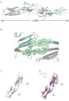
Figure 2. Structure of mB7H3
(A) Crystals of mB7H3 contain the dimeric form of the protein. Ribbon representation of the crystallographic dimer of mB7H3 with each protomer drawn in different colors (gray and cyan) with N and C-termini labeled for one of the protomers. Unmodeled residues 152–156 of the IgC domain of each protomer are shown with dots of corresponding color. The glycans (2 on each protomer) are drawn in stick representation. Strands making up the immunoglobulin fold in each domain are labeled in yellow, while the strand exchanged between the two protomers (G*) is labeled in pink. (B) Electron density of the extended FG-loop bridging sequence. FoFc electron density map, calculated with the FG-loop sequence omitted, contoured at +3σ is shown in green near the location of the strand exchange highlighted by a box in Fig. 2A. Ribbon diagrams representing the structure for each mB7H3 protomer are shown. The sidechains of the FG-loop residues are shown in gray stick-figure representation. See also Fig. S1. (C) Structure-based model of monomeric mB7H3. Model of the mB7H3 monomer was constructed by combining residues 35–125 from one protomer and residues 130–238 from its 2-fold-related symmetry mate. (D) Monomeric model ofmB 7H3 corresponds well to the PD-L1 structure. Structural alignment (overall Cα r.m.s.d. ~2.7 Å) of the monomeric mB7H3 model (gray) and the structure of human PD-L1 (purple).
Figure 3
Figure 3. Functional characterization of mB7H3 wild-type and mutants
(A) CD4+ T-cell proliferation is inhibited by monomeric and dimeric mB7H3 in a dose-dependent manner. CD4+OT-II cells were cultured in plasticware pretreated with the proteins indicated below the x-axis. T-cell proliferation was assayed by pulsing with [3H]-thymidine during the last 18 hours of a 3-day incubation. Average values obtained from 4 replicates are shown with error bars representing the standard error of mean. (B) Mapping of “front-sheet” and FG-loop mutations on the structure of mB7H3. The N-terminal IgV domain of mB7H3 monomer is shown in ribbon diagram (gray). Residues of the FG loop (red) and mutated “front-sheet” positions (blue) are labeled and shown with sidechains represented as stick figure. (C) T-cell proliferation is inhibited by wild-type, but not the FG-loop chimera of mB7H3. CD4+OT-II T cells were cultured in 96-well plates precoated with 0.5 μg/mL anti-CD3 and 5 μg/mL proteins indicated along the x-axis. T-cell proliferation was assayed by pulsing with [3H]-thymidine during the last 18 hours of a 3-day incubation. Data were normalized to the mean value obtained for anti-CD3 alone (PBS). Average values obtained from 2 experiments (4 replicates per sample per experiment) are shown with error bars representing the standard error of mean. Statistically significant differences (p < 0.05, ANOVA) with respect to the wild-type mB7H3 sample (double dagger) are labeled with asterisks. See also Fig. S2.
Figure 4
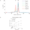
Figure 4. mB7H3 forms stable dimers in solution
(A) Purified mB7H3 dimerizes during incubation. Protein samples were concentrated to ~1.6 mM (60 mg/mL) and incubated at 4°C. SEC traces represent experiments carried out at the timepoints indicated. Monomer (M) and dimer (D) peaks are labeled. (B) Wild-type mB7H3 dimerizes over time in a concentration-dependent manner. SEC timecourse data similar to those shown in (A) were obtained for the indicated concentrations of wild-type mB7H3. Each data point represents the proportion of dimer observed (as measured by dimer peak height) compared to the total (monomer peak height + dimer peak height).
Figure 5
Figure 5. Sequence conservation of the FG loop
(A) Sequence of the B7-H3 FG loop is conserved. ClustalW (Larkin et al., 2007; Goujon et al., 2010) was used to align the mB7H3 sequences from 8 mammalian species and the resulting alignment is presented using ESPript (Gouet et al., 1999). Secondary structural elements of the mB7H3 structure are labeled (top line). Sequence differences are shown as red lettering on black background. The 4 FG-loop residues are highlighed with yellow background. (B) Sequence of the FG loop is divergent between B7 family members. ClustalW (Larkin et al., 2007; Goujon et al., 2010) sequence alignment of murine and human sequences of B7-H3 and PD-L1 is shown. Positions identical in all sequences are shown in green lettering with gray background. The 4 FG-loop residues are highlighted with yellow background.
Figure 6
Figure 6. Functional characterization of mB7H3 monomer and dimer
(A) Mapping FG-loop mutations on the structure of mB7H3. The N-terminal IgV domain of mB7H3 monomer is shown in ribbon diagram (gray). Residues of the FG loop (black) are labeled and shown with sidechains represented as stick figure. (B) Mutations at the FG loop disrupt dimerization of mB7H3. Dimer formation was measured for mB7H3 FG-loop mutants using experiments similar to those shown in Fig. 4A. (C) T-cell proliferation is inhibited by wild-type, but not the FG-loop chimera of mB7H3. CD4+OT -II cells were cultured in 96-well plates precoated with 0.5 μg/mL anti-CD3 and 5 μg/mL of the proteins indicated along the x-axis. T-cell proliferation was assayed by pulsing with [3H]-thymidine during the last 18 hours of a 3-day incubation. Data were normalized to the mean value obtained for the treatment with anti-CD3 alone (PBS). Average values obtained from 2 experiments (4 replicates per sample per experiment) are shown with error bars representing the standard error of mean. Statistically significant differences (p < 0.05, ANOVA) with respect to the wild-type mB7H3 sample (double dagger) are labeled with asterisks.
Figure 7
Figure 7. Strand-swapped dimer formation among the members of the immunoglobulin superfamily
Structures of strand-swapped dimers of IgV domains. N-terminal IgV domain dimer of mB7H3 (A), CD47 (B), CTLA-4 (C) and llama VHH-R9 immunoglobulin (D). Each protomer is labeled in different colors (green and red), with N- termini labeled. Strands making up the the two β-sheets of the immunoglobulin fold are labeled (yellow) and the swapped strand (G*) is labeled in black.

References

    1. Barach YS, Lee JS, Zang X. T cell coinhibition in prostate cancer: new immune evasion pathways and emerging therapeutics. Trends in Molecular Medicine. 2010;17:47–55. - PMC - PubMed
    1. Battye TGG, Kontogiannis L, Johnson O, Powell HR, Leslie AGW. iMOSFLM: a new graphical interface for diffraction-image processing with MOSFLM. Acta Crystallogr D Biol Crystallogr. 2011;67:271–281. - PMC - PubMed
    1. Bennett MJ, Sawaya MR, Eisenberg D. Deposition diseases and 3D domain swapping. Structure. 2006;14:811–824. - PubMed
    1. Chapoval AI, Ni J, Lau JS, Wilcox RA, Flies DB, Liu D, Dong H, Sica GL, Zhu G, Tamada K, et al. B7-H3: a costimulatory molecule for T cell activation and IFN-gamma production. Nat Immunol. 2001;2:269–274. - PubMed
    1. Chattopadhyay K, Lázár-Molnár E, Yan Q, Rubinstein R, Zhan C, Vigdorovich V, Ramagopal UA, Bonanno J, Nathenson SG, Almo SC. Sequence, structure, function, immunity: structural genomics of costimulation. Immunol Rev. 2009;229:356–386. - PMC - PubMed

Publication types