. 2013 May 9;50(3):379-93.
doi: 10.1016/j.molcel.2013.03.010. Epub 2013 Apr 11.

Principles of cotranslational ubiquitination and quality control at the ribosome

Affiliations

Principles of cotranslational ubiquitination and quality control at the ribosome

Stefanie Duttler et al. Mol Cell. .

Abstract

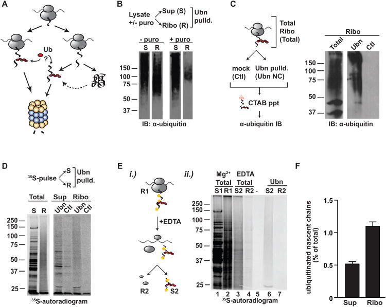
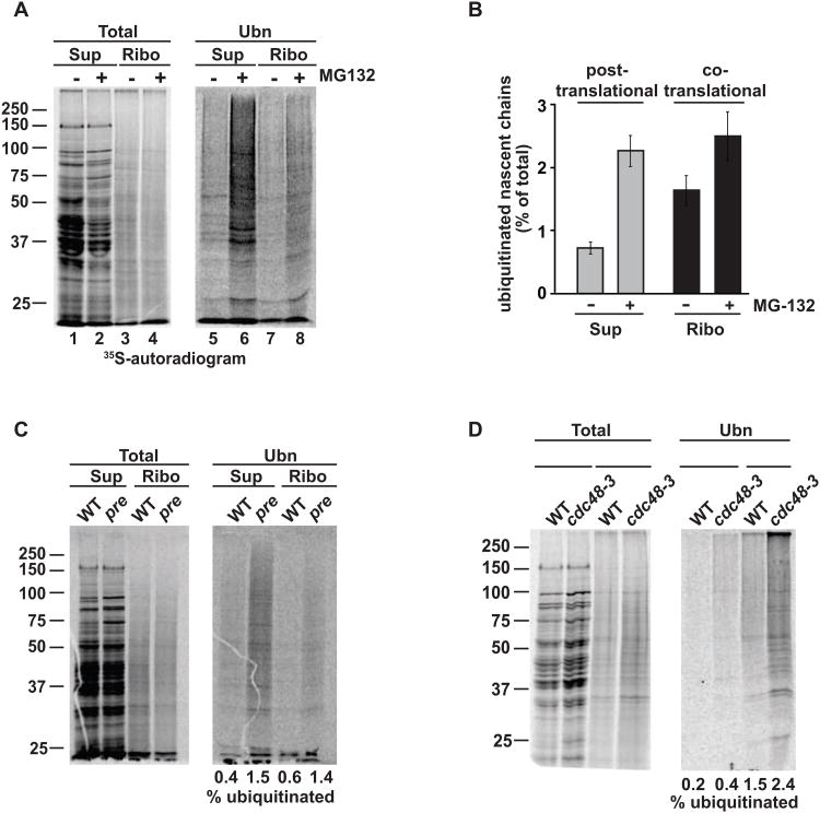
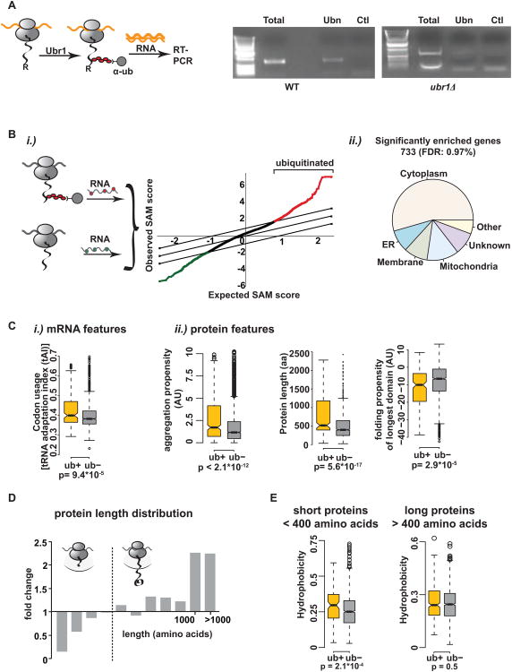
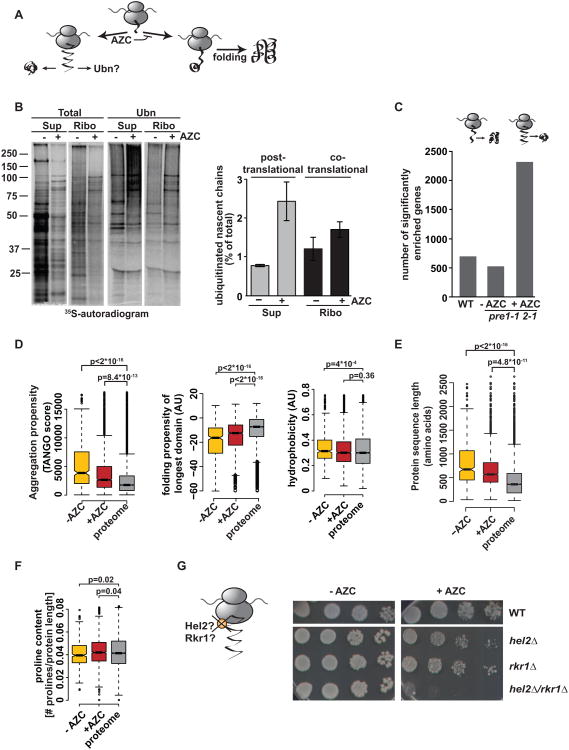
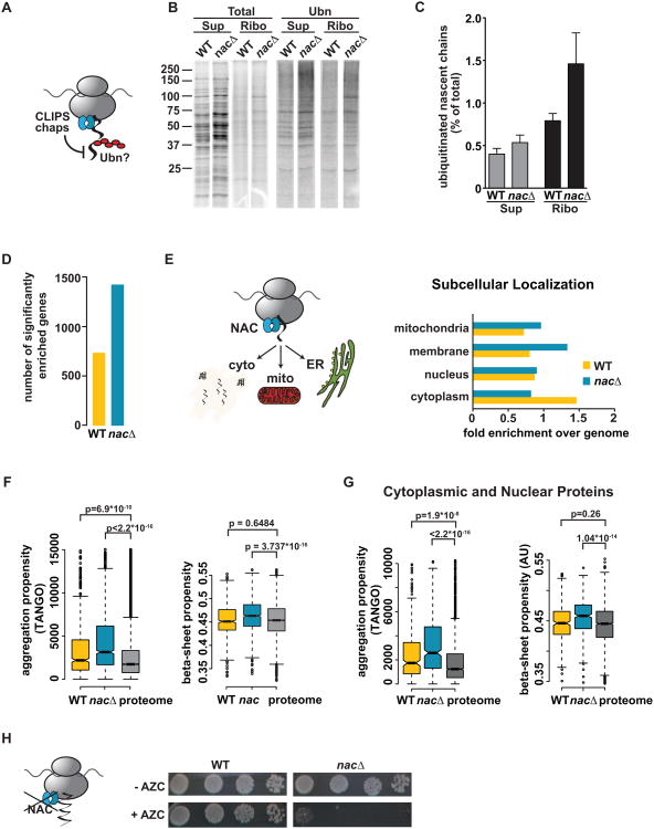
Achieving efficient cotranslational folding of complex proteomes poses a challenge for eukaryotic cells. Nascent polypeptides that emerge vectorially from the ribosome often cannot fold stably and may be susceptible to misfolding and degradation. The extent to which nascent chains are subject to cotranslational quality control and degradation remains unclear. Here, we directly and quantitatively assess cotranslational ubiquitination and identify, at a systems level, the determinants and factors governing this process. Cotranslational ubiquitination occurs at very low levels and is carried out by a complex network of E3 ubiquitin ligases. Ribosome-associated chaperones and cotranslational folding protect the majority of nascent chains from premature quality control. Nonetheless, a number of nascent chains whose intrinsic properties hinder efficient cotranslational folding remain susceptible for cotranslational ubiquitination. We find that quality control at the ribosome is achieved through a tiered system wherein nascent polypeptides have a chance to fold before becoming accessible to ubiquitination.


Figures

Figure 1
Figure 1. Cotranslational ubiquitination occurs at low levels in vivo
(A) Nascent chains may be ubiquitinated as they emerge from the ribosome or post-translationally once the protein has been fully synthesized (B) Ribosome-nascent chain complexes (RNCs) contain ubiquitinated material that is released by puromycin. Ultrasedimentation with or without puromycin to release nascent chains separated the supernatant (S) from the ribosome-nascent chain complexes (R), which were subjected to polyUb-affinity isolation (Ubn). Totals and pulldowns were analyzed by SDS-PAGE and ubiquitin immunoblot. (C) Peptidyl-tRNA isolation through CTAB precipitation shows direct ubiquitination of nascent chains. Isolated RNCs were incubated with polyUb-affinity resin or GST-only and the nascent chain-tRNA complexes precipitated with CTAB. Samples run on SDS-PAGE were immunoblotted for ubiquitin. (D) 35S–pulse-labeled nascent chains are ubiquitinated while still ribosome-bound. Cells were pulse-labeled for 1 min with 35S-Met/Cys; fractionated into supernatant (S or Sup), containing newly made, full-length 35S-polypeptides and RNCs (R or Ribo), containing ribosome bound 35S-nascent chains and pulled-down either with GST (Ctl) or polyUb-affinity resin (Ubn). (E) 35S-labeled nascent chains are directly ubiquitinated and can be isolated independently of the ribosome. i) Cells were pulse-labeled and fractionated as before; S: supernatant; R: ribosomal pellet. EDTA was added to the RNCs (R1), which was resuspended and spun again to yield empty ribosomes (R2) and released nascent chains (S2). S2 and R2 were then subject to polyUb-affinity pulldown. Samples were analyzed by SDS-PAGE and autoradiography. ii.) Autoradiogram showing totals of S1/R1 and S2/R2, as well as the polyUb-affinity pulldown of S2 and R2 (Ubn). (F) A small fraction of nascent chains and newly made proteins is cotranslationally ubiquitinated; calculated as the ratio of 35S-labeled nascent chains isolated by Dsk2 over total 35S-labeled nascent chains in the ribosomal or in the supernatant fraction (mean ± SEM, n=59). See also Figure S1.
Figure 2
Figure 2. Co- and post-translationally ubiquitinated nascent chains are degraded by the proteasome
(A) Ubiquitinated nascent chains and newly made proteins accumulate upon proteasome inhibition. Cells were pulse-labeled with 35S-Met/Cys with or without MG132. RNC isolation and polyUb-affinity isolation were performed as above and analyzed by SDS-PAGE and autoradiography. (B) Quantification of co- and post-translational ubiquitination in the presence and absence of proteasome inhibitors (mean ± SEM, n=3). (C) Genetic proteasome impairment in pre1-1 2-1 leads to accumulation of ubiquitinated nascent chains. Pulse-labeling and ubiquitin pulldown as described in (A). (D) Impairment of CDC48 increases levels of ubiquitinated nascent chains and newly made proteins. Ubiquitin-pulldown after radiolabeling in WT and cdc48-3 was carried out as in (A). See also Figure S2
Figure 3
Figure 3. A network of ubiquitin mediates cotranslational ubiquitination
(A) WT and E3 ubiquitin ligase mutant strains were pulse-labeled with 35S-Met/Cys, and RNCs were isolated and subjected to polyUb-affinity isolation as before. Shown is a representative autoradiogram for pulldowns in different E3 ligase deletion strains. (B) Quantification indicates the strongest effect is observed for hel2Δ/rkr1Δ (red bar). * p < 0.05, (mean ± SEM, n=3). (C) Differential role of Rkr1, Hel2 and Not4 on the degradation of translation products of stalled or non-stop mRNAs. rkr1Δ abrogates degradation, while hel2Δ has a partial effect and not4Δ is without effect. (Left) Schematic of non-stop (NS) and stalled (K12) reporters (Bengtson and Joazeiro, 2010; Ito-Harashima et al., 2007). (Right) The indicated constructs were expressed in the presence or absence of MG132 and equal amounts of lysate analysed by SDS-PAGE and immunoblot anti-GFP. (D) Impairing mRNA quality control increases cotranslational ubiquitination. WT and indicated CCR4/NOT mutant strains were pulse-labeled with 35S-Met/Cys, RNCs isolated and subjected to polyUb-affinity isolation as described before. (E) Quantification: mean and SEM of 3 experiments. * p < 0.05. (F) not4Δ deletion is hypersensitive to AZC. Dilution series on YPD plates with or without 1 mg/ml AZC. Plates were imaged after 3 days at 30°C. See also Figure S3
Figure 4
Figure 4. Identification and characterization of ubiquitinated nascent chains
(A) (Left) Experimental setup: R-β-galactosidase is ubiquitinated cotranslationally by Ubr1. After RNC isolation with polyUb-affinity resin, RNA is extracted and reverse transcribed. (Right) RT-PCR detects cotranslational ubiquitination of R-β-galactosidase in WT but not in ubr1Δ cells. T = total, Ubn = polyUb-affinity isolation, Ctl = GST-pulldown. (B)i.) Globally identification of ubiquitinated nascent chains in vivo. After RNC isolation and ubiquitin-pulldown, RNA was extracted. An aliquot of mRNAs from the initial RNCs serves as total translation reference. Both RNA samples are reverse transcribed, labeled with Cy3 and Cy5, respectively and labeled cDNAs hybridized to DNA microarrays. The graph shows a representative SAM plot for 10 WT replicates with a false discovery rate (FDR) of 1%. Red = significantly positively enriched genes; green = significantly negatively enriched genes in the data set. A set of 733 proteins is consistently ubiquitinated cotranslationally (ub+ set). ii.) Subcellular distribution of cotranslationally ubiquitinated proteins in WT yeast. (C) Features in the mRNA and polypeptide distinguishing the ub+ set from cytosolic proteins not in the dataset (ub-): i.) mRNA: tRNA adaptation index. ii.) protein features: aggregation propensity, protein length and folding propensity of the longest domain. (D) Cotranslational ubiquitination is disfavored in shorter proteins. Size analysis of the identified genes from WT experiments. Fold change of amino acid length compared to the genome. (E) Shorter ubiquitinated nascent chains (encoding proteins <400 amino acids) tend to be more hydrophobic. The cytosolic ub+ dataset was split into proteins shorter and longer than 400 amino acids and the overall hydrophobicity was compared separately to proteins of similar size distribution in the ub-data set. See also Figure S4 and Table S1.
Figure 5
Figure 5. Impairing cotranslational folding increases cotranslational ubiquitination
(A) The proline analogue AZC impairs cotranslational folding. (B) AZC treatment leads to increased ubiquitination of 35S-labeled nascent chains and newly made proteins. (Left) Cells were pulse-labeled in the presence or absence of 1 mg/ml AZC. Supernatant (Sup) and RNC fractions (Ribo) were subject to polyUb-affinity isolation (Ubn), and analyzed by SDS-PAGE and autoradiography. (Right) Mean and SEM of 3 experiments. (C) Addition of AZC increases the number of mRNAs encoding cotranslationally ubiquitinated nascent chains. Number of identified genes in, pre1-1 2-1 − AZC and pre1-1 2-1 + AZC at 30°C. (D) Analysis of ub+ datasets in pre1-1 2-1 −/+ AZC identified as shown in (C). Comparison of aggregation propensity (TANGO score), folding propensity of the longest domain and hydrophobicity. (E) Size distribution of pre1-1 2-1 −/+ AZC datasets as in Figure 2. (F) AZC treatment increases the proline content of ubiquitinated nascent chains. (G) Role of Rkr1 and Hel2 in cotranslational quality control. hel2/rkr1Δ cells are hypersensitive to AZC. Plates were grown for 3 days at 30 °C.
Figure 6
Figure 6. The ribosome-associated chaperone NAC protects nascent chains from cotranslational ubiquitination
(A) Hypothesis: Ribosome-bound chaperones such as NAC protect nascent chains from ubiquitination (B) Pulse-labeling followed by polyUb-affinity isolation in egd1/2Δbtt1Δ (nacΔ) shows increased ubiquitination compared to WT. (C) Quantification: Mean ± SEM (n=4). (D) Deletion of NAC increases in the number of mRNAs encoding cotranslationally ubiquitinated nascent chains. (E) Deletion of NAC enhances ubiquitination of nascent chains trafficked to the secretory pathway and mitochondria. (Left) Role of NAC in targeting to the ER and mitochondria. (Right) Subcellular localization of ub+ proteins in WT and nacΔ datasets. (F) Deletion of NAC causes ubiquitination of nascent chains with very high aggregation- and beta-sheet propensity compared to the proteome. (G) As in (F) but analysis was restricted to cytoplasmic and nuclear proteins. (H) Synthetic effect of loss of NAC with impaired cotranslational folding: nacΔ cells are hypersensitive to AZC. Plates were grown for 3 days at 30 °C. See also Figure S5.
Figure 7
Figure 7. Balancing protein folding and cotranslational ubiquitination at the ribosome
Upon translation, most nascent chains are protected from degradation through cotranslational folding and interaction with NAC (shown here) and likely other ribosome-bound chaperones, which create a protected folding environment in the vicinity of the ribosomal exit site (shaded area). Damaged or non-stop mRNAs that escape mRNA quality control produce translation products eliminated by the ubiquitin ligase Rkr1. Most nascent chains fold quickly and efficiently with the assistance of chaperones or spontaneously. Nascent chains with features challenging efficient folding are susceptible to cotranslational ubiquitination by ribosome-bound and non-ribosome bound quality control ligases. See also Figure S6.

Comment in

References

    1. Albanese V, Yam AY, Baughman J, Parnot C, Frydman J. Systems analyses reveal two chaperone networks with distinct functions in eukaryotic cells. Cell. 2006;124:75–88. - PubMed
    1. Balch WE, Morimoto RI, Dillin A, Kelly JW. Science. Vol. 319. New York, NY: 2008. Adapting proteostasis for disease intervention; pp. 916–919. - PubMed
    1. Belle A, Tanay A, Bitincka L, Shamir R, O'Shea E. Quantification of protein half-lives in the budding yeast proteome. Proc Natl Acad Sci USA. 2006;103:13004–13009. - PMC - PubMed
    1. Bengtson MH, Joazeiro CAP. Role of a ribosome-associated E3 ubiquitin ligase in protein quality control. Nature. 2010;467:470–473. - PMC - PubMed
    1. Brandman O, Stewart-Ornstein J, Wong D, Larson A, Williams CC, Li GW, Zhou S, King D, Shen PS, Weibezahn J, et al. A ribosome-bound quality control complex triggers degradation of nascent peptides and signals translation stress. Cell. 2012;151:1042–1054. - PMC - PubMed

Publication types