. 2013 May 9;50(3):368-78.
doi: 10.1016/j.molcel.2013.03.009. Epub 2013 Apr 11.

A cotranslational ubiquitination pathway for quality control of misfolded proteins

Affiliations

A cotranslational ubiquitination pathway for quality control of misfolded proteins

Feng Wang et al. Mol Cell. .

Abstract

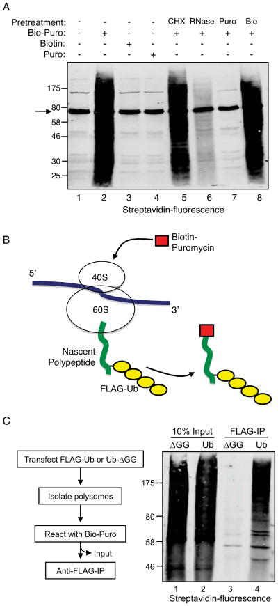
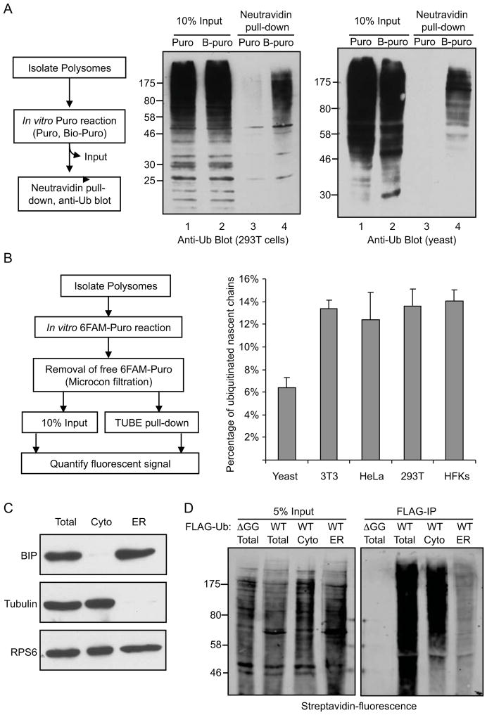
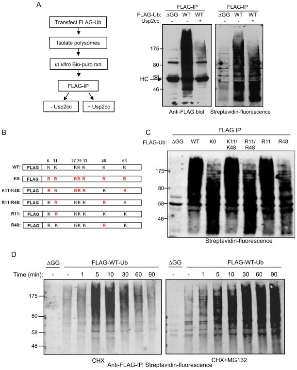
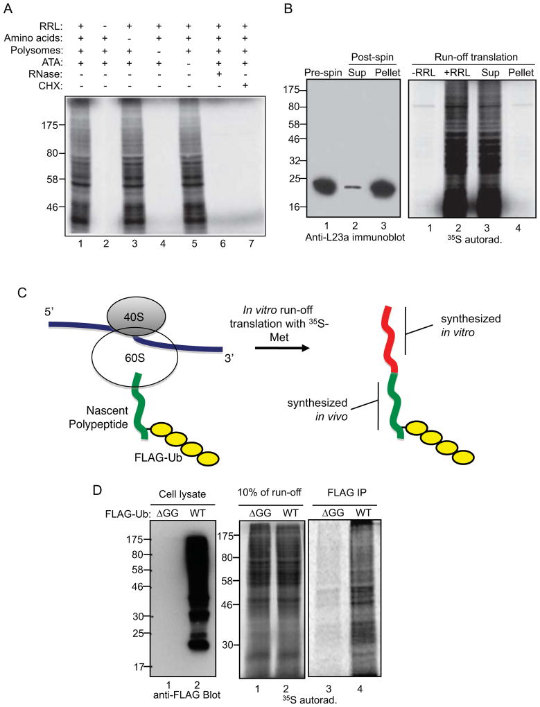
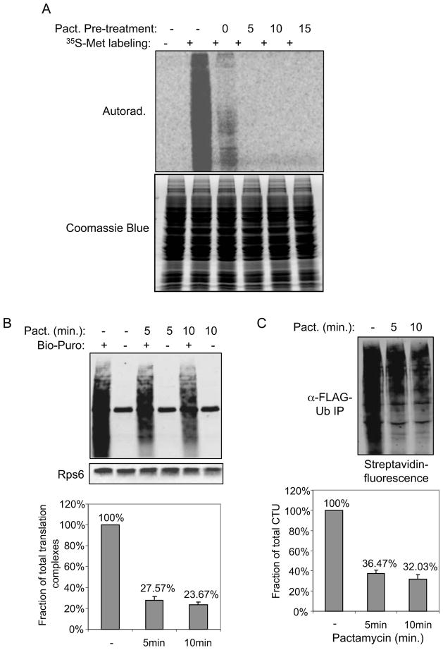
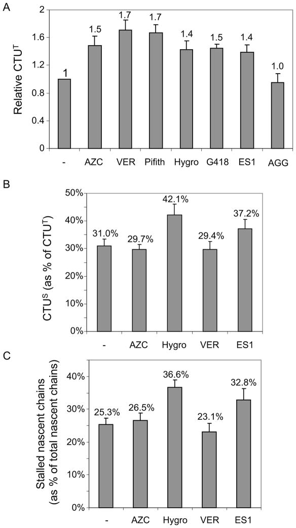
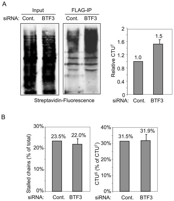
Previous studies have indicated that 6%-30% of newly synthesized proteins are rapidly degraded by the ubiquitin-proteasome system; however, the relationship of ubiquitination to translation for these proteins has been unclear. We report that cotranslational ubiquitination (CTU) is a robust process, with 12%-15% of nascent polypeptides being ubiquitinated in human cells. CTU products contained primarily K48-linked polyubiquitin chains, consistent with a proteasomal targeting function. While nascent chains have been shown previously to be ubiquitinated within stalled complexes (CTU(S)), the majority of nascent chain ubiquitination occurred within active translation complexes (CTU(A)). CTU(A) was increased in response to agents that induce protein misfolding, while CTU(S) was increased in response to agents that lead to translational errors or stalling. These results indicate that ubiquitination of nascent polypeptides occurs in two contexts and define CTU(A) as a component of a quality control system that marks proteins for destruction while they are being synthesized.


Figures

Figure 1
Figure 1. Co-translational ubiquitination (CTU) in human cells
A. Validation of in vitro biotin-puromycin (Bio-Puro) conjugation reaction. Polysomes from HEK293T cells were used in all reactions. Reaction with Bio-Puro is shown in lane 2; Bio-Puro was deleted in lane 1, and replaced by free biotin (Bio) in lane 3 or by untagged puromycin (Puro) in lane 4. Polysomes were pretreated with cycloheximide (CHX), RNase, untagged puromycin or biotin in lane 5, 6, 7, and 8 respectively. Bio-Puro was detected with fluorescently-labeled streptavidin. Background bands seen, for example, in lane 1, represent endogenous biotin-conjugated proteins (e.g., arrow at 74 kD represents priopionyl coA carboxylase). B. Scheme for detection of CTU. If CTU occurs in vivo, polysomes should contain nascent polypeptides modified with both FLAG-ubiquitin and Bio-Puro. C. Polysome-associated nascent polypeptides are conjugated to FLAG-Ub. Cells were transfected with plasmids expressing wild type FLAG-Ub (Ub) or FLAG-Ub-ΔGG (ΔGG). Nascent chains were labeled with Bio-Puro in vitro, then immunoprecipitated with anti-FLAG antibody, and blotted with fluorescent streptavidin (lanes 3 and 4). Lanes 1 and 2 represent 10% of input levels of Bio-Puro labeled nascent chains (prior to FLAG immunoprecipitation). See also Figure S1.
Figure 2
Figure 2. Polysome-associated nascent polypeptides are conjugated to endogenous ubiquitin in human and yeast cells
A. Polysomes from untransfected HEK293T or S. cerevisiae cells were incubated with either Bio-Puro or untagged Puro to label associated nascent chains. Biotin labeled polypeptides were isolated by neutravidin pull down and analyzed by anti-ubiquitin immunoblotting. B. Quantitation of CTU in different cell types. Polysomes were isolated from the indicated cell types, and polysome-associated nascent chains were labeled with fluorescently-tagged puromycin (6-FAM-dC-Puro). 10% of the total reactions were used to estimate the amount of total nascent chains (input); the remainder was subject to TUBE pull down to isolate ubiquitinated proteins, and fluorescence intensity was measured. Error bars indicate standard error of the mean (SEM) of three independent experiments. C. CTU occurs predominantly on cytosolic rather than ER-associated polysomes. Fractionation of cytosolic and ER-associated polysomes was validated by immunoblotting with antibodies that recognize tubulin (cytosolic protein), Bip (ER luman protein) and RPS6 (small ribosomal protein). D. Cytosolic and ER-associated polysomes were isolated from HEK293T cells expressing FLAG-Ub. Polysome-associated nascent chains were labeled with Bio-Puro and then immunoprecipitated with anti-FLAG antibody. Left panel is 5% input of total Bio-Puro conjugation products, and right panel represents immunoprecipitation of nascent proteins that were modified with both FLAG-Ub and Bio-Puro. See also Figure S2.
Figure 3
Figure 3. CTU products contain primarily K48-linked polyubiquitin chains
A. CTU target proteins are multubiquitinated. Ubiquitinated nascent polypepetides were labeled with Bio-Puro, and immunoprecipitated with anti-FLAG antibody. The immunoprecipitated products were treated with the catalytic domain of ubiquitin-specific protease 2 (USP2cc) to strip ubiquitin (see schematic, left). Middle panel is the anti-FLAG-Ub blot analysis of samples before and after USP2cc treatment (HC is IgG heavy chain). Right panel shows the fluorescent-streptavidin blot of the FLAG IPs. The molecular weight change of CTU products after USP2cc treatment indicates that the majority of the CTU products contained multiple ubiquitin moieties. B. Schematic diagram of mutant forms of FLAG-ubiquitin expressed in HEK293T cells. C. Polysomes were isolated from cells expressing the indicated form of ubiquitin. Nascent chains were labeled with Bio-Puro in vitro, and then immunoprecipitated with anti-FLAG antibody. The immunoprecipitated products were subject to SDS-PAGE, and detection was with fluorescent streptavidin. All exogenously expressed ubiquitin mutants were expressed at similar levels (not shown). D. HEK293T cells were transfected with plasmids expressing FLAG-Ub or FLAG-Ub-ΔGG. Prior to lysis, cells were treated with CHX alone or simultaneously with CHX and MG132 for the indicated times. Polysomes were isolated from these cells and reacted with Bio-Puro. Nascent polypeptides were analyzed by anti-FLAG immunoprecipitation and detection with fluorescent streptavidin. See also Figure S3.
Figure 4
Figure 4. CTU occurs within active translation complexes
A. Requirements for in vitro run-off translation reactions. A complete run-off reaction is shown in lane 1. A single component was deleted in lane 2 (rabbit reticulocyte lysate; RRL), lane 3 (exogenous amino acids), lane 4 (polysomes), and lane 5 (aurintricarboxylic acid; ATA). RNase or CHX was added to the reactions in lanes 6 and 7. B. Ribosomes present in RRL are not required for the run-off reaction. Ribosomes were depleted from RRL by ultracentrifugation, and untreated RRL or ribosome-depleted supernatant (Sup) was used in run-off reactions. Left panel shows an immunoblot for the L23a ribosomal protein in RRL (pre-spin), the supernatant (Sup), and the pellet. C. The CTU model predicts that if ubiquitin is conjugated to nascent chains on actively translating polysomes and translation can proceed following CTU, then in vitro run-off products will contain both ubiquitin and 35S-methionine. Green represents the portion of the polypeptide chain translated in vivo, and red represents the 35S-methionine-containing polypeptide translated in vitro. D. Cells were transfected with plasmids expressing FLAG-Ub (WT) or FLAG-Ub-ΔGG (ΔGG), and polysomes from these cells were used in run-off reactions. Left panel shows an anti-FLAG immunoblot of total cell extracts, center panel shows 10% of the run-off reactions, and right panel shows the anti-FLAG immunoprecipitates of the run-off reactions.
Figure 5
Figure 5. CTUA accounts for the majority of the total ubiquitination activity against nascent chains
A. A brief treatment of cells with pactamycin was sufficient to allow run off of actively translating nascent chains. Cells were treated with pactamycin for the indicated times, followed by a 5 min. labeling with 35S-methionine. In lane 3, pactamycin was added simultaneously with the 35S-Met. Total cell lysate was subject to SDS-PAGE, and analyzed by autoradiography. Coomassie Blue staining shows proteins were loaded equally in each lane. B. A significant amount of nascent chains are in stalled ribosomes. Cells were either untreated or pretreated with pactamycin for 5 or 10 min. to run off of actively translating ribosomes. Polysomes were isolated from these cells and incubated with Bio-Puro in vitro to label associated nascent chains. Biotin-labeled nascent chains were subject to SDS-PAGE, and blotted with fluorescent streptavidin. A blot for a ribosomal protein, Rps6, is shown as a loading control. Error bars indicate SEM of three independent replicates. C. CTUS accounts for approximately one-third of CTUT. Cells expressing FLAG-Ub were treated with pactamycin for 10 min. and polysome-associated nascent chains were labeled with Bio-Puro in vitro and then immunoprecipitated with anti-FLAG antibody. The immunoprecipitated products were subject to SDS-PAGE analysis, and the fluorescent streptavidin signal was quantitated relative to cells that were not treated with pactamycin. Error bars represent SEM of three independent experiments.
Figure 6
Figure 6. CTU was enhanced under conditions that promoted translational errors or protein misfolding
A. CTUT was analyzed after treating cells for 60 min. with either AZC, the Hsp70 chaperone inhibitors VER155008 (VER) or Pifithrin (Pfith), hygromycin B (Hygro), G418, eeyarestatin 1 (ES1) or the Hsp90 inhibitor 17-AGG (AAG). CTU products were quantified by measuring fluorescent-streptavidin signal on blots, and relative signals were normalized in all panels to signal in the absence of inhibitor. Error bars indicate SEM of three independent replicates. B. Cells expressing FLAG-Ub were treated with the indicated agents for 60 min., and CTUS was quantitated as in Figure 6. Error bars indicate SEM of three independent experiments. C. Cells were treated with the indicated agents for 60 min., and the stalled nascent chains were quantitated relative to total nascent chains. Error bars indicate SEM of three independent experiments. See also Figure S4.
Figure 7
Figure 7. Depletion of NAC activity enhances CTUT
A. 293T cells were transfected with siRNAs targeting the mRNA for BTF3 (the β subunit of the NAC) or a control siRNA, and the FLAG-Ub plasmid DNA was transfected 15 hours later. Cells were harvested 48 hours post-siRNA transfection, and CTU products were analyzed by anti-FLAG immunoprecipitation followed by quantitation of Bio-Puro incorporation with fluorescent-streptavidin. Signal was normalized in all panels to that of the control siRNA. Error bar indicates SEM of three independent experiments. BTF3 siRNA knockdown was confirmed by immunoblotting (Figure S5). B. Total stalled nascent chains and CTUS was analyzed in BTF3 or control siRNA-treated cells, using a 10 min. pactamycin treatment (as in Figure 6). See also Figure S5. Error bars indicate SEM of three independent replicates.

Comment in

References

    1. Balch WE, Morimoto RI, Dillin A, Kelly JW. Adapting proteostasis for disease intervention. Science (New York, NY. 2008;319:916–919. - PubMed
    1. Behrends C, Harper JW. Constructing and decoding unconventional ubiquitin chains. Nat Struct Mol Biol. 2011;18:520–528. - PubMed
    1. Bengtson MH, Joazeiro CA. Role of a ribosome-associated E3 ubiquitin ligase in protein quality control. Nature. 2010;467:470–473. - PMC - PubMed
    1. Brandman O, Stewart-Ornstein J, Wong D, Larson A, Williams CC, Li GW, Zhou S, King D, Shen PS, Weibezahn J, et al. A ribosome-bound quality control complex triggers degradation of nascent peptides and signals translation stress. Cell. 2012;151:1042–1054. - PMC - PubMed
    1. Brodersen DE, Clemons WM, Jr, Carter AP, Morgan-Warren RJ, Wimberly BT, Ramakrishnan V. The structural basis for the action of the antibiotics tetracycline, pactamycin, and hygromycin B on the 30S ribosomal subunit. Cell. 2000;103:1143–1154. - PubMed

Publication types

MeSH terms