. 2013 Apr 10;78(1):109-23.
doi: 10.1016/j.neuron.2013.01.036.

Transglutaminase and polyamination of tubulin: posttranslational modification for stabilizing axonal microtubules

Affiliations

Transglutaminase and polyamination of tubulin: posttranslational modification for stabilizing axonal microtubules

Yuyu Song et al. Neuron. .

Abstract

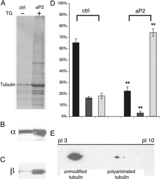
Neuronal microtubules support intracellular transport, facilitate axon growth, and form a basis for neuronal morphology. While microtubules in nonneuronal cells are depolymerized by cold, Ca(2+), or antimitotic drugs, neuronal microtubules are unusually stable. Such stability is important for normal axon growth and maintenance, while hyperstability may compromise neuronal function in aging and degeneration. Though mechanisms for stability are unclear, studies suggest that stable microtubules contain biochemically distinct tubulins that are more basic than conventional tubulins. Transglutaminase-catalyzed posttranslational incorporation of polyamines is one of the few modifications of intracellular proteins that add positive charges. Here we show that neuronal tubulin can be polyaminated by transglutaminase. Endogenous brain transglutaminase-catalyzed polyaminated tubulins have the biochemical characteristics of neuronal stable microtubules. Inhibiting polyamine synthesis or transglutaminase activity significantly decreases microtubule stability in vitro and in vivo. Together, these findings suggest that transglutaminase-catalyzed polyamination of tubulins stabilizes microtubules essential for unique neuronal structures and functions.


Figures

Figure 1
Figure 1. Effects of Difluoromethylornithine (DFMO) on microtubule stability in 3-month-old rat optic nerve
A) Scheme for fractionation of axonal tubulin in Soluble (S1), Cold Stable (S2) and Cold/Ca2+ Stable (P2, CST) tubulin. B) Fluorographs of tubulin fractions from optic nerve labeled by slow axonal transport (21 days after injection of 35S-methionine in the eye of control or DFMO treated rats (7D pretreatment). Tubulin and neurofilament triplet proteins are the major labeled protein species in the P2 fraction of control nerves. Tubulin consistently shifted from P2 to S1 with DFMO treatment, whereas neurofilaments remain in P2. C) Compared with control, DFMO treatments for both 7 and 21 days significantly decreased stable tubulin levels without affecting neurofilament fractionation (see suppl. table 1). Statistical analysis was by Student T-test. **denotes P<0.001. See also Table S1 and fig S1.
Figure 2
Figure 2. Covalent incorporation of radiolabeled PUT into rat optic nerve with tubulin as a putative substrate
A) A protein of tubulin size was labeled with 3H-PUT (left) while 35S-Methionine labeled total proteins (right). 3H-PUT was incorporated into axonal tubulin mainly in cold/Ca2+ stable tubulin fractions (P2). 14C-PUT modified axonally transported P2 tubulin in a similar pattern (not shown). B) Upper panel showed gel filtration chromatography of 14C-labeled P2 proteins in rat optic nerve at 21-day ISI indicating a single broad peak of radioactivity. Lower panel showed that the peak of eluted radioactivity coincided with tubulin immunoreactivity, consistent with covalent incorporation of polyamines into tubulin in vivo.
Figure 3
Figure 3. In vitro polyamination of neuronal tubulin and MTs and transglutaminase activity
A) SDS-PAGE showed a fluorescent band at tubulin MW indicating covalent addition of Monodansylcadaverine (MDC) to purified mouse tubulin and MTs with transglutaminase (gpTG) activity. This modification required Ca2+ to activate transglutaminase and was also seen with pig brain tubulin and MTs. B) Spermine (SPM) and Spermidine (SPD) were also incorporated into neuronal tubulin. Immunoblotting for polyamine (red) and tubulin (green) showed polyaminated tubulins in yellow (merge), also see fig S2. Neuronal MTs polymerized in vitro showed comparable tubulin modifications (not shown). C) gpTG-catalyzed covalent addition of MDC was comparable for tubulin and taxol-stabilized MTs. Polyaminated proteins showed decreased solubility and were enriched in pellets. D) Coomassie blue stained gel (left) showed similar amounts of tubulin and taxol-stabilized MTs as substrates for polyamination in C. In the absence of polyamines, transglutaminase catalyzed cross-linking of tubulins (right), but cross-linked tubulins were primarily in supernatants and may not enter the stacking gel (not shown). E) Negative stain electron microscopy (EM) showed polymerized MTs with polyaminated tubulin. Several different magnification images show that many MTs are present in this fraction. F) Negative stain EM showed cross-linked tubulin aggregates by transglutaminase in the absence of polyamines.
Figure 4
Figure 4. Polyamination sites mapped on tubulins by Liquid Chromatography-Tandem Mass Spectrometry
Trypsin-digested peptides from in vitro modified mouse brain tubulin and in vivo stable and labile MT fractions were subjected to LC-MS. Several putative modification sites from different tubulin isoforms were identified based on the mass shift. A representative modification site on a conserved glutamine residue (Q) in N-terminal β-tubulins is illustrated. A) A diagram showing sequence ions for peptide EIVHIQAGQCGNQIGAK (PEIVH). Based on accurate mass for sequence ions (see below), the peptide contained unmodified Qs at positions 6 and 9, leaving only Q at position 13 (in red, Q15 in the β-tubulin sequence) as a modification site with PUT in vitro. B) Targeted tandem MS spectrum on Ions derived from PUT modified PEIVH (PUT- PEIVH) with m/z 632.0043+ (peptide mass 1892.989, 1.06 ppm error) showed an accurate mass shift of 199.1321 from y4 to y5, suggesting an additional shift of 71.07 as a result of one PUT (mass: 88.15148) added to Q15 with a loss of one NH3 (mass: 17.0306) C, D) Chromatograms confirmed the presence of PUT- PEIVH in brain stable MT fraction (P8), suggesting the same modification occurs in vivo and that the modification is sequence specific. Detailed modification patterns on the precursor and its ion series were shown in D, peaks corresponding to SPM modified peptide (SPM- PEIVH) were observed in the same fractions. E) A summary of peak areas for polyaminated PEIVH from mouse brain MT fractions showed significantly higher levels of PUT- PEIVH and SPM- PEIVH in stable MT fractions (P) compared with labile MT fractions (S). F) Putative polyaminated sites on tubulins were shown with regard to the protein structure. Using coordinates from the Protein Structure database (MMDB ID: 8900; PDB ID: 1TUB) for predicted structures for taxotere-bound tubulin dimer (Nogales et al., 1998) the conserved Q15 residue is located adjacent to phosphates on the exchangeable GTP in β-tubulin. The structure was generated using Cn3D software (National Library of Medicine). This location suggests that positively charged polyamine might stabilize GTP and affect hydrolysis, consistent with a role in modulating microtubule dynamics. Circles are superimposed at Q15 to illustrate approximate dimensions of putrescine (red dashed circle) and spermine (grey circle). Note the potential for interacting with β-tubulin GTP (see also fig S3 and table S2–S4).
Figure 5
Figure 5. Biochemical similarities between in vitro polyaminated tubulins and in vivo neuronal cold stable tubulins
Mouse brain soluble tubulins were polyaminated in vitro by endogenous mouse brain transglutaminase/polyamine and subjected to cold Ca2+ fractionation (fig S4). A) Coomassie blue stained gel; B) Immunoblots with DM1A (α-tubulin); and C) Tu27 (β-tubulin). Compared to controls without transglutaminase activation by addition of Ca2+, modified tubulins showed a remarkable increase in the cold/Ca2+ stable fraction (P2a). D) Quantitation of α-tubulin showed >70% soluble tubulins were converted to cold/Ca2+ stable tubulins (P fraction in aP2 group) after polyamination while most tubulin remained soluble with cold (ctrl S1) or Ca2+ (ctrl S2) in the control group. Statistical analysis was by Student T-test. **denotes P<0.001. E) 2D PAGE showed a shift towards a basic pI for polyaminated tubulins, consistent with added positive charge on in vivo cold/Ca2+ stable tubulin (see also fig S2).
Figure 6
Figure 6. Characterization of transglutaminase activity and TG2 protein levels in the nervous system
A) Cold/Ca2+ stable tubulin was enriched in brain but was at or below levels of detection in non-neuronal tissues other than testes. The stable tubulin fraction in testes is presumably due to the stable microtubules in sperm flagella and may not be equivalent to the brain fraction. However, polyamines and TG2 are present in testes. B) Transglutaminase activity was present in various neural tissues including cerebral cortex (CC), brain stem (BS), spinal cord (SC), optic nerve (ON) and sciatic nerve (SN) from 5-week-old mice. C) Quantitative plots showed that transglutaminase activity was higher in axon-enriched compartments, like ON and SN, correlated with higher MT stability in axons. D) TG2 protein was also expressed differentially in various neural tissues. The same amount of total protein was loaded in each lane. Actin and GAPDH were loading controls. Actin was higher in axon-rich domains (ON and SN), while GAPDH was lower in axon-rich tissues as a fraction of total protein. E) Quantitative comparison shows TG2/actin ratio was lower in SN than ON but SN exhibited comparable levels of TG activity (C), thus other transglutaminase isoforms may contribute to axonal transglutaminase activity, particularly in the PNS. Statistical analysis was by Student T-test. **denotes P<0.001.
Figure 7
Figure 7. Effects of transglutaminase inhibition on neurite extension in SH-SY5Y cells
To determine functions of polyamination and MT stability in neuronal development, we differentiated SH-SY5Y neuroblastoma in the presence of IR072 (fig S5), an irreversible transglutaminase inhibitor. A) Control SH-SY5Y cells were differentiated, fixed and stained with beta III tubulin antibody. Normal neurite extension and neuron-like phenotypes were observed. B) SH-SY5Y cells differentiated in the presence of the IR072 showed significant inhibition of neurite outgrowth and a phenotype more like undifferentiated SH-SY5Y cells. C) Length distribution of neurites for control SH-SY5Y cells showed an average of 76μm with some neurites >150μm. D) Length distribution of neurites for differentiated SH-SY5Y cells treated with IR072 showed a significant shift towards shorter neurites with an average length of 29μm with few neurites >90μm. The difference in mean neurite length for control and IR072 treated cells was statistically significant (P<0.00001). This suggests that transglutaminase activity is required for efficient elongation of neurites during differentiation of SH-SY5Y cells. E) Cold/Ca2+ fractionations were done on SH-SY5Y cells under 3 conditions (undifferentiated, differentiated, differentiated with IR072). Western blots with DM1A antibody (α-tubulin) showed that: 1) in undifferentiated cells with low transglutaminase activity and TG2 protein levels, <10% of total tubulin was cold stable, with no cold/Ca2+ stable tubulin. 2) In fully differentiated cells where both transglutaminase activity and TG2 proteins level peaked, cold stable tubulin level increased significantly to >40% of total tubulin and cold/Ca2+stable tubulin was >20% of total tubulin); 3) In differentiated cells treated with IR072 where transglutaminase activity was significantly reduced, cold/Ca2 stable tubulin was dramatically lowered <10%. This suggests a strong correlation between transglutaminase activity and microtubule stability, both of which may contribute to neurite development.
Figure 8
Figure 8. Neuronal MT stability in 5wk and 5mo TG2 KO mouse brains
A) Immunoblots documented absence of TG2 immunoreactivity inTG2 KO mouse brain and spinal cord. In WT controls, brain TG2 was expressed at similar levels in both age groups but spinal cord TG2 levels dropped at 5mo relative to 5wk mice. B) transglutaminase activity was significantly reduced in TG2 KO mouse brains compared with age matched WT. Brain transglutaminase activities were similar between two ages paralleling TG2 protein levels. C) Spinal cord transglutaminase activity was much lower in TG2 KO mice than WT at 5wk, but transglutaminase activity was comparable in 5mo TG2 KO and WT spinal cord. D) Brain cold and cold/Ca2+ stable tubulin levels decreased drastically in TG2 KO mice for both age groups, consistent with changes in TG2 protein and transglutaminase activity. Residual transglutaminase activity due to other transglutaminase isoforms may contribute to cold/Ca2+ stable tubulin formation. E) Spinal cord cold stable tubulin and cold/Ca2+ stable tubulin levels decreased in 5wk TG2 KO mice as compared to age matched WT, but levels of stable tubulins in 5mo TG2 KO and WT groups were not significantly different, consistent with TG2 protein expression and transglutaminase activity. Other transglutaminase isoforms may make a greater contribution to MT stability in adult spinal cord. Statistical analysis was by Student T-test. **denotes P<0.001, *denotes P<0.005.
Figure 9
Figure 9. Characterization of transglutaminase activity, TG2 protein and microtubule stability in postnatal mouse brain development
To evaluate changes in transglutaminase activity and TG2 protein as neurons develop and mature, mouse brain was analyzed at 10d (pre myelination); 3wk (active myelination) and 3mo (mature adult neurons). A) Fluorescent intensity of MDC incorporated into casein shows significantly increased transglutaminase activity during postnatal brain development. During this interval, neurite outgrowth plays a minimal role, but consolidation of synaptic connections and myelination progresses toward adult levels. Changes in transglutaminase activity during this time may be related to axonal maturation, correlated with myelination, B) Quantitative analysis show that transglutaminase activity increased 1.5 and 3 fold in 3wk and 3mo brains respectively, as compared with 10d mouse brain. C) Consistent with changes in transglutaminase activity, TG2 protein levels in western blots increased. Kinesin (H2) immunoreactivity was a loading control. D) Similar changes in TG2 protein and activity levels occurred during postnatal mouse brain development. E) Alteration of MT stability during postnatal mouse brain development. Cold/Ca2+ fractionations were performed on mouse brain from the 3 age groups and analyzed by immunoblot with DM1A antibody, which recognizes α-tubulin. Cold stable and cold/Ca2+ stable tubulin levels are 45% and 25% of total tubulin at 10d, respectively. At 3wk, cold stable tubulin increased to 58% of total tubulin, of which cold/Ca2+ stable tubulin was 32%. At 3mo, cold stable tubulin increased to 64%, while cold/Ca2+ tubulin increased to 36%. Differences are statistically significant (** P< 0.001 between 10-day and 3-week groups, * P<0.05 between 3-week and 3-month groups) n = 6.

Comment in

References

    1. Ambron RT, Kremzner LT. Post-translational modification of neuronal proteins: Evidence for transglutaminase activity in R2, the giant cholinergic neuron of Aplysia. Proc Nat Acad Sci USA. 1982;79:3442–3446. - PMC - PubMed
    1. Bailey CD, Johnson GV. Developmental regulation of tissue transglutaminase in the mouse forebrain. J Neurochem. 2004;91:1369–1379. - PubMed
    1. Bailey CD, Tucholski J, Johnson GV. Transglutaminases in neurodegenerative disorders. Prog Exp Tumor Res. 2005;38:139–157. - PubMed
    1. Basso M, Berlin J, Xia L, Sleiman SF, Ko B, Haskew-Layton R, Kim E, Antonyak MA, Cerione RA, Iismaa SE, et al. Transglutaminase inhibition protects against oxidative stress-induced neuronal death downstream of pathological ERK activation. J Neurosci. 2012;32:6561–6569. - PMC - PubMed
    1. Brady ST. Axonal Dynamics and Regeneration. In: Gorio A, editor. Neuroregeneration. Raven Press; New York City, NY: 1993. pp. 7–36.

Publication types

MeSH terms