. 2013 Apr 10;78(1):124-37.
doi: 10.1016/j.neuron.2013.01.037.

Developmental regulation and activity-dependent maintenance of GABAergic presynaptic inhibition onto rod bipolar cell axonal terminals

Affiliations

Developmental regulation and activity-dependent maintenance of GABAergic presynaptic inhibition onto rod bipolar cell axonal terminals

Timm Schubert et al. Neuron. .

Abstract

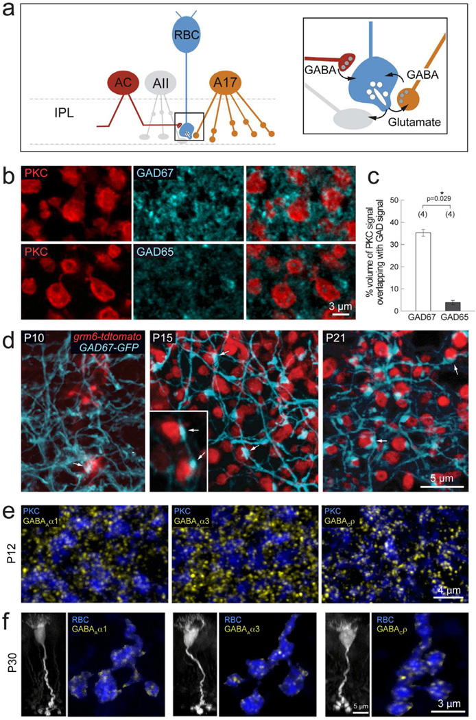
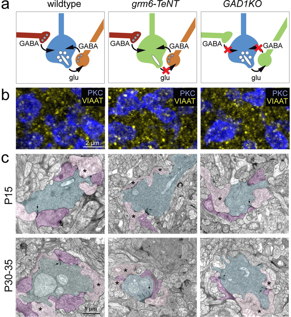
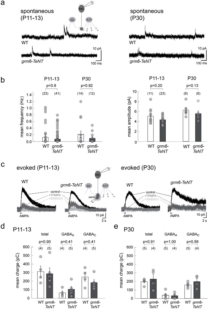
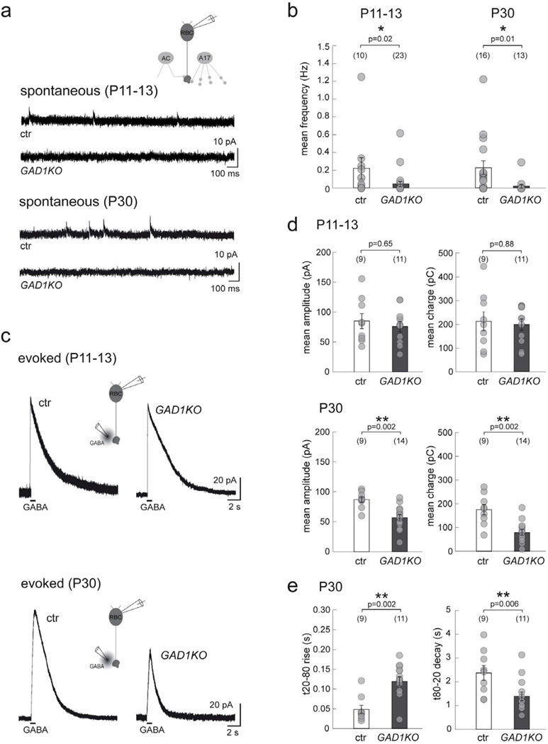
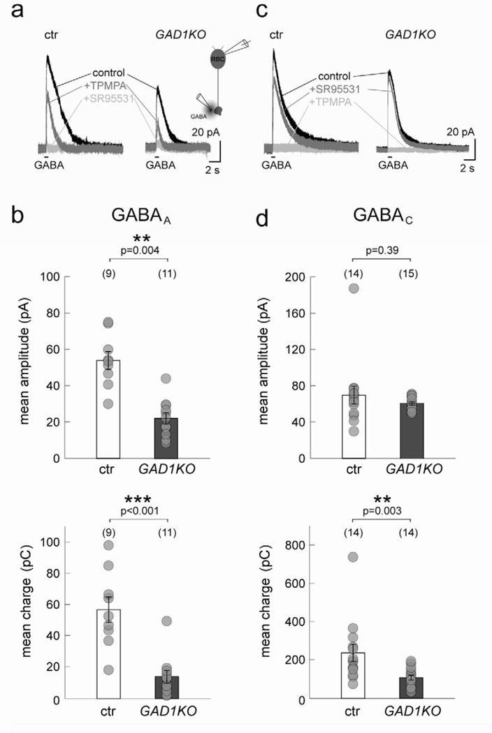
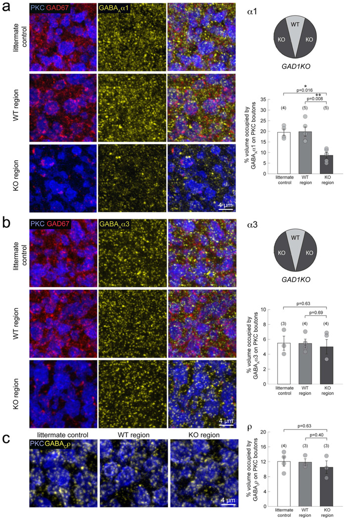
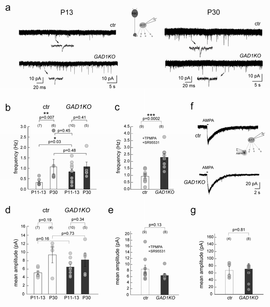
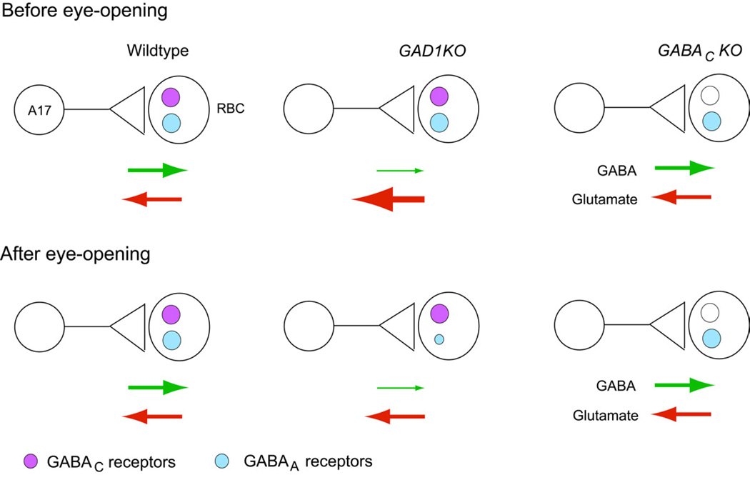
Presynaptic inhibition onto axons regulates neuronal output, but how such inhibitory synapses develop and are maintained in vivo remains unclear. Axon terminals of glutamatergic retinal rod bipolar cells (RBCs) receive GABAA and GABAC receptor-mediated synaptic inhibition. We found that perturbing GABAergic or glutamatergic neurotransmission does not prevent GABAergic synaptogenesis onto RBC axons. But, GABA release is necessary for maintaining axonal GABA receptors. This activity-dependent process is receptor subtype specific: GABAC receptors are maintained, whereas GABAA receptors containing α1, but not α3, subunits decrease over time in mice with deficient GABA synthesis. GABAA receptor distribution on RBC axons is unaffected in GABAC receptor knockout mice. Thus, GABAA and GABAC receptor maintenance are regulated separately. Although immature RBCs elevate their glutamate release when GABA synthesis is impaired, homeostatic mechanisms ensure that the RBC output operates within its normal range after eye opening, perhaps to regain proper visual processing within the scotopic pathway.


Figures

Figure 1
Figure 1. Development of GABAergic synapses onto RBC axon terminals
(a) Schematic showing RBC connections with amacrine cells in retina IPL. RBCs (blue) provide excitatory glutamatergic drive (glu) onto glycinergic AII (grey) and GABAergic A17 (orange) amacrine cells. In turn, they receive feedback inhibition from A17 cells and additional GABAergic input from other as yet unidentified amacrine cells (AC, red). (b) PKC labeled RBC terminals (red) colabeled with GAD67 or GAD65 (cyan), reveal more abundant immunoreactivity of GAD67 as compared to GAD65 in the IPL lamina where RBC axons stratify. (c) Quantification of % volume of GAD signal overlapping with PKC immunoreactivity revealed a significantly higher % overlap with GAD67 positive processes as compared to GAD65. Asterisk marks significant difference. (d) Development of contacts (arrows) between RBCs (red) and GAD67-GFP positive amacrine processes (cyan) in the grm6-tdtomato×GAD67-GFP double transgenic line. By P15, large varicosities can be seen at sites of contact between RBC axon terminals and GAD67-GFP amacrine processes. (e) Axonal terminals of P12 PKC-labeled RBCs (blue) express GABAAα1, GABAAα3, and GABACρ receptor clusters (yellow), indicating the presence of these three GABAergic postsynapses on RBC terminals before eye-opening. (f) P30 RBCs in the grm6-tdtomato transgenic line (left, vertical view, grayscale). Axonal terminals of individual P30 RBCs (horizontal view, blue) express all three GABA receptor cluster types: GABAAα1, GABAAα3, and GABACρ (yellow),
Figure 2
Figure 2. Inhibitory synapses onto RBC axons in the grm6-TeNT and GAD1KO retina
(a) Schematic showing RBC axon terminal circuitry in wildtype and transgenic lines with suppressed glutamatergic transmission from ON-bipolar cells (grm6-TeNT) and reduced GABAergic transmission from amacrine cells (GAD1KO). (b) Immunolabeling for VIAAT (yellow) at the level of PKC positive RBC boutons (blue) in P30 wildtype, grm6-TeNT and GAD1KO retina. (c) Electron micrographs showing dyad synapses between RBC boutons (cyan) and AII (purple) and A17 (pink) amacrine cell processes in the wildtype (WT), grm6-TeNT and GAD1KO retinae at P15 and P30–35. Presynaptic ribbons are indicated by arrows. Asterisks mark examples of A17 contact. Note that RBCs in the P30 grm6-TeNT retina have multiple ribbons at a single dyad synapse.
Figure 3
Figure 3. Spontaneous and GABA-evoked currents of RBCs in the grm6-TeNT retina
(a) Spontaneous GABAergic currents recorded in grm6-TeNT and wildtype (WT) RBCs at P11–13 and P30. (b) Quantification revealed no significant differences in either the mean frequency or mean amplitude of GABAergic sIPSCs from grm6-TeNT RBCs as compared to WT. (c) Example traces showing chloride-mediated outward currents in RBCs evoked by AMPA puffs in grm6-TeNT and WT RBCs at both ages. The GABAC component was isolated by application of TPMPA, and residual GABAA-mediated current blocked by SR95531. Scatter plots show that the mean charge transfer of the total, GABAA-mediated and GABAC-mediated currents are comparable in grm6-TeNT RBCs and WT at P11–13 (d) and P30 (e). Numbers in brackets in b, d, and e represents number of cells. Error bars represent S.E.M.
Figure 4
Figure 4. Spontaneous and GABA-evoked currents of RBCs in GAD1KO retina
(a) Spontaneous GABAergic IPSCs at RBC axon terminals recorded from GAD1KO and littermate control (ctr) at P11–13 and P30. (b) Scatter plots show that the mean frequency of these sIPSCs are reduced in GAD1KO as compared to ctr. (c) Puffs of GABA evoke similar chloride-mediated outward currents in RBCs from ctr and GAD1KO at P11–13, which are markedly reduced for GAD1KO RBCs at P30. (d) Quantification reveals a significant reduction for both the mean peak amplitude and charge of GABA-evoked currents from GAD1KO RBCs at P30. (e) In addition, the rise time for the P30 evoked response from GAD1KO RBCs is longer while the decay is faster as compared to control. Numbers in brackets in b, d, and e represents number of cells. Asterisks mark significant difference. Error bars represent S.E.M.
Figure 5
Figure 5. GABAA and GABAC receptor-mediated currents from P30 RBCs in GAD1KO retina
(a) Example traces of P30 GAD1KO and littermate control (ctr) RBC responses to GABA puffs. TPMPA was applied to isolate the GABAA-mediated component. Further addition of SR95531 blocked all evoked response. (b) Scatter plots show that mean peak amplitude and charge of GABAA-mediated currents in GAD1KO RBCs are significantly reduced as compared to ctr. (c) Application of SR95531 alone isolates the GABAC-mediated component of evoked responses in RBCs. (d) Quantification of GABAC-mediated currents indicates a significant reduction of the mean charge in GAD1KO RBCs as compared to ctr. However, the mean amplitude remained unchanged. Numbers in brackets in b and d represents number of cells. Asterisks mark significant difference. Error bars represent S.E.M.
Figure 6
Figure 6. GABAA receptor subsets on mature RBC boutons in GAD1KO retina
(a) Triple immunolabeling of PKC immunopositive RBCs (blue), GAD67 (red) and GABAAα1 subunits (yellow), revealed a visible reduction of α1-containing GABAA receptor clusters in the KO region of GAD1KO retina, compared to the WT region or littermate control. Quantification of the % volume occupied by GABAAα1 clusters on PKC positive boutons confirmed a significant reduction for the KO region. (b) Immunostaining for the GABAAα3 receptor subunit revealed no significant differences in the % volume occupied by these receptor clusters on KO region RBC boutons. (c) Immunolabeling and % volume occupancy of GABACρ receptor subunits were comparable across littermate control, WT region and KO region. Numbers in brackets represent number of animals. Asterisks mark significant difference. Error bars represent S.E.M.
Figure 7
Figure 7. RBC output is transiently increased in GAD1KO retina during circuit development
(a) sEPSCs recorded from A17 amacrine cells at P13 and P30 in GAD1KO and littermate control retina shows an increase during normal development (ctr). (b) The sEPSC frequency was increased for A17 amacrine cells in GAD1KO as compared to ctr early in development at P13, but was comparable to control at P30. (c) Mean frequency of sEPSCs from P11–13 A17 amacrine cells in GAD1KO and ctr in the presence of GABA receptor antagonists TPMPA and SR95531, also showed increased frequency in the GAD1KO. (d) Mean sEPSC amplitudes recorded in A17s from littermate control and GAD1KO at P11–13 and P30 revealed no differences. (e) Mean amplitude of sEPSCs recorded from P11-P13 A17 amacrine cells in GAD1KO and ctr in the presence of GABA receptor antagonists TPMPA and SR95531 showed no difference. (f) Example traces showing AMPA puff evoked cation-mediated inward currents from A17 cells clamped at −60 mV evoked by AMPA puffs in ctr and GAD1KO at P11–13. Scatter plots show that the mean amplitude (g) is comparable between genotypes. Numbers in brackets represents number of cells. Asterisk marks significant difference. Error bars represent S.E.M.
Figure 8
Figure 8. Summary of the alterations in RBC-A17 synapses in GABAergic transmission-defective mutants before and after eye-opening
Schematic illustrating the A17-RBC synapse before and after eye-opening in the wildtype, GAD1KO and GABACKO retina. GABA and glutamate release is similar between wildtype and GABACKO (see also Eggers and Lukasiewicz, 2006a), but is altered in the GAD1KO. There is a transient upregulation of glutamate release from developing RBCs in the GAD1KO before eye-opening. However, after eye-opening, glutamate release from RBCs in the GAD1KO becomes comparable to wildtype. GABAA and not GABAC receptors are selectively reduced at maturity in the GAD1KO. This reduction of GABAA receptors on RBCs does not occur in GABAC receptor mutants.

Similar articles

Cited by

References

    1. Bloomfield SA, Dacheux RF. Rod vision: pathways and processing in the mammalian retina. Prog Retin Eye Res. 2001;20:351–384. - PubMed
    1. Bu DF, Erlander MG, Hitz BC, Tillakaratne NJ, Kaufman DL, Wagner-McPherson CB, Evans GA, Tobin AJ. Two human glutamate decarboxylases, 65-kDa GAD and 67-kDa GAD are each encoded by a single gene. Proc Natl Acad Sci U S A. 1992;89:2115–2119. - PMC - PubMed
    1. Burrone J, Murthy VN. Synaptic gain control and homeostasis. Curr Opin Neurobiol. 2003;13:560–567. - PubMed
    1. Chattopadhyaya B, Di Cristo G, Higashiyama H, Knott GW, Kuhlman SJ, Welker E, Huang ZJ. Experience and activity-dependent maturation of perisomatic GABAergic innervation in primary visual cortex during a postnatal critical period. J Neurosci. 2004;24:9598–9611. - PMC - PubMed
    1. Chattopadhyaya B, Di Cristo G, Wu CZ, Knott G, Kuhlman S, Fu Y, Palmiter RD, Huang ZJ. GAD67-mediated GABA synthesis and signaling regulate inhibitory synaptic innervation in the visual cortex. Neuron. 2007;54:889–903. - PMC - PubMed

Publication types

MeSH terms

LinkOut - more resources