. 2013 Apr 10;78(1):191-204.
doi: 10.1016/j.neuron.2013.02.007.

Circuits for grasping: spinal dI3 interneurons mediate cutaneous control of motor behavior

Affiliations

Circuits for grasping: spinal dI3 interneurons mediate cutaneous control of motor behavior

Tuan V Bui et al. Neuron. .

Abstract

Accurate motor performance depends on the integration in spinal microcircuits of sensory feedback information. Hand grasp is a skilled motor behavior known to require cutaneous sensory feedback, but spinal microcircuits that process and relay this feedback to the motor system have not been defined. We sought to define classes of spinal interneurons involved in the cutaneous control of hand grasp in mice and to show that dI3 interneurons, a class of dorsal spinal interneurons marked by the expression of Isl1, convey input from low threshold cutaneous afferents to motoneurons. Mice in which the output of dI3 interneurons has been inactivated exhibit deficits in motor tasks that rely on cutaneous afferent input. Most strikingly, the ability to maintain grip strength in response to increasing load is lost following genetic silencing of dI3 interneuron output. Thus, spinal microcircuits that integrate cutaneous feedback crucial for paw grip rely on the intermediary role of dI3 interneurons.


Figures

Figure 1
Figure 1. dI3 INs are multipolar spinal interneurons located in the deep dorsal horn and the intermediate laminae of the spinal cord
(A) Transverse distribution of YFP and ChAT labeled spinal neurons in a lumbar spinal cord section. Note the expected expression in dorsal root ganglion neurons as seen by fluorescence in axons in the dorsal columns (arrows) and dorsal horns. (B) Laminar distribution of dI3 INs based upon cell counts from ten transverse L4/L5 sections. Laminae divisions based upon Watson et al. (2008). (C) dI3 INs are multipolar neurons with multiple dendrites. The somata of dI3 INs were of intermediate size (17.5 ± 3.7 μm, n = 95) and multipolar in shape with, on average, 3.9 ± 3.7 (n = 95) primary dendritic trees. (D) dI3 IN with dendrites (arrows) extending into lamina IV-VI of the dorsal horn. Dorsal column (DC), central canal (oval) and ventral white matter are delineated by dashed curves. (E) Neurobiotin-filled dI3 IN (arrow) with dendritic process (arrowheads) extended towards motor pools (mp). All images from Isl1-YFP mice. See also Figure S1.
Figure 2
Figure 2. dI3 INs are excitatory premotor interneurons
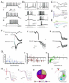
(A) In-situ hybridization for vGluT2 mRNA colocalizes with YFP+ interneurons in lamina V-VII. Quantitative analysis was restricted to laminae V-VII to avoid sampling YFP+ somatic motoneurons in laminae IX, and revealed that 118/139 YFP+ neurons clearly expressed vGluT2. (B) YFP+ motoneuron in lamina IX with YFP+ bouton-like structures (arrow) from putative dI3 INs in a spinal cord of a Isl1+/Cre; Thy1.lox-stop-lox.mGFP mouse. vGluT1 is labeled in red, demonstrating that these processes are not from primary afferents. (C and D) YFP+ motoneurons in lamina IX motor pool with bouton-like processes from neurobiotin-labeled dI3 IN. Thick red processes are blood vessels. White dashed boxes highlight clusters of filled boutons. (E) YFP+/vGluT2+ boutons (arrows) apposed on motoneuron (labeled with ChAT). (F) YFP+/vGluT2+ boutons (arrows) apposed to motoneuron processes in chronically transected spinal cord. (G) Diagram representing dI3 INs as last-order, excitatory interneurons. Unless otherwise noted, all images from Isl1-YFP mice. See also Figure S2.
Figure 3
Figure 3. Anatomical evidence that primary afferents project to dI3 INs
(A) Left: YFP+ dI3 IN with vGluT1+ boutons apposed (labeled by arrows). Right: Orthogonal sections confirming apposition of boutons labeled 1-3. (B) vGluT1+ boutons that are PV+ and PVnull on a YFP+ dI3 IN from a P7 spinal cord. Boutons in dashed boxes are magnified in Bii-Bvii. Inset in 3Bii depicts orthogonal sections of the PV+/YFP+ bouton in the Y-Z plane. (C) vGluT1+ boutons (arrowheads) on YFP+ dI3 IN from chronically transected spinal cord confirm that they do not originate from supraspinal descending inputs. All images from Isl1-YFP mice.
Figure 4
Figure 4. Response of dI3 INs to sensory afferent stimulation
(A) Three types of firing behaviors. i. Tonic firing. ii. Initial bursting. iii. Delayed firing. (B) Current-clamp recording of two dI3 INs showing response to DR stimulation (20 μA, just under 3 × T, 250 μs) during current steps of three different magnitudes. i. Cell responding with an action potential followed by a prolonged hyperpolarization. ii. Cell responding with a prolonged depolarization. Arrowheads mark the time of stimulation. Top row is a scaled version of area marked by dashed box in second row. (C) Voltage-clamp recording of L5 dI3 IN depicting an EPSC in response to DR stimulation (20 μA, just under 3 × T, 250 μs) with accompanying extracellular ventral root (L5) recording. Arrowhead marks the stimulation artifact while the arrow marks the monosynaptic ventral root response. (D) EPSCs in response to DR stimulation (20 μA, just under 3 × T) reversibly blocked by CNQX. (E) drEPSCs at different holding potentials showing reversal of EPSC at depolarized potential. (F) Demonstration of monosynaptic nature of sensory input. i. Onset of drEPSC (15 μA, 3 × T) in dI3 IN as compared to onset of monosynaptic EPSC in a motoneuron from the same preparation. These two cells were recorded separately. The ventral root recording was concomitant with the motoneuron recording. The timing of the ventral root response during dI3 IN recording was the same. Blue dashed line marks the onset of the motoneuron EPSC. Red dashed line marks the onset of the dI3 IN EPSC. The difference was below 0.2 ms. ii. Top: drEPSCs (15 μA, 3 × T) in dI3 IN with low jitter (0.002 ms2). Jitter calculated on 20 responses. Bottom: drEPSCs (20 μA, 4 × T) in dI3 IN with high-jitter (0.47 ms2). iii. Low-latency, low-jitter drEPSCs in dI3 IN with no failures in response to 0.25 and 2.5 Hz stimulation frequency (n = 3), confirming these are monosynaptic responses. (G) Shift towards monosynaptic sensory inputs with age. Relation between i. Onset of drEPSC in dI3 INs and age, ii.Variance of drEPSC onset and age, and iii.Variance of drEPSC onset and onset of drEPSC. (H) Response to different strengths of DR stimulation. i. dI3 IN with increases in drEPSC magnitude with shift from low to medium strength but not to high threshold dorsal root stimulation. ii. dI3 IN with increases in drEPSC magnitude with shift from low to medium to high threshold dorsal root stimulation. T refers to the threshold at which a monosynaptic ventral root reflex was elicited. iii. Distribution of sets of responses in dI3 INs to DR stimulation (single 250 μs pulses). Low threshold fibers were considered to be recruited by stimulation between 1-2 × T, medium threshold fibers were recruited by 2-5 × T, and high threshold fibers were recruited by stimulation above 5 × T. T is the earliest stimulation strength at which a monosynaptic ventral root reflex was elicited in ventral roots. Number of cells in parentheses. (I) Diagram representing dI3 INs as part of a disynaptic pathway between sensory input and motoneurons. See also Figure S3.
Figure 5
Figure 5. Loss of vGluT2 expression in dI3OFF mice
(A) Breeding strategy to generate silencing of the output of dI3 INs (dI3OFF) by conditionally knocking out vGluT2. fsYFP refers to lox-stop-lox.YFP. Only the genotypes that were used are shown. (B) Fluorescent in-situ hybridization shows reduced presence of vGluT2 mRNA to 28% of YFP+ dI3 INs, (n = 92 out of 330 neurons from 2 mice) in dI3OFF animals. Asterisks denote dI3 INs with presence of vGluT2 mRNA, arrowheads denote dI3 INs with absence of vGluT2 mRNA. (C) Following neurobiotin injection in a dI3 IN from a dI3OFF animal, bouton-like appositions are present on a putative YFP+ motoneuron in lamina IX. (D) Loss of vGluT2+/YFP+ boutons on ChAT+ motoneuron. (E) vGluT1+ inputs to YFP+ dI3 INs are still present in dI3OFF mice (8/9 dI3 INs). (F) Ventral-root reflex and monosynaptic dorsal root-evoked EPSCs in dI3 INs are present in dI3OFF mice (8 out of 10 dI3 INs showed dorsal-root evoked EPSCs; 4/10 dI3 INs met strict short–latency, low jitter thresholds as above). Interestingly, these mice became pruritic (Figure S4B), as might be expected from loss of vGluT2 from high threshold afferents (Lagerström et al., 2010; Liu et al., 2010). All images from lumbar spinal cord of P13-16 dI3OFF mice. See also Figure S4.
Figure 6
Figure 6. Conditional silencing of the output of dI3 INs abolishes short latency response to cutaneous nerve stimulation
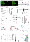
(A) Tetramethylrhodamine (TMRD)+ boutons on YFP+ dI3 IN in a Isl1-YFP spinal cord three days after TMRD labelling of sural nerve, a predominantly cutaneous nerve (Peyronnard and Charron, 1982). (B) Schematic describing the estimated latencies of monosynaptic and disynaptic ventral root reflexes in response to tibial and sural nerves, respectively. Estimates were calculated for experiments with postnatal isolated spinal-cord preparation with sural nerve left in continuity and for adult in-vivo recordings. They were based upon observed latencies between stimulation, DR volley or CDP, and ventral root reflexes or EMG recordings. (C) Scheme of isolated spinal-cord preparation with sural nerve left in continuity. Stimulating electrodes were placed on the sural nerve and the tibial nerve distal to the sural nerve branchpoint. Recording electrodes were placed on the ipsilateral L5 dorsal and ventral roots. (D) L5 dorsal root potentials in response to sural nerve or tibial nerve stimulations. In this example, the earliest DR volley to sural nerve and tibial nerve stimulations were seen at 3 and 2 μA, respectively. (E) 20 traces of L5 ventral root reflexes elicited by two 250 μs shocks (500 Hz) applied to the sural nerve and the tibial nerve. Note the low-jitter monosynaptic reflex in response to tibial nerve stimulation (arrowhead) versus the high-jitter short-latency oligosynaptic reflex in response to sural nerve stimulation (arrow). (F) i. Recordings of L5 ventral root ENGs to multiple stimulation pulses applied to the sural nerve. Putative disynaptic reflex responses are highlighted in dashed box. ii. Methodology for quantification of short-latency response in 8-ms time window, 14 ms from low-threshold, earliest L5 DR volley from sural nerve stimulation (not shown). iii. Summary of short-latency L5 ventral root ENG reflex response to sural nerve stimulation. Data point shows the ratio of the time-integrated ENG response to sural nerve stimulation in the short-latency ENG time-window over the time-integrated ENG response in a randomly selected 8-ms time-window prior to the application of the stimulus trains. Clear circles represent each isolated spinal cord, filled circles represent group average. Error bars represent SD. Dashed line represents reflex threshold of 2. (G) Scheme of EMG recordings of chronically implanted electrodes into Gs and TA muscles of control and dI3OFF mice. (H) Recordings of cord dorsum potentials (CDPs) in response to tibial nerve stimulation. i. Recording set up. ii. (top) CDP showing activation of proprioceptive and cutaneous potentials with increasing current stimulation, (bottom) corresponding EMG recording of Gs. (I) i. Recordings of Gs EMG to multiple stimulation pulses applied to the sural nerve. Putative disynaptic reflex responses are highlighted in dashed box. ii. Summary of short-latency synaptic EMG response reflex response measured in a 4-ms time-window, 4 ms after the onset of the last stimulation pulse to sural nerve stimulation. Data point shows the ratio of the time-integrated EMG response to sural nerve stimulation in the short-latency EMG time-window over the time-integrated EMG response in a randomly selected 4-ms time-window prior to the application of the stimulus trains. Clear circles represent each animal, filled circles represent group average. Error bars represent SD. Dashed line represents reflex threshold of 2. (J) Diagram representing dI3 INs as part of a disynaptic pathway between low threshold mechanoreceptors from the skin and motoneurons. See also Figure S5.
Figure 7
Figure 7. Reduced performance in several motor tasks involving cutaneous afferents from the paw, including loss of functional grip in dI3OFF mice
(A) During ladder walking with rung spacing of 2 cm, forelimb errors are similar in controls and mutants, whereas the mutants have more hindlimb errors than controls. Error bars represent SD. (B) Comparison of hindlimb grip of a metal bar between control and dI3OFF mice. (C) In the wire hang test, control animals grip onto the cage top, while upside down, for close to one minute whereas the dI3OFF mice are unable to hang onto the cage top while inverted. Diagram depicting minimal angle from horizontal axis at which dI3OFF mice are unable to hang onto the cage top. The grey cone represents the pooled SD. (D) Performance of control and dI3OFF mice during the wire hang test. The maximal duration of the test was one minute. Every control animals would hang on for periods longer than a minute in at least one of three trials. Similar results were observed when mice were tested a second time one or two days later. Error bars represent SD. See also Movies S1 and S2. (E) Forepaw grasp reflex in control and dI3OFFpostnatal (P1-P7) mice. Chi-square test indicated. (F) Diagram representing dI3 INs as part of a disynaptic pathway between low threshold mechanoreceptors from the skin and motoneurons involved with regulating grip strength.

Similar articles

Cited by

References

    1. Abdala V, Manzano AS, Tulli MJ, Herrel A. The tendinous patterns in the palmar surface of the lizard manus: functional consequences for grasping ability. Anat. Rec. (Hoboken) 2009;292:842–853. - PubMed
    1. Akay T, Acharya HJ, Fouad K, Pearson KG. Behavioral and electromyographic characterization of mice lacking EphA4 receptors. J. Neurophysiol. 2006;96:642–651. - PubMed
    1. Alstermark B, Pettersson LG, Nishimura Y, Yoshino-Saito K, Tsuboi F, Takahashi M, Isa T. Motor command for precision grip in the macaque monkey can be mediated by spinal interneurons. J. Neurophysiol. 2011;106:122–126. - PubMed
    1. Alvarez FJ, Villalba RM, Zerda R, Schneider SP. Vesicular glutamate transporters in the spinal cord, with special reference to sensory primary afferent synapses. J. Comp. Neurol. 2004;472:257–280. - PubMed
    1. Amendola J, Verrier B, Roubertoux P, Durand J. Altered sensorimotor development in a transgenic mouse model of amyotrophic lateral sclerosis. Eur. J. Neurosci. 2004;20:2822–2826. - PubMed

Publication types

MeSH terms

LinkOut - more resources