Review
. 2013 Jun;98(3):476-83.
doi: 10.1016/j.antiviral.2013.03.021. Epub 2013 Apr 9.

Broadly neutralizing antibodies against influenza viruses

Affiliations
Review

Broadly neutralizing antibodies against influenza viruses

Nick S Laursen et al. Antiviral Res. 2013 Jun.

Abstract

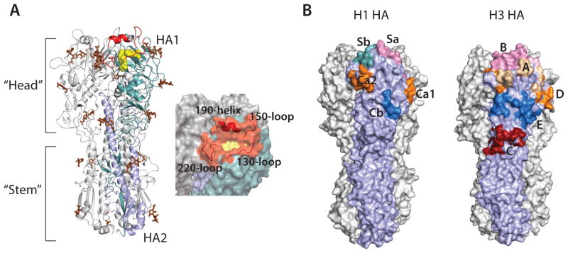
Despite available antivirals and vaccines, influenza continues to be a major cause of mortality worldwide. Vaccination generally induces an effective, but strain-specific antibody response. As the virus continually evolves, new vaccines have to be administered almost annually when a novel strain becomes dominant. Furthermore, the sporadic emerging resistance to neuraminidase inhibitors among circulating strains suggests an urgent need for new therapeutic agents. Recently, several cross-reactive antibodies have been described, which neutralize an unprecedented spectrum of influenza viruses. These broadly neutralizing antibodies generally target conserved functional regions on the major influenza surface glycoprotein hemagglutinin (HA). The characterization of their neutralization breadth and epitopes on HA could stimulate the development of new antibody-based antivirals and broader influenza vaccines. This article forms part of a symposium in Antiviral Research on "Treatment of influenza: targeting the virus or the host."


Conflict of interest statement

The authors have no financial or personal relationships that could be viewed as a potential conflict of interest.

Figures

Fig. 1
Fig. 1
Crystal structure of HA. (A) Structure of the trimeric HA spike (PDB code; 4FNK) (Ekiert et al., 2012). One protomer is colored in cyan (HA1) and light blue (HA2). The receptor binding site is colored in yellow and the surrounding loops and helix in red. Glycans are colored brown (left). Surface representation of the receptor binding site and its surroundings (right). (B) The antigenic sites on HA. Antigenic sites Sa (pink), Sb (cyan), Ca1 and Ca2 (orange), and Cb (blue) on H1 HAs (left) (PDB code; 3LZG) (Xu et al., 2010). Antigenic sites A (wheat), B (pink), D (orange), E (blue) and C (red) on H3 HAs (right) (PDB code; 4FNK) (Ekiert et al., 2012).
Fig. 2
Fig. 2
Structure of HA in complex with head binding antibodies. (A) Structure of Fab CH65 in complex with H1 HA (PDB code; 3SM5) (Whittle et al., 2011). HA is shown as surface representation with one protomer colored light blue. Residues in the epitope that are part of the receptor binding site colored in red. Residues in the epitope adjacent to the receptor-binding site are colored in blue. The Fab is shown in cartoon with the light chain (LC) in yellow and the heavy chain (HC) in green (top). CDR loops from the heavy chain (green) and the light chain (yellow) involved in the interaction with HA are shown. (B) Structure of Fab S139/1 in complex with H3 HA (PDB code; 4GMS) (Lee et al., 2012). Colored as in Fig. 2A. (C) Structure of Fab C05 in complex with H3 HA (PDB code 4FQR) (Ekiert et al., 2012).
Fig. 3
Fig. 3
Structures of HA in the pre-fusion and post-fusion conformation, and HA in complex with stem reactive Fab fragments. (A) Pre-fusion conformation of HA with helices A (red), C (yellow), and D (blue) and B loop (orange) in one protomer colored distinctly (PDB code; 4FNK) (Ekiert et al., 2012). (B) Post-fusion conformation of HA (PDB code; 1HTM) (Bullough et al., 1994). Colored as in (A). Note that most of HA1 is not present in the structure and the B loop folds into helix B. (C) Structure of Fab CR6261 in complex with H5 HA (PDB code; 3GBM) (Ekiert et al., 2009). HA is colored as in (A) shown as surface representation and in the same orientation (left). The epitope recognized by CR6261 is shown on the right. CDR loops from the heavy chain (green) and the light chain (yellow) involved in the interaction with HA are shown. The position of the glycan at Asn38 in group 2 viruses is indicated by #. Trp21 is shown by *. The side chain of the Phe residue interacting with Trp21 is shown (D) Structure of Fab CR8020 in complex with H3 HA (PDB code; 3SDY) (Ekiert et al., 2011). The glycan at Asn21 in most group 2 viruses is shown by # and Tyr34 as *, both in red. (E) Structure of Fab CR9114 in complex with H5 HA (PDB code; 4FQI) (Dreyfus et al., 2012). Colored as in (A). (F) Structure of Fab FI6v3 in complex with H3 HA (PDB code; 3ZTJ) (Corti et al., 2011). The glycan at Asn38 is shown in brown.
Fig. 3
Fig. 3
Structures of HA in the pre-fusion and post-fusion conformation, and HA in complex with stem reactive Fab fragments. (A) Pre-fusion conformation of HA with helices A (red), C (yellow), and D (blue) and B loop (orange) in one protomer colored distinctly (PDB code; 4FNK) (Ekiert et al., 2012). (B) Post-fusion conformation of HA (PDB code; 1HTM) (Bullough et al., 1994). Colored as in (A). Note that most of HA1 is not present in the structure and the B loop folds into helix B. (C) Structure of Fab CR6261 in complex with H5 HA (PDB code; 3GBM) (Ekiert et al., 2009). HA is colored as in (A) shown as surface representation and in the same orientation (left). The epitope recognized by CR6261 is shown on the right. CDR loops from the heavy chain (green) and the light chain (yellow) involved in the interaction with HA are shown. The position of the glycan at Asn38 in group 2 viruses is indicated by #. Trp21 is shown by *. The side chain of the Phe residue interacting with Trp21 is shown (D) Structure of Fab CR8020 in complex with H3 HA (PDB code; 3SDY) (Ekiert et al., 2011). The glycan at Asn21 in most group 2 viruses is shown by # and Tyr34 as *, both in red. (E) Structure of Fab CR9114 in complex with H5 HA (PDB code; 4FQI) (Dreyfus et al., 2012). Colored as in (A). (F) Structure of Fab FI6v3 in complex with H3 HA (PDB code; 3ZTJ) (Corti et al., 2011). The glycan at Asn38 is shown in brown.

Similar articles

Cited by

References

    1. Ambrose CS, Levin MJ. The rationale for quadrivalent influenza vaccines. Hum Vaccin Immunother. 2012;8:81–88. - PMC - PubMed
    1. Bommakanti G, Citron MP, Hepler RW, Callahan C, Heidecker GJ, Najar TA, Lu X, Joyce JG, Shiver JW, Casimiro DR, ter Meulen J, Liang X, Varadarajan R. Design of an HA2-based Escherichia coli expressed influenza immunogen that protects mice from pathogenic challenge. Proc Natl Acad Sci U S A. 2010;107:13701–13706. - PMC - PubMed
    1. Bright RA, Medina MJ, Xu X, Perez-Oronoz G, Wallis TR, Davis XM, Povinelli L, Cox NJ, Klimov AI. Incidence of adamantane resistance among influenza A (H3N2) viruses isolated worldwide from 1994 to 2005: a cause for concern. Lancet. 2005;366:1175–1181. - PubMed
    1. Bullough PA, Hughson FM, Skehel JJ, Wiley DC. Structure of influenza haemagglutinin at the pH of membrane fusion. Nature. 1994;371:37–43. - PubMed
    1. Carrat F, Flahault A. Influenza vaccine: the challenge of antigenic drift. Vaccine. 2007;25:6852–6862. - PubMed

Publication types

MeSH terms