Review
. 2013 Nov;11(11):1374-84.
doi: 10.1016/j.cgh.2013.03.019. Epub 2013 Apr 10.

Management of gastric polyps: an endoscopy-based approach

Affiliations
Review

Management of gastric polyps: an endoscopy-based approach

Yasser H Shaib et al. Clin Gastroenterol Hepatol. 2013 Nov.

Abstract

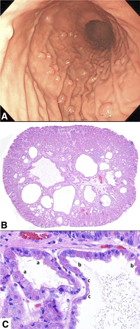
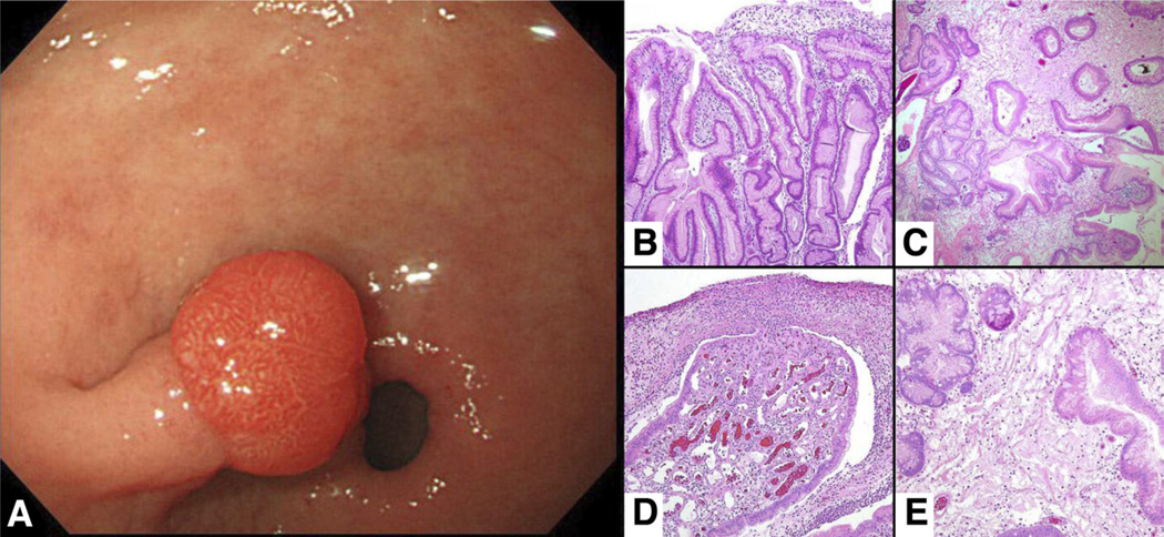
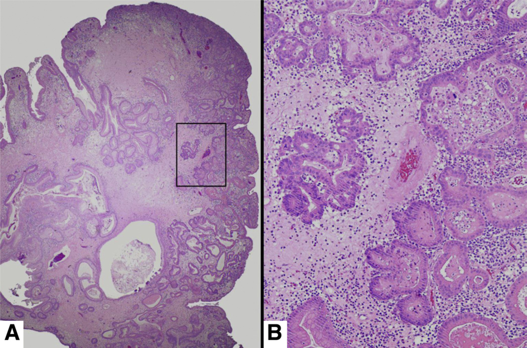
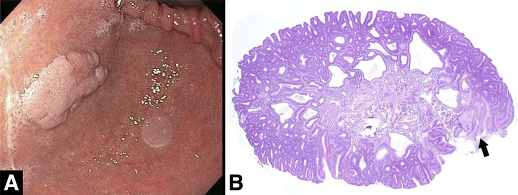
The endoscopic finding of a gastric polyp and the histopathologic report that follows may leave clinicians with questions that have not been addressed in formal guidelines: do all polyps need to be excised, or can they just be sampled for biopsy? If so, which ones and how many should be sampled? What follow-up evaluation is needed, if any? This review relies on the existing literature and our collective experience to provide practical answers to these questions. Fundic gland polyps, now the most frequent gastric polyps in Western countries because of widespread use of proton pump inhibitors, and hyperplastic polyps, the second most common polyps notable for their association with gastritis and their low but important potential for harboring dysplastic or neoplastic foci, are discussed in greater detail. Adenomas have had their name changed to raised intraepithelial neoplasia and are decreasing in parallel with Helicobacter pylori infection; however, they do retain their importance as harbingers of gastric cancer, particularly in East Asia. Gastrointestinal stromal tumors have low incidence and no known associations, but their malignant potential is high; early diagnosis and proper management are crucial. Although rare and benign, inflammatory fibroid polyps need to recognized, particularly by pathologists, to avoid misdiagnosis. Gastric neuroendocrine tumors (carcinoids) are important because of their association with either atrophic gastritis or the multiple endocrine neoplasia syndromes; those that do not arise in these backgrounds have high malignant potential and require aggressive management. The review concludes with some practical suggestions on how to approach gastric polyps detected at endoscopy.

Keywords: ECL; EGD; Endoscopic Management; GIST; Gastric Polyps; OLGA; Operative Link for Gastritis Assessment; PPI; enterochromaffin-like; esophagogastroduodenoscopy; gastrointestinal stromal tumors; proton pump inhibitor.


Conflict of interest statement

Conflicts of interest

The authors disclose no conflicts.

Figures

Figure 1
Figure 1
Fundic gland polyp. (A) Endoscopic view of multiple fundic gland polyps in the body of the stomach in a patient taking PPIs. (B) Low-power photomicrograph showing the characteristic dilatations of oxyntic glands. There is only minimal stroma, and the surface foveolar epithelium is either normal or focally flattened. (C) High-power photomicrograph showing a dilated oxyntic gland lined by cuboidal parietal cells (a); as the gland dilate more (right), both mucous and parietal cells progressively flatten (b and c).
Figure 2
Figure 2
Hyperplastic polyp. (A) Endoscopic view of a hyperplastic polyp on a stalk in the antrum. Histologically, (B) hyperplastic polyps are characterized by marked foveolar hyperplasia, and (C) a mixoid stroma are characterized with dilated tortuous glands lined by normal or reactive foveolar epithelium. (D) Larger polyps have prominent erosions covered with fibrinopurulent material with underlying granulation tissue, (E) often with areas of edematous stroma and oddly shaped glands.
Figure 3
Figure 3
(A) Hyperplastic polyp with focus of high-grade dysplasia. (B) Multiple sections from this 3-cm hyperplastic polyp revealed an area with dysplastic epithelial cells forming complex glandular structures. These represent high-grade dysplasia or possibly a focus of intramucosal carcinoma.
Figure 4
Figure 4
Adenoma. (A) Flat gastric adenoma with a velvety appearance in the distal body of the stomach. (B) Gastric adenomas consist of dysplastic columnar epithelium indistinguishable from colonic adenoma. In resected specimens, the only clue to their gastric origin is often a small remnant of gastric tissue from which they originate (arrow).
Figure 5
Figure 5
GIST. (A) Endoscopic view of an ulcerated submucosal mass in the body of the stomach in a patient who presented with upper-gastrointestinal bleeding. (B) The stroma consists of compact bundles of spindle cells, (C) which stain uniformly with CD117. Staining with DOG-1 (not shown) would have an identical appearance to the staining in panel B.
Figure 6
Figure 6
Inflammatory fibroid polyp. (A) Endoscopic view of an inflammatory fibroid polyp in the antrum showing a firm, well-circumscribed submucosal lesion. (B) Histologically, a flattened, often eroded, gastric epithelium lines a compact aggregate of fibrous tissue mixed with inflammatory cells. (C) Vessels usually are surrounded by a characteristic circumferential deposition of fibroblasts (onion skin), and the stroma contains myriad eosinophils.
Figure 7
Figure 7
Gastric neuroendocrine tumor. (A) A small gastric carcinoid with surface ulceration seen on retroflexion in the distal body. (B) Merging nests of ECL cells arranged in cords in the deeper part of carcinoids are characteristic of carcinoid tumors. (C) The neuroendocrine origin of their cells can be confirmed by a positive synaptophysin immunohistochemical stain.
Figure 8
Figure 8
Ki-67 for carcinoids. When stained with Ki-67 (an immunohistochemical stain that selectively highlights proliferating cells), the indolent carcinoids type I neuroendocrine tumors) found in patients with atrophic gastritis show that less than 2% of the neuroendocrine cells are in a proliferative status (A). In contrast, the usually malignant sporadic carcinoids (type III) show a proliferation index of a 20% or higher (B).

References

    1. Carmack SW, Genta RM, Schuler CM, et al. The current spectrum of gastric polyps: a 1-year national study of over 120,000 patients. Am J Gastroenterol. 2009;104:1524–1532. - PubMed
    1. Omori T, Kamiya Y, Tahara T, et al. Correlation between magnifying narrow band imaging and histopathology in gastric protruding/or polypoid lesions: a pilot feasibility trial. BMC Gastroenterol. 2012;12:17. - PMC - PubMed
    1. Elster K. Histologic classification of gastric polyps. Curr Top Pathol. 1976;63:77–93. - PubMed
    1. el-Zimaity HM, Jackson FW, Graham DY. Fundic gland polyps developing during omeprazole therapy. Am J Gastroenterol. 1997;92:1858–1860. - PubMed
    1. Raghunath AS, O’Morain C, McLoughlin RC. Review article: the long-term use of proton-pump inhibitors. Aliment Pharmacol Ther. 2005;22(Suppl 1):55–63. - PubMed