. 2013 Apr 18;38(4):805-17.
doi: 10.1016/j.immuni.2013.02.020. Epub 2013 Apr 11.

Distinct memory CD4+ T cells with commitment to T follicular helper- and T helper 1-cell lineages are generated after acute viral infection

Affiliations

Distinct memory CD4+ T cells with commitment to T follicular helper- and T helper 1-cell lineages are generated after acute viral infection

J Scott Hale et al. Immunity. .

Abstract

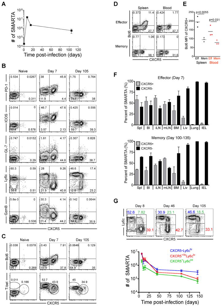
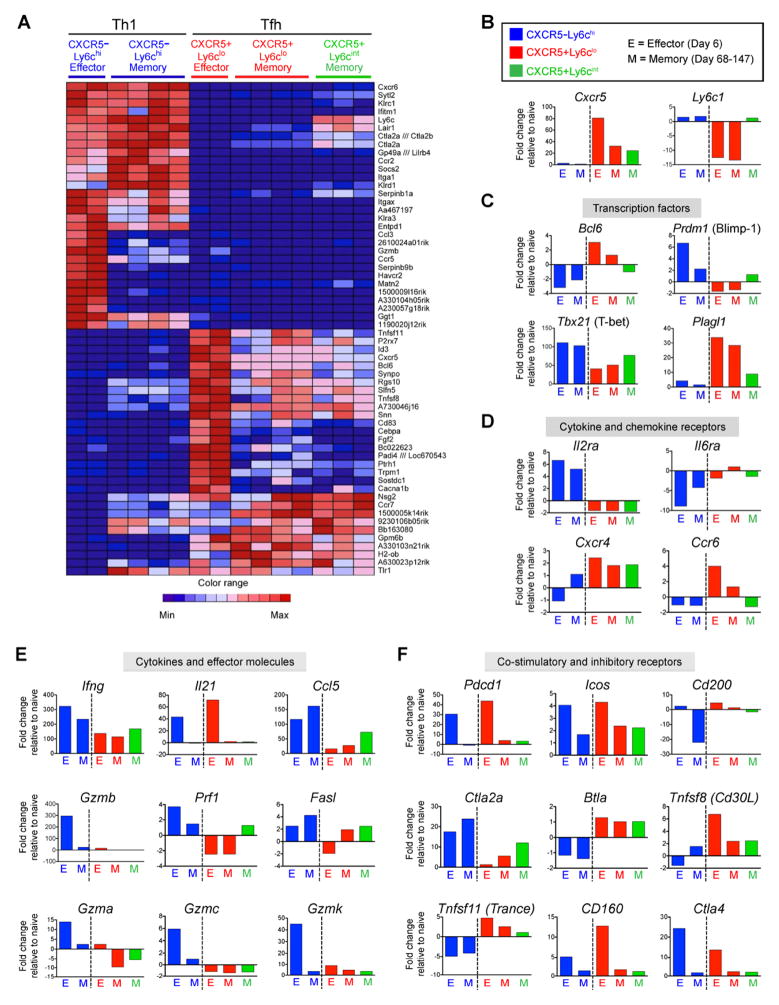
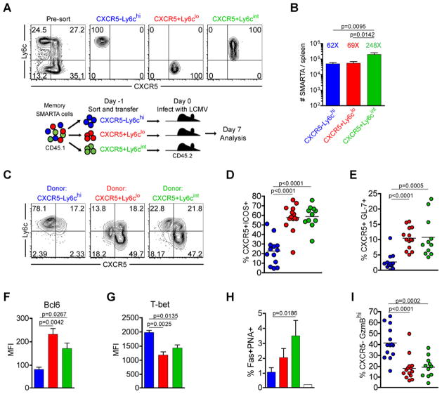
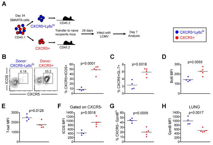
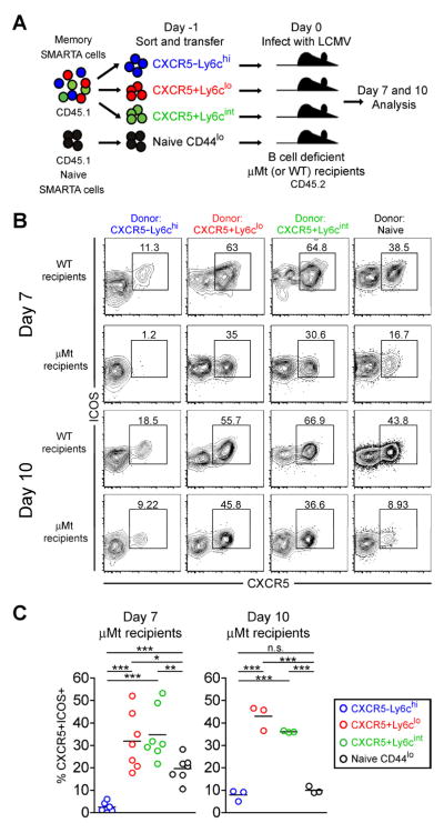
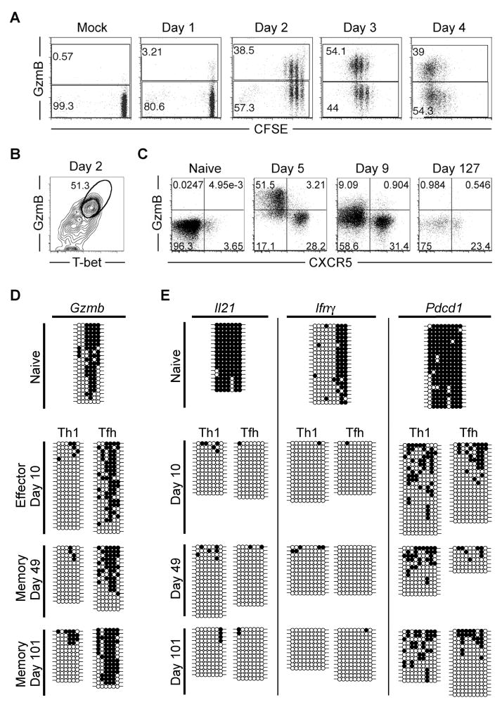
CD4(+) T follicular helper (Tfh) cells provide the required signals to B cells for germinal center reactions that are necessary for long-lived antibody responses. However, it remains unclear whether there are CD4(+) memory T cells committed to the Tfh cell lineage after antigen clearance. By using adoptive transfer of antigen-specific memory CD4(+) T cell subpopulations in the lymphocytic choriomeningitis virus infection model, we found that there are distinct memory CD4(+) T cell populations with commitment to either Tfh- or Th1-cell lineages. Our conclusions are based on gene expression profiles, epigenetic studies, and phenotypic and functional analyses. Our findings indicate that CD4(+) memory T cells "remember" their previous effector lineage after antigen clearance, being poised to reacquire their lineage-specific effector functions upon antigen reencounter. These findings have important implications for rational vaccine design, where improving the generation and engagement of memory Tfh cells could be used to enhance vaccine-induced protective immunity.


Conflict of interest statement

There are no financial conflicts of interest.

Figures

Figure 1
Figure 1. Phenotypic heterogeneity of virus-specific CD4+ T cells is maintained during effector and memory differentiation
2×10⁵ CD45.1+ LCMV-specific naive SMARTA transgenic CD4+ T cells were adoptively transferred into CD45.2+ naïve recipients that were then infected with 2×10⁵ PFU of LCMV Armstrong. FACS plots are gated on CD4+CD45.1+ SMARTA cells at the indicated timepoints relative to infection. A) Kinetics of splenic SMARTA CD4+ T cells. B) CXCR5, PD-1, ICOS, GL-7, Ly6c, and granzyme B analysis of naïve, effector, and memory SMARTA CD4+ T cells. C) Analysis of T-bet and Bcl6 expression. D) Analysis of Bcl6 and CXCR5 on effector (Day 8) and memory (Day 100–133) SMARTA cells in blood and spleen. E) Bcl6 MFI of CXCR5+ gated effector and memory SMARTA cells. Statistically significant p values are shown, and were determined using a two-tailed unpaired Students t test. F) The frequency of effector and memory SMARTA cells that are CXCR5+ and CXCR5 in the following tissues: Spl=spleen; Bl=blood; iLN=inguinal lymph node; mLN=mesenteric lymph node; BM=bone marrow; Liv=liver; Lung, and IEL=intestinal intraepithelial lymphocytes. Graphs show the mean and s.e.m. N=3 mice at each timepoint. G) Plots of CD4+CD45.1+ gated SMARTA cells with gates indicating the Ly6c+CXCR5 (blue), CXCR5+Ly6clo (red), and CXCR5+Ly6cint (green) subsets. Chart indicates the number of each subset in spleen following infection (N≥3 at each timepoint). Error bars represent the SEM. See also Figure S1 and Table S1.
Figure 2
Figure 2. Transcriptional profiling suggests a lineage relationship between Tfh effector and memory cells and between Th1 effector and memory cells
Microarray analysis of CD4+ SMARTA CXCR5Ly6chi (blue), CXCR5+Ly6clo (red), and CXCR5+Ly6cint (green) subsets sorted from chimeric mice at effector (day 6 post-infection) and memory time points (day 68–147 p.i.). A) The 30 most up-regulated and 30 most down-regulated genes in the Day 6 Ly6hiCXCR5 (Th1) relative to the day 6 CXCR5+Ly6clo (Tfh) SMARTA CD4+ T cells were identified and their expression in all subsets at effector and memory stages is shown as a heatmap. B–F) Expression of select genes as determined by microarray analysis, including: B) Cxcr5 and Ly6c; C) transcription factors; D) cytokine and chemokine receptors; E) cytokines and cytotoxic effector molecules; and F) costimulatory and inhibitory receptors. Data are shown as fold change relative to naïve SMARTA CD4+ T cells. Bars show the average value for each indicated population; effector CXCR5Ly6chi (n=2), effector CXCR5+Ly6clo (n=2), memory CXCR5Ly6chi (n=4), memory CXCR5+Ly6clo (n=4), and memory CXCR5+Ly6cint (n=3). See also Figure S2.
Figure 3
Figure 3. Th1 and Tfh memory CD4+ T cells are committed for recall of lineage-specific functions
CD45.1 congenically marked CXCR5Ly6chi (blue), CXCR5+Ly6clo (red), and CXCR5+Ly6cint (green) memory CD4+ subsets (between days 56 and 101 post-infection) were FACS purified to >97%. 8×10³ sorted cells were adoptively transferred into naïve CD45.2 recipients, and recipient mice were then infected with LCMV Armstrong 16–20 hours later. In (C–I), the phenotype and function of transferred SMARTA cells was analyzed 7 days post-infection. A) Representative post-sort analysis of memory SMARTA subsets and cartoon of experimental setup. B) Absolute number of transferred CD4+ CD45.1+ SMARTA splenocytes 7 days following rechallenge with LCMV. The relative fold increase of each population, assuming a 10% take of transferred SMARTA cells, is shown above each bar. C) Representative CXCR5 and Ly6c analysis. D) Chart shows the percent of transferred SMARTA cells that are CXCR5+ICOS+ Tfh cells. E) The percent of SMARTA cells that are CXCR5+GL-7+ germinal center Tfh cells. F) Bcl6 MFI of SMARTA cells. G) T-bet MFI of SMARTA cells. H) Chart shows the percent of B220+ gated splenic B cells that are Fas+PNA+ germinal center B cells. Data were combined from 2 experiments. I) Chart shows the percent of transferred SMARTA cells that are CXCR5GranzymeBhi cells. Data in B, D, E and I are combined from 4 independent experiments for a total of N=11–14 mice per group. Data in F and G were from a single experiment (N=3 per group) and were representative of 2–4 independent experiments. Statistically significant p values are shown, and were determined using a two-tailed unpaired Students t test. Error bars represent the SEM. See also Figure S3.
Figure 4
Figure 4. Lineage commitment of Th1 and Tfh memory subsets is maintained in the absence of antigen
CD45.1 congenically marked CXCR5Ly6chi (blue) and CXCR5+ (red) memory SMARTA CD4+ T cell subsets 34 days post-infection were FACS purified to >97%. 8×10⁴ purified cells were adoptively transferred into naïve CD45.2 recipients. 28 days following cell transfer, naïve recipients were infected with 2×10⁵ p.f.u. LCMV Armstrong 16–20 hours later. The phenotype (B–H) of transferred CD4+CD45.1+ gated SMARTA cells were analyzed 7 days post-infection. A) Cartoon of experimental setup. B) CXCR5 and ICOS analysis. Chart shows the percent of SMARTA cells that are CXCR5+ICOS+ Tfh cells. C) The percent of transferred SMARTA cells that are CXCR5+GL-7+ germinal center Tfh cells. D) Bcl6 MFI of SMARTA cells. E) T-bet MFI of SMARTA cells. F) ICOS MFI on CXCR5 gated effector SMARTA cells 7 days post rechallenge. G) Chart shows the percent of transferred SMARTA cells that are CXCR5Granzyme Bhi cells 7 days post rechallenge. H) Chart shows the Granzyme B MFI of SMARTA cells in lung 7 days post rechallenge. For A–H: CXCR5Ly6chi (N=4); CXCR5+ (N=5). Statistically significant p values of <0.05 are indicated, and were determined using a two-tailed unpaired Students t test. See also Figure S4.
Figure 5
Figure 5. Tfh memory CD4+ T cells recall a Tfh-like response even in B cell deficient recipient mice
CD45.1 congenically marked CXCR5Ly6chi (blue), CXCR5+Ly6clo (red), and CXCR5+Ly6cint (green) SMARTA memory CD4+ T cell subsets (days 68 and 88 post-infection), and naïve SMARTA cells sorted as CD44lo (black) were FACS purified to >97%. 8×10³ purified cells were adoptively transferred into naïve CD45.2+ WT and μMT (B cell deficient) B6 recipients, and recipient mice were then infected with LCMV Armstrong. The phenotype and function of SMARTA cells were analyzed 7 and 10 days post-infection. A) Cartoon of experimental setup. B) Representative CXCR5 and ICOS analyses of CD4+ CD45.1+ gated SMARTA cells 7 and 10 days post-infection. C) Charts show the frequency of SMARTA cells that are CXCR5+ICOS+ Tfh-like cells in B cell deficient mice at each time point following infection with LCMV. Data for day 7 were combined from 2 independent experiments (N=7 per experimental group) and day 10 were from one experiment (N=3 per group). Statistically significant p values were determined using a two-tailed unpaired Students t test (*** p<0.001; ** p<0.01; * p<0.05). See also Figure S5.
Figure 6
Figure 6. Epigenetic modifications of the granzyme B locus distinguish Tfh memory from Th1 memory CD4+ T cells
A–B) 1-2×10⁶ CFSE-labeled SMARTA cells were transferred into host mice and infected one day later. A) Plots show granzyme B expression and CFSE dilution analysis of SMARTA cells at the indicated time points post-infection. B) Plot shows Granzyme B and T-bet expression analysis of SMARTA cells 2 days post-infection. C–E) 2×10⁵ SMARTA cells were transferred into host mice that were infected 1 day later. C) Flow cytometry plots show granzyme B and CXCR5 expression of SMARTA cells at the indicated time points post-infection. D and E) Sorted antigen-specific SMARTA CD4+ Tfh and Th1 cells were isolated from the spleen and purified by FACS at naïve, effector, and memory stages of differentiation. Tfh subsets were defined as CXCR5+ Ly6clo, while Th1 subsets were defined as CXCR5Ly6chi. DNA methylation status of D) Gzmb and E) Il21, Ifnγ, and Pdcd1 loci were determined by bisulfite sequencing the genomic DNA from the purified cells. Each horizontal line corresponds to the sequence of an individual clone. Filled circles = methylated cytosine. Open circles = non-methylated cytosine. Representative dot plots from one complete data set are shown. Data are representative of at least 3 indepentently isolated populations of naïve (uninfected CD44lo), effector (days 5–10), and memory (days 49–101) SMARTA cells. See also Figure S6.

References

    1. Abdollahi A. LOT1 (ZAC/PLAGL1) and its family members: mechanisms and functions. Journal of Cellular Physiology. 2007;210:16–25. - PubMed
    1. Ansel KM, McHeyzer-Williams LJ, Ngo VN, McHeyzer-Williams MG, Cyster JG. In vivo-activated CD4 T cells upregulate CXC chemokine receptor 5 and reprogram their response to lymphoid chemokines. Journal of Experimental Medicine. 1999;190:1123–1134. - PMC - PubMed
    1. Breitfeld D, Ohl L, Kremmer E, Ellwart J, Sallusto F, Lipp M, Forster R. Follicular B helper T cells express CXC chemokine receptor 5, localize to B cell follicles, and support immunoglobulin production. Journal of Experimental Medicine. 2000;192:1545–1552. - PMC - PubMed
    1. Burton DR, Ahmed R, Barouch DH, Butera ST, Crotty S, Godzik A, Kaufmann DE, McElrath MJ, Nussenzweig MC, Pulendran B, et al. A Blueprint for HIV Vaccine Discovery. Cell Host Microbe. 12:396–407. - PMC - PubMed
    1. Cannons JL, Qi H, Lu KT, Dutta M, Gomez-Rodrigquez J, Cheng J, Wakeland EK, Germain RN, Schwartzberg PL. Optimal germinal center responses require a multistage T cell:B cell adhesion process involving integrins, SLAM-associated protein, and CD84. Immunity. 2010;32:253–265. - PMC - PubMed

Publication types

MeSH terms