. 2013 Jul:60:27-35.
doi: 10.1016/j.yjmcc.2013.04.001. Epub 2013 Apr 11.

Endothelial nitric oxide signaling regulates Notch1 in aortic valve disease

Affiliations

Endothelial nitric oxide signaling regulates Notch1 in aortic valve disease

Kevin Bosse et al. J Mol Cell Cardiol. 2013 Jul.

Erratum in

Abstract

The mature aortic valve is composed of a structured trilaminar extracellular matrix that is interspersed with aortic valve interstitial cells (AVICs) and covered by endothelium. Dysfunction of the valvular endothelium initiates calcification of neighboring AVICs leading to calcific aortic valve disease (CAVD). The molecular mechanism by which endothelial cells communicate with AVICs and cause disease is not well understood. Using a co-culture assay, we show that endothelial cells secrete a signal to inhibit calcification of AVICs. Gain or loss of nitric oxide (NO) prevents or accelerates calcification of AVICs, respectively, suggesting that the endothelial cell-derived signal is NO. Overexpression of Notch1, which is genetically linked to human CAVD, retards the calcification of AVICs that occurs with NO inhibition. In AVICs, NO regulates the expression of Hey1, a downstream target of Notch1, and alters nuclear localization of Notch1 intracellular domain. Finally, Notch1 and NOS3 (endothelial NO synthase) display an in vivo genetic interaction critical for proper valve morphogenesis and the development of aortic valve disease. Our data suggests that endothelial cell-derived NO is a regulator of Notch1 signaling in AVICs in the development of the aortic valve and adult aortic valve disease.


Conflict of interest statement

5. Conflict of interest

None.

Figures

Figure 1
Figure 1
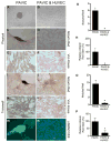
Endothelial cells inhibit calcification of porcine aortic valve interstitial cells (AVICs) via a secreted signal. Porcine AVICs spontaneously calcify in culture (A,C,E,G,H), however co-culturing porcine AVICs with HUVECs reduced calcification as measured by the lack of nodule formation (B,G) and significant decrease in Alizarin Red (D,H) and Von Kossa (F) staining. Inhibitory effects on calcification by HUVECs were demonstrated using a transwell co-culture system (I–P). The addition of HUVECs decreased nodule formation as well as Alizarin Red (J,O,P), von Kossa (K,L) and phospho-Smad1/5/8 (M,N) as compared to porcine AVICs alone (I,K,M,O,P), suggesting the effect is caused by a secreted signal and is contact independent. Alizarin Red, reddish brown; Von Kossa, brown; phospho-Smad, bright green; DAPI, blue. PAVIC, porcine AVIC; HUVEC, human umbilical vein endothelial cell; * p value < 0.05; scale bar 200 μm.
Figure 2
Figure 2
Nitric oxide signaling affects calcification of porcine aortic valve interstitial cells (AVICs). Porcine AVICs cultured in transwell with wild type mouse lung endothelial cells (MLEC) displayed decreased calcification as determined by nodule counts (D), phospho-Smad1/5/8 (A–C, green) and Alizarin red (E–H, red) staining as compared to porcine AVICs alone (A,E). Transwell culture with NOS3−/− MLECS did not attenuate calcification of AVICs (B,F,D,H). Alizarin Red, reddish brown; phospho-Smad, bright green; DAPI, blue. PAVIC, porcine AVIC; EC, endothelial cell; * p value < 0.05; scale bar 200 μm.
Figure 3
Figure 3
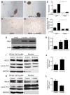
Nitric oxide regulates Notch1 signaling in porcine AVICs. Porcine AVICs transfected with NICD alone demonstrate an inhibition of calcification as shown by a decrease in nodule formation (C,E) and Alizarin red staining (C,F) compared to control GFP-transfected porcine AVICs (A). L-NAME treatment (2 mM) of GFP-transfected porcine AVICs results in increased calcification (B), that is inhibited by the transfection of NICD (D) as shown by a decrease in nodule formation (E) and Alizarin red staining (F). (A–D) are stained with Alizarin Red. (G,H) Co-culturing of porcine AVICs with HUVECs or adding NO donor (20 μM DETA-NONOate) results in increased Hey1 protein expression by immunoblot and densitometry. Actin expression is shown as loading control. (I–L) Cell fractionation experiments of treated PAVICs demonstrate increased nuclear expression of NICD with NO donor (I,J) and decreased nuclear expression of NICD with L-NAME (K,L) by immunoblotting and densitometry analysis. Lamin A/C and GAPDH are shown as loading controls.The graphs in J and L are representative of 3 independent experiments in which NICD expression was normalized to Lamin A/C. PAVIC, porcine AVIC; HUVEC, human umbilical vein endothelial cell; * p value <0.05; scale bar 200 μm.
Figure 4
Figure 4
Abnormal aortic valve morphology in NOS3−/−;Notch1+/− animals. (A) Table summarizing aortic valve morphology in different genotypes. (B–I) Representative images of aortic valves of 8–16 week old wild type (B,C), Notch1+/− (D,E), NOS3−/− (F,G) and NOS3−/−;Notch1+/− (H,I) animals. AoV, aortic valve; BAV, bicuspid aortic valve; scale bar 200 μm.
Figure 5
Figure 5
Aortic valve disease in adult NOS3−/−;Notch1+/− mice. Echocardiographic evaluation displayed aortic valve stenosis in NOS3−/−;Notch1+/− animals (D) compared to normal flow in wild type, NOS3−/− and Notch1+/− mice (A,B,C). Histological examination demonstrated thickened valves of compound mutants (H) compared to wild type, NOS3−/− and Notch1+/− mice (E, F, G). NOS3−/−;Notch1+/− animals display increased Alcian blue staining (I–L), SMA expression (M–P) and decreased Sox9 expression (Q–T) compared to NOS3−/− and Notch1+/− mice. (wild type, n=2; NOS3−/−, n=3; Notch1+/−, n=2; NOS3−/−;Notch1+/−, n=4) WT, wild type; scale bar 200 μm.

References

    1. Roger VL, Go AS, Lloyd-Jones DM, Benjamin EJ, Berry JD, Borden WB, et al. Heart disease and stroke statistics--2012 update: a report from the american heart association. Circulation. 2012;125:e2–e220. - PMC - PubMed
    1. Miller JD, Weiss RM, Heistad DD. Calcific aortic valve stenosis: methods, models, and mechanisms. Circ Res. 2011;108:1392–412. - PMC - PubMed
    1. Nkomo VT, Gardin JM, Skelton TN, Gottdiener JS, Scott CG, Enriquez-Sarano M. Burden of valvular heart diseases: a population-based study. Lancet. 2006;368:1005–11. - PubMed
    1. Rajamannan NM. Bicuspid aortic valve disease: the role of oxidative stress in Lrp5 bone formation. Cardiovasc Pathol. 2011;20:168–76. - PMC - PubMed
    1. Ward C. Clinical significance of the bicuspid aortic valve. Heart. 2000;83:81–5. - PMC - PubMed

Publication types

MeSH terms