. 2013 Jul 24;425(14):2509-28.
doi: 10.1016/j.jmb.2013.04.001. Epub 2013 Apr 10.

Ligand-induced dynamic changes in extended PDZ domains from NHERF1

Affiliations

Ligand-induced dynamic changes in extended PDZ domains from NHERF1

Shibani Bhattacharya et al. J Mol Biol. .

Abstract

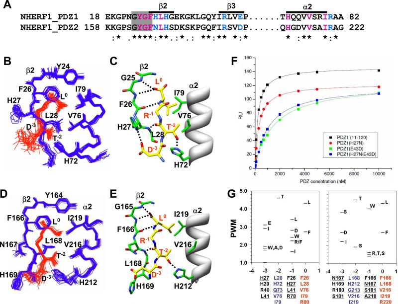
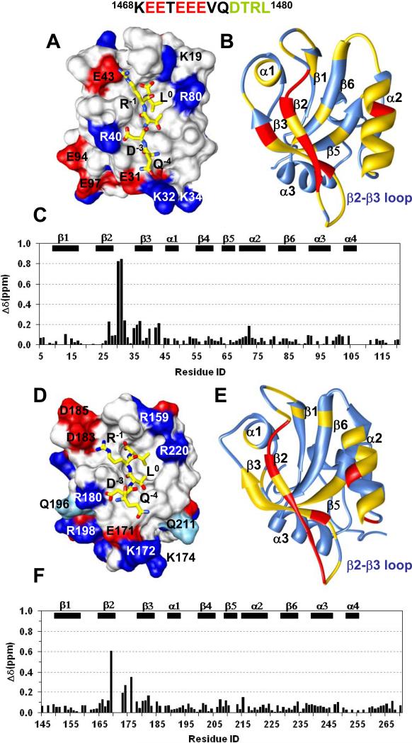
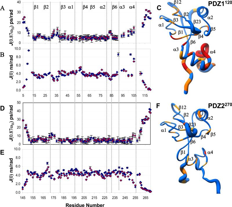
The multi-domain scaffolding protein NHERF1 modulates the assembly and intracellular trafficking of various transmembrane receptors and ion-transport proteins. The two PDZ (postsynaptic density 95/disk large/zonula occluden 1) domains of NHERF1 possess very different ligand-binding capabilities: PDZ1 recognizes a variety of membrane proteins with high affinity, while PDZ2 only binds limited number of target proteins. Here using NMR, we have determined the structural and dynamic mechanisms that differentiate the binding affinities of the two PDZ domains, for the type 1 PDZ-binding motif (QDTRL) in the carboxyl terminus of cystic fibrosis transmembrane regulator. Similar to PDZ2, we have identified a helix-loop-helix subdomain coupled to the canonical PDZ1 domain. The extended PDZ1 domain is highly flexible with correlated backbone motions on fast and slow timescales, while the extended PDZ2 domain is relatively rigid. The malleability of the extended PDZ1 structure facilitates the transmission of conformational changes at the ligand-binding site to the remote helix-loop-helix extension. By contrast, ligand binding has only modest effects on the conformation and dynamics of the extended PDZ2 domain. The study shows that ligand-induced structural and dynamic changes coupled with sequence variation at the putative PDZ binding site dictate ligand selectivity and binding affinity of the two PDZ domains of NHERF1.


Figures

Figure 1
Figure 1. NHERF1 multiple sequence alignment
(A) Schematic representation of human NHERF1 consisting of tandem PDZ1 and PDZ2 domains with a mostly disordered carboxy-terminal (CT) domain. The CT domain has overlapping Ezrin-binding (EB) and PDZ-binding motifs. The lengths of the putative PDZ domains are indicated in the box and the extended structure by solid bars. (B) Multiple sequence alignment of PDZ1, and (C) PDZ2 domains of NHERF1 from various species generated by the program ClustalX . The conserved sequence in secondary structure from the canonical domain is highlighted by grey boxes.
Figure 2
Figure 2. Structural comparison of PDZ1120 and PDZ2270 domains
(A) Stereoview of an ensemble of extended PDZ1120 structures determined by NMR with the canonical PDZ domain (residues 13-91) indicated in blue and the HLG subdomain in gold. (B) Backbone representation of PDZ1120 domain with annotated secondary structure. (C) Backbone superposition of the canonical domain from PDZ1120 (blue, residues 13-91) and PDZ2270 (pink, residues 153-231). The corresponding RMSD of 0.95 Å is significantly worse with RMSD of 1.5 Å when the HLG extensions are included in the alignment. The unstructured amino- and carboxy-termini residues were excluded from the figure. (D) Hydrophobic and aliphatic side-chain contacts (green sticks) in the HLG extension from PDZ1120, and (E) PDZ2270 domain.
Figure 3
Figure 3. Overview of electrostatic interactions in the HLG subdomains.
(A) Intra-helical and long-range electrostatic interactions involving charged and polar side-chains in PDZ1120, and (B) PDZ2270 domain represented by sticks color coded by type of heteroatom. (C) Chemical shift difference plot between wildtype PDZ1120 domain and the Glu61->Gly61 mutant at 15°C. The weighted difference was calculated using the relation Δ √ ( (δHN)2 + (δN/5)2 ). The N-terminal residues and secondary structure (β4, β5, α3 and α4) with significant chemical shift perturbation (>0.2 ppm, dotted line) in the mutant protein are indicated by different box pattern.
Figure 4
Figure 4. CFTR-C peptide (QDTRL) bound PDZ1120 and PDZ2270 structures
(A) Stereoview of the ensemble of 20 best NMR structures of PDZ1120 in complex with CFTR-C peptide (red). (B) Ribbon representation of single PDZ1120 complex structure. (C) Backbone superposition of PDZ1 canonical domain (residues 13-91) in the presence (pink) and absence of peptide (blue). The corresponding backbone RMSD (13-91) of 1.7 Å increases to 1.9 Å when additional residues (13-110) from the HLG subdomain are included in the alignment. (D) Stereoview of the CFTR-C peptide (red) bound PDZ2270 complex structure. (E) Ribbon representation of single PDZ2270 complex structure. (F) Backbone superposition of PDZ2 canonical domain (residues 153-231) in the presence (pink) and absence of peptide (blue). The corresponding backbone RMSD (153-231) of 1.0 Å increases to 1.2 Å when residues (153-252) from the HLG subdomain are included in the alignment. The stereoviews were generated in MOLMOL 2.1 and ribbons using UCSF Chimera package .
Figure 5
Figure 5. Overview of intermolecular interactions in the PDZ1 and PDZ2 peptide complexes
(A) Partial sequence alignment of the binding site residues from PDZ1 and PDZ2 domains color-coded based on hydrophobic (magenta), H-bond (magenta), and variable electrostatic (blue) interactions. Residues in the carboxylate binding loop are highlighted by the grey box. (B) Structural ensemble of PDZ1120 (blue) binding site with CFTR-C peptide (red), and (C) the corresponding intermolecular H-bonds (dotted lines) in a single structure. As per established convention for annotating PDZ-binding motifs, the carboxy-terminal residue of the CFTR-C peptide is ‘0’ and the amino-terminal Asp is ‘-3’ . For visual clarity the side-chain of Arg-1 is not shown in the figure. (D) Structural ensemble of PDZ2270 (blue) binding site with CFTR-C peptide (red), and (E) the corresponding intermolecular H-bonds (dotted lines) in a single structure. (F) Binding curves of CFTR-C domain with wildtype and mutant PDZ1120 domains from SPR measurements with the affinities reported in Table 1. (G) Graphical representation of the position weight matrix (PWM) of amino acid propensities at various positions (0 to -3) along the Type 1 PDZ motif calculated using published protocol with details provided in Supplementary Material . PWM reported along the Y-axis is effectively the logarithm of probabilities and hence no units are required. Residues associated with each binding pocket identified from sequence alignment are listed below the plot with the natural mutations underlined.
Figure 6
Figure 6. Electrostatic complementarity at peptide/protein complex interface
(A) The annotated electrostatic binding surface of PDZ1120 with peptide (yellow) showing charged side-chain interactions. (B) Chemical shift perturbation of PDZ1120 bound to CFTR-C domain painted yellow (>0.2 ppm) and red (>1.00 ppm) on the ribbon representation of the protein backbone. (C) The weighted difference in amide chemical shifts between the peptide and the CFTR-C domain bound to PDZ1120. (D) The annotated electrostatic surface of PDZ2270 with bound peptide (yellow) showing charged side-chain interactions. Polar residues (cyan) at the binding site with significant chemical shift perturbation are also labeled. (E) Chemical shift mapping of the CFTR-C binding site of PDZ2270 domain using identical cutoffs described above for panel (B). (F) The weighted difference in amide chemical shifts between the peptide and CFTR-C domain bound to PDZ2270.
Figure 7
Figure 7. Ligand induced dynamic changes in PDZ1 and PDZ2 domains
Backbone profile of the spectral density function calculated from relaxation measurements at 500 MHz, for PDZ1120 in the presence (red) and absence (blue) of CFTR-C peptide. (A) J(0.87ωH) and (B) J(0). (C) The radius-of-worm representation of the backbone of PDZ1120 in the absence of ligand is scaled by the amplitude of the J(0.87ωH) values. The fast timescale picosecond motions are reflected by a thicker tube with residues undergoing slow conformational exchange painted yellow. The presence of slower motions were detected from the ratio of J(0)900/J(0)500 > 1.2. Resonances broadened beyond detection are colored in red. In the panels (D)-(F) the corresponding graphs for PDZ2270 are displayed.

References

    1. Harris BZ, Lim WA. Mechanisms and role of PDZ domains in signalling complex assembly. J. Cell. Sci. 2001;114:3219–3231. - PubMed
    1. Zhang M. Scaffold proteins as dynamic switches. Nat. Chem. Biol. 2007;3:756–757. - PubMed
    1. Doyle DA, Lee A, Lewis J, Kim E, Sheng M, MacKinnon R. Crystal structures of a complexed and peptide-free membrane protein-binding domain: molecular basis of peptide recognition by PDZ. Cell. 1996;85:1067–76. - PubMed
    1. Tochio H, Mok YK, Zhang Q, Kan HM, Bredt DS, Zhang M. Formation of nNOS/PSD-95 PDZ dimer requires a preformed beta-finger structure from the nNOS PDZ domain. J. Mol. Biol. 2000;303:359–370. - PubMed
    1. Gallardo R, Ivarsson Y, Schymkowitz J, Rousseau F, Zimmermann P. Structural diversity of PDZ-lipid interactions. Chembiochem. 2010;11:456–467. - PubMed

Publication types