
Whole-genome sequencing identifies genetic alterations in pediatric low-grade gliomas

Jinghui Zhang et al. Nat Genet. 2013 Jun.

Abstract

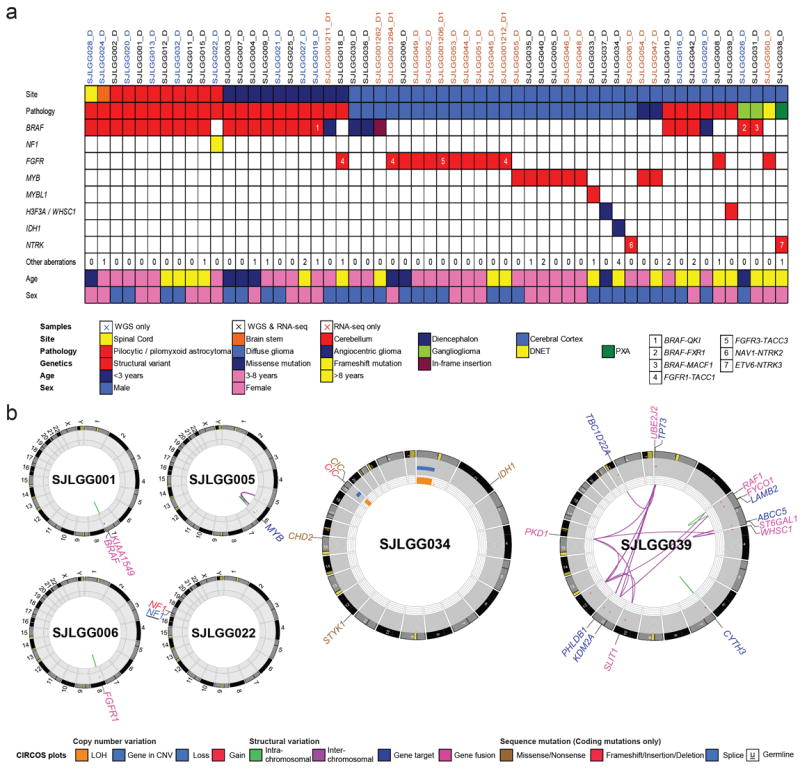
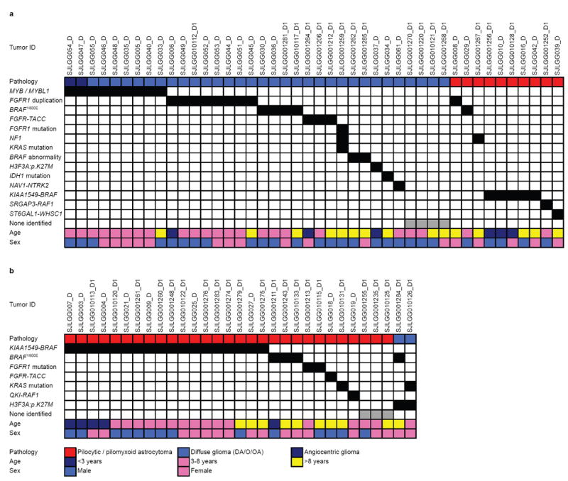
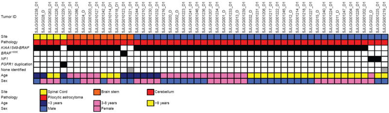
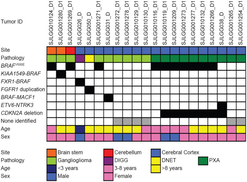
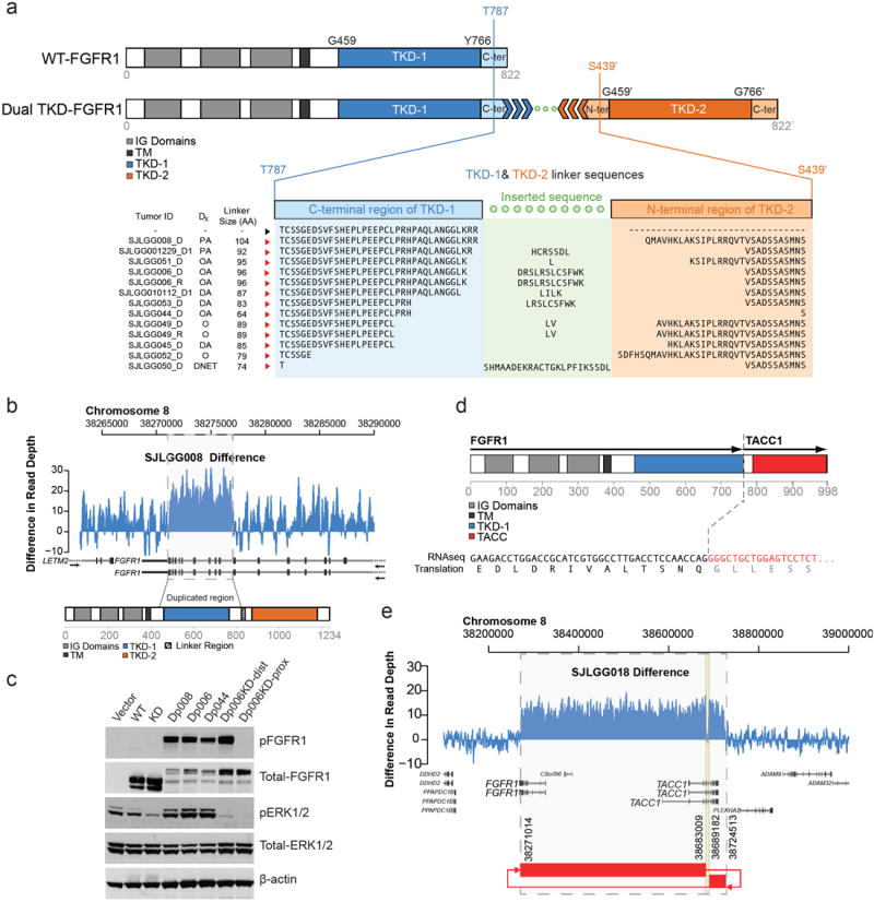
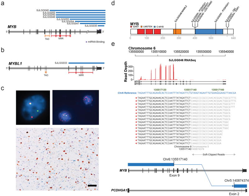
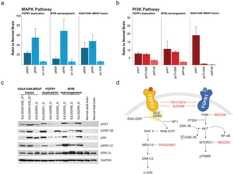
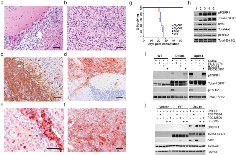
The most common pediatric brain tumors are low-grade gliomas (LGGs). We used whole-genome sequencing to identify multiple new genetic alterations involving BRAF, RAF1, FGFR1, MYB, MYBL1 and genes with histone-related functions, including H3F3A and ATRX, in 39 LGGs and low-grade glioneuronal tumors (LGGNTs). Only a single non-silent somatic alteration was detected in 24 of 39 (62%) tumors. Intragenic duplications of the portion of FGFR1 encoding the tyrosine kinase domain (TKD) and rearrangements of MYB were recurrent and mutually exclusive in 53% of grade II diffuse LGGs. Transplantation of Trp53-null neonatal astrocytes expressing FGFR1 with the duplication involving the TKD into the brains of nude mice generated high-grade astrocytomas with short latency and 100% penetrance. FGFR1 with the duplication induced FGFR1 autophosphorylation and upregulation of the MAPK/ERK and PI3K pathways, which could be blocked by specific inhibitors. Focusing on the therapeutically challenging diffuse LGGs, our study of 151 tumors has discovered genetic alterations and potential therapeutic targets across the entire range of pediatric LGGs and LGGNTs.


Figures

Figure 1
Figure 1. Clinicopathological characteristics and genetic alterations in tumors from series 1 examined by WGS or mRNA-seq
a Summary of clinicopathological characteristics (Tumor site, Pathology, Age at surgery, Sex) and key genetic alterations identified in 56 LGGs/LGGNTs by WGS (n=38) or mRNA-seq (n=44). Very few SVs or SNVs are found across the WGS tumor series. Numbers of aberrations for tumor samples highlighted in beige were based on analysis of mRNA-seq alone. Numbers in colored cells refer to gene fusions listed in box at right below table. b Selected CIRCOS plots summarizing aspects of WGS data, including a solitary KIAA1549-BRAF fusion (SJLGG001), MYB rearrangement (SJLGG005), FGFR1 TKD-duplication (SJLGG006), and NF1 mutation (SJLGG022). The genomic profiles of two oligodendroglial tumors, SJLGG006 and SJLGG034, illustrate some key differences between this type of tumor in children and in adults, the latter demonstrating IDH1 and CIC mutations alongside 1p/19q co-deletion. SJLGG039 exemplifies five LGGs with complex rearrangements.
Figure 2
Figure 2. Genetic alterations in supratentorial LGGs
Associations between genetic alterations and clinicopathological characteristics for LGGs in the (a) cerebral hemispheres or (b) diencephalon.
Figure 3
Figure 3. Genetic alterations in infratentorial LGGs
Associations between genetic alterations and clinicopathological characteristics for LGGs in the cerebellum, brain stem or spinal cord.
Figure 4
Figure 4. Genetic alterations in LGGNTs
Associations between genetic alterations and clinicopathological characteristics for LGGNTs across the tumor cohort. Abbreviations: DIGG – desmoplastic infantile ganglioglioma, DNET – dysembryoplastic neuroepithelial tumor, PXA – pleomorphic xanthoastrocytoma
Figure 5
Figure 5. FGFR1 aberrations in LGGs/LGGNTs
a FGFR1 – WT and with TKD duplication. With TKD duplication, two full length TKDs (TKD-1 & TKD-2) are separated by a linker sequence between the end (TKD-1) and beginning (TKD-2) of the auto-inhibited, activation-competent kinase domain spanning amino acids 459-766. Linker length varies between 74 and 104 amino acids. b Normalized (tumor minus germline) WGS read-coverage of FGFR1 in sample SJLGG008, with a 5kb duplication encompassing exons 10-18. c Western blot showing autophosphorylation of TKD-duplicated FGFR1 (Dp006, Dp008, Dp044) in 293T cells. FGFR1 autophosphorylation is associated with activation of the MAPK pathway, as indicated by raised p-ERK1/2 levels. Introducing a kinase-dead (KD) mutation (D623A) into either TKD-1 (Dp006KD-prox) or TKD-2 (Dp006KD-dist) of Dp006 abrogates MAPK pathway upregulation, but an active proximal TKD will still autophosphorylate FGFR1, while one with an active distal TKD will not. d FGFR1-TACC1 fusion protein detected in SJLGG018 preserving the TKD. e Normalized (tumor minus germline) WGS read coverage plot and SV connection plot for FGFR1/TACC1 locus suggesting formation of an episome. Abbreviations: WT – wild-type, TKD – tyrosine kinase domain, IG – Ig-like, TM – transmembrane domain, Dx – diagnosis, PA – pilocytic astrocytoma, OA – oligoastrocytoma, DA – diffuse astrocytoma, O – oligodendroglioma, DNET – dysembryoplastic neuroepithelial tumor, KD – kinase-dead, TACC – TACC domain.
Figure 6
Figure 6. MYB and MYBL1 aberrations in diffusely infiltrating LGGs
a, b Deletion through re-arrangement (blue bars) of MYB in 8 diffuse cerebral gliomas (a) or of MYBL1 in 1 diffuse cerebral glioma (b). c FISH - ‘break-apart’ probes were used to screen the study cohort for MYB, MYBL1, and MYBL2 rearrangements: (top left) probes targeting MYB are contiguous (yellow) at the normal locus, but split (green/red) where MYB is rearranged, (top right) MYB amplification (red probe), (bottom) cerebral diffuse astrocytoma with overexpression (brown reaction product) of MYB protein in the nuclei of neoplastic cells, but not normal glial cells (blue/gray hematoxylin counterstain). Scale bar = 50μm. d MYB protein showing breakpoints for rearrangements with other genes. The MYB transactivation domain is intact in all fused proteins, except in SJLGG048, where fusion with MAML2 may cause hyper-activation. In other fusions, the negative regulatory region or regulatory miRNA binding site is disrupted, or there is episome formation. e mRNA-seq and WGS data for SJLGG046 demonstrating MYB and PCDHGA1 fusion and over-expression of fused MYB. Abbreviations: TAD – transactivating domain, NRR – negative regulatory region.
Figure 7
Figure 7. Activation of MAPK and PI3K pathways in LGGs/LGGNTs with FGFR1, MYB and BRAF abnormalities
a, b Multiplex immunoassay levels for three MAPK (a) and three PI3K (b) pathway components are given relative to levels in normal brain for tumors with either FGFR1 duplication (n=11), or MYB rearrangement (n=6), or KIAA1549-BRAF fusion (n=18); bars represent the standard error of the mean. Gene expression profiling showed no significant elevation in the levels of corresponding mRNAs, with the exception of c-JUN. c Western blot data showing upregulation of the MAPK pathway (pERK1/2) and PI3K pathway (pAKT/pS6) in three groups of LGGs characterized by KIAA1549-BRAF fusion, FGFR1 duplication, or MYB rearrangement (AKT inhibits GSK-3β by phosophorylation). d Diagrams of MAPK (left) and PI3K (right) pathway components downstream of FGFR1, showing targets of pharmaceuticals used to inhibit the actions of TKD-duplicated FGFR1 (see Fig. 8).
Figure 8
Figure 8. TKD-duplicated FGFR1 in neonatal astrocytes generates gliomas in vivo
a-f Histopathology of TKD-duplicated FGFR1 tumors in mouse brain. Diffuse gliomas could demonstrate either an astrocytic (a) or oligodendroglial (b) phenotype, and were focally immunopositive for GFAP (c). Tumor cells, but not normal brain, showed overexpression of FGFR1 (d), pMAPK (e), or pAKT (f). Scale bar = 100μm. g Survival curves for mice transplanted with neonatal astrocytes containing empty vector (MIG), wild-type FGFR1 (WT), or two variants of TKD-duplicated FGFR1 (Dp006, Dp008). All Dp006/8 mice died from the effects of a glioma with a median survival of 23 days. h Western blot of proteins from normal brain (1) or tumor cell lysates (separate mice: 2,3 – Dp006; 4,5 – Dp008). TKD-duplicated FGFR1 is associated with increased levels of pFGFR1 (Y463/464), pAKT (S473), and pMAPK (T202/204). i Inhibitor studies showing that FGFR1 inhibitors PD173074 and BGJ398 block both autophosphorylation of duplicated FGFR1 (Dp044 & Dp006) and downstream activation of the MAPK pathway. The MEK1 inhibitor PD0325901 does not block FGFR1 autophosphorylation, but inhibits MAPK pathway activation. j Inhibitor studies showing that the FGFR1 inhibitor PD173074 blocks autophophorylation of duplicated FGFR1 and downstream activation of the PI3K pathway. The PI3K/mTOR inhibitor BEZ235 does not block FGFR1 autophosphorylation, but inhibits PI3K pathway activation.

References

    1. Armstrong GT, et al. Survival and long-term health and cognitive outcomes after low-grade glioma. Neuro-oncology. 2011;13:223–34. - PMC - PubMed
    1. Arora RS, et al. Age-incidence patterns of primary CNS tumors in children, adolescents, and adults in England. Neuro Oncol. 2009;11:403–13. - PMC - PubMed
    1. Qaddoumi I, Sultan I, Gajjar A. Outcome and prognostic features in pediatric gliomas: a review of 6212 cases from the Surveillance, Epidemiology, and End Results database. Cancer. 2009;115:5761–70. - PMC - PubMed
    1. Bouffet E, et al. Phase II study of weekly vinblastine in recurrent or refractory pediatric low-grade glioma. Journal of clinical oncology : official journal of the American Society of Clinical Oncology. 2012;30:1358–63. - PubMed
    1. Duffner PK, Cohen ME, Myers MH, Heise HW. Survival of children with brain tumors: SEER Program, 1973-1980. Neurology. 1986;36:597–601. - PubMed

Publication types

MeSH terms