. 2013 Jul;9(7):985-95.
doi: 10.4161/auto.24406. Epub 2013 Apr 12.

Host and bacterial factors that regulate LC3 recruitment to Listeria monocytogenes during the early stages of macrophage infection

Affiliations

Host and bacterial factors that regulate LC3 recruitment to Listeria monocytogenes during the early stages of macrophage infection

Grace Y Lam et al. Autophagy. 2013 Jul.

Abstract

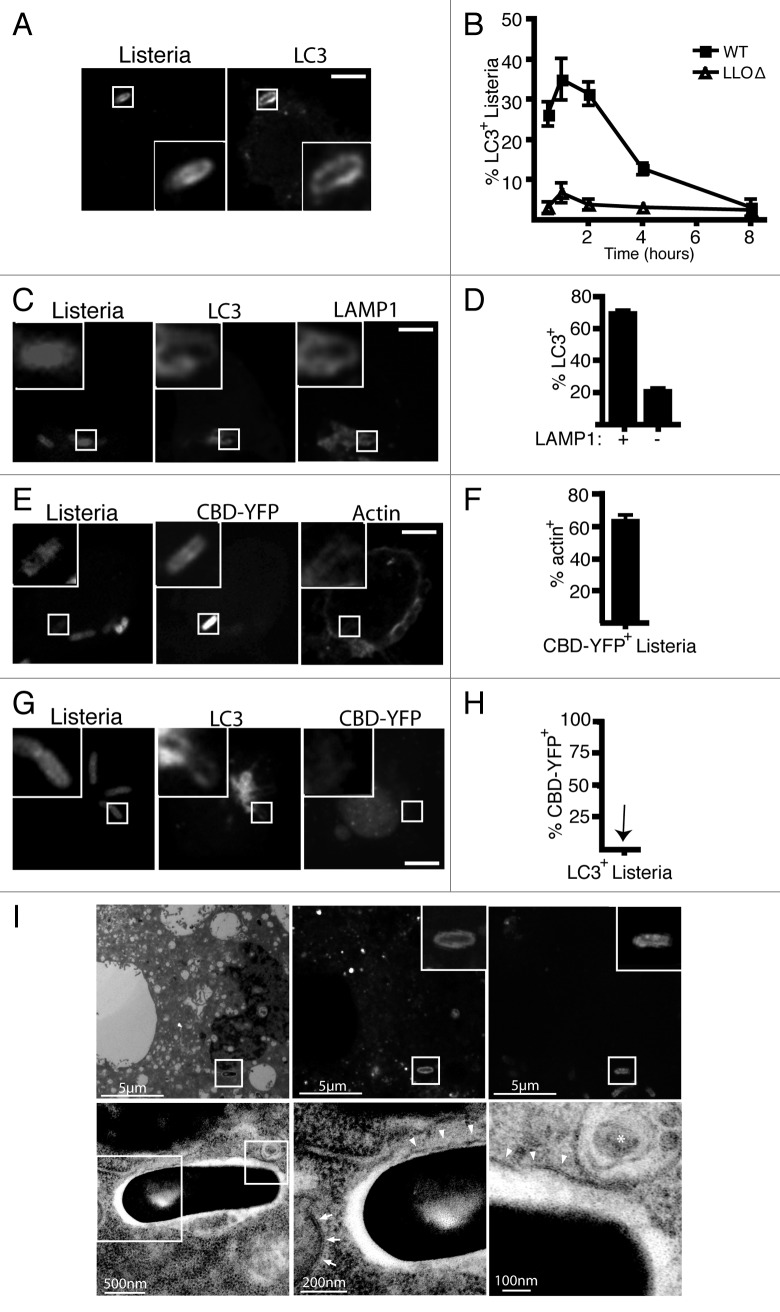
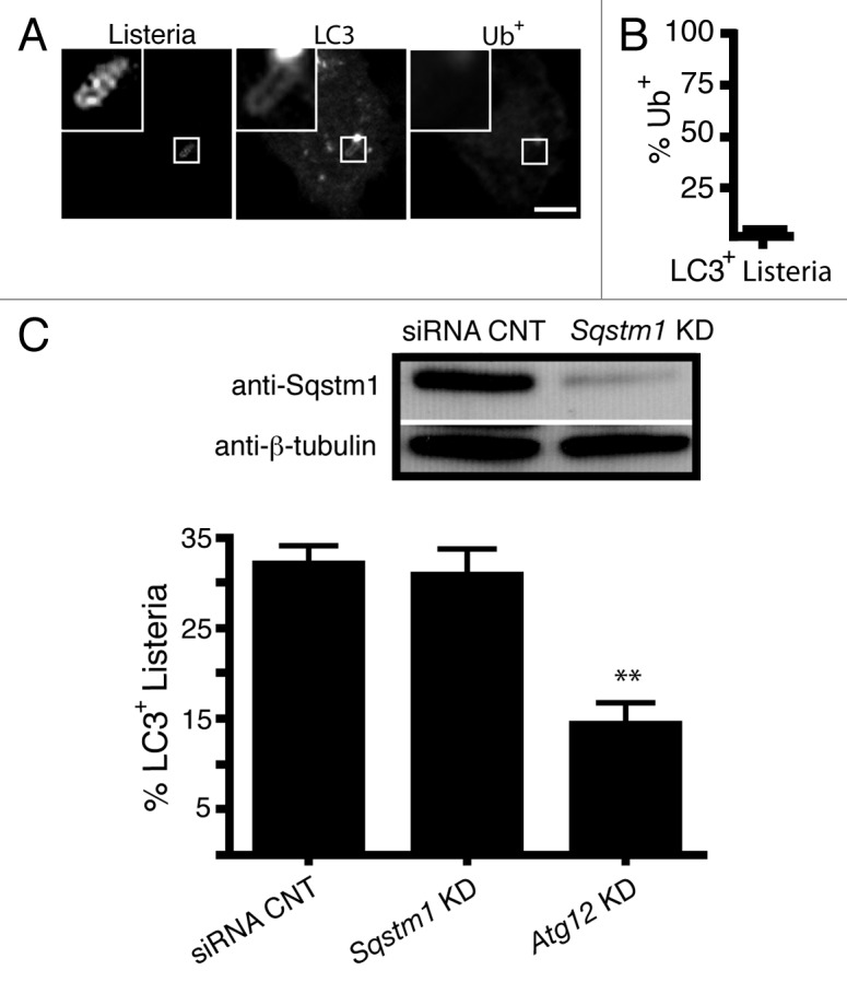
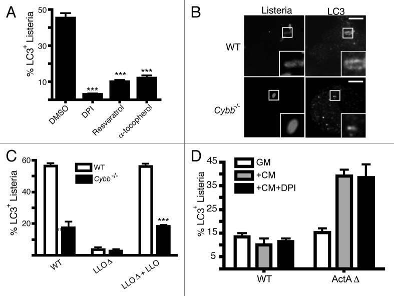
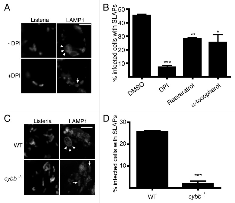
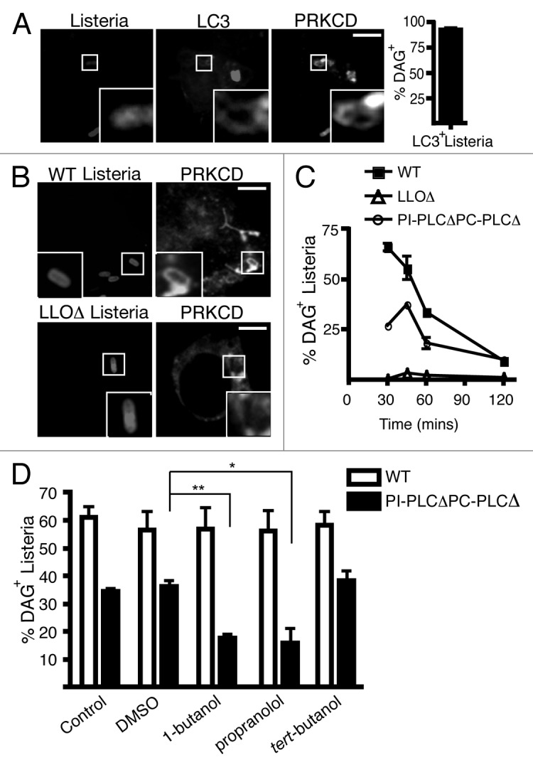
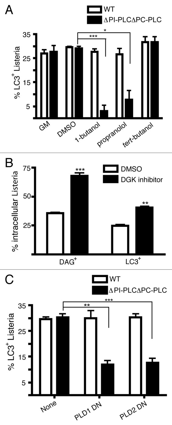
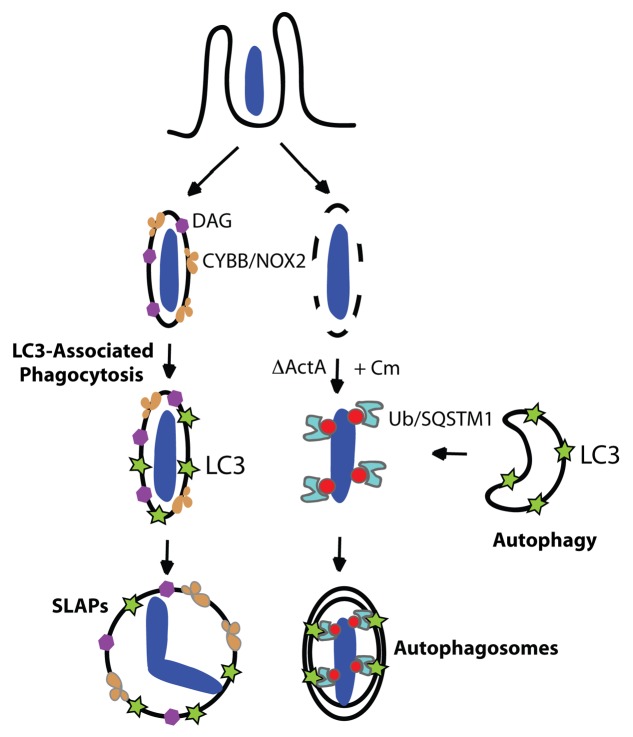
Listeria monocytogenes is a bacterial pathogen that can escape the phagosome and replicate in the cytosol of host cells during infection. We previously observed that a population (up to 35%) of L. monocytogenes strain 10403S colocalize with the macroautophagy marker LC3 at 1 h postinfection. This is thought to give rise to spacious Listeria-containing phagosomes (SLAPs), a membrane-bound compartment harboring slow-growing bacteria that is associated with persistent infection. Here, we examined the host and bacterial factors that mediate LC3 recruitment to bacteria at 1 h postinfection. At this early time point, LC3(+) bacteria were present within single-membrane phagosomes that are LAMP1(+). Protein ubiquitination is known to play a role in targeting cytosolic L. monocytogenes to macroautophagy. However, we found that neither protein ubiquitination nor the ubiquitin-binding adaptor SQSTM1/p62 are associated with LC3(+) bacteria at 1 h postinfection. Reactive oxygen species (ROS) production by the CYBB/NOX2 NADPH oxidase was also required for LC3 recruitment to bacteria at 1 h postinfection and for subsequent SLAP formation. Diacylglycerol is an upstream activator of the CYBB/NOX2 NADPH oxidase, and its production by both bacterial and host phospholipases was required for LC3 recruitment to bacteria. Our data suggest that the LC3-associated phagocytosis (LAP) pathway, which is distinct from macroautophagy, targets L. monocytogenes during the early stage of infection within host macrophages and allows establishment of an intracellular niche (SLAPs) associated with persistent infection.

Keywords: LC3; LC3-associated phagocytosis; Listeria monocytogenes; autophagy; diacylglycerol; innate immunity; reactive oxygen species; ubiquitin.


Figures

None
Figure 1. LC3 is recruited to wild-type L. monocytogenes strain 10403S in macrophage phagosomes during early stages of infection. (A) Confocal images of RAW 264.7 macrophages transfected with GFP-LC3 and infected for 1 h with wild-type L. monocytogenes. (B) Quantification of LC3 colocalization with intracellular wild-type or LLOΔ bacteria over time. (C) Confocal images of RAW 264.7 macrophages transfected with GFP-LC3 and infected for 1 h with wild-type L. monocytogenes. Cells were then stained for LAMP1. (D) Quantification of the percentage of LAMP1+ bacteria that are LC3+ or percentage of LAMP1- bacteria that are LC3+. (E) Confocal images of RAW 264.7 macrophages transfected with CBD-YFP and infected for 1 h with wild-type L. monocytogenes. Cells were then stained with phalloidin to label F-actin. (F) Quantification of the percentage of CBD-YFP+ bacteria that are actin+. (G) Confocal images of RAW 264.7 macrophages expressing RFP-LC3 and CBD-YFP and infected for 1 h with wild-type L. monocytogenes. (H) Quantification of the percentage of LC3+L. monocytogenes that are CBD-YFP+. The inner panels represent a higher magnification of the boxed areas. Size bars: 5 µm. The brightness and contrast for the image (but not the inset) was enhanced to allow visualization of fluorescence inside the cell. (I) Correlative imaging of RAW 264.7 macrophage expressing GFP-LC3 infected with NHS-647 labeled wild-type L. monocytogenes. Images of the same cell were captured using either electron microscopy (upper row) or fluorescence microscopy (lower row). Size bars as indicated. Arrowheads indicate single membrane while arrows indicate double membrane. The asterix indicates a multivesicular body.
None
Figure 2. Protein ubiquitination and the ubiquitin-adaptor protein p62/SQSTMI do not mediate LC3 recruitment to L. monocytogenes at 1 h p.i. (A) Confocal images of RAW 264.7 macrophages transfected with GFP-LC3 and infected for 1 h with wild-type L. monocytogenes. Cells were then stained for ubiquitinated proteins (Ub+). (B) Quantification of the percentage of LC3+L. monocytogenes that are Ub+. The inner panels represent a higher magnification of the boxed areas. Size bars: 5 µm. (C) Quantification of LC3 colocalization to wild-type L. monocytogenes in RAW 264.7 macrophages with siRNA silencing of Sqstm1 or Atg12 (positive control). Knockdown was confirmed using sodium dodecyl sulfate polyacrylamide gel electrophoresis (SDS-PAGE).
None
Figure 3. ROS production by the CYBB/NOX2 NADPH oxidase is required for LC3 recruitment to L. monocytogenes during early stages of infection. (A) RAW 264.7 cells were infected with wild-type L. monocytogenes for 1 h in the presence or absence of DPI, resveratrol or α-tocopherol, as indicated. The percentage of intracellular bacteria that are LC3+ upon treatment with each agent is shown. (B) Confocal images of wild-type or cybb−/− bone marrow-derived macrophages transfected with GFP-LC3 and infected for 1 h with wild-type bacteria. Size bar: 5 µm. (C) Quantification at 1 h p.i. of the percentage of intracellular L. monocytogenes that are LC3+ in bone marrow-derived macrophages isolated from wild-type or cybb-/- mice. Wild-type bacteria were compared with LLOΔ bacteria (negative control), as well as a LLOΔ mutant complemented with LLO. (D) Quantification of the percentage of L. monocytogenes that are LC3+ in RAW 264.7 macrophages infected with either wild-type or ActAΔ bacteria for: 8 h (GM); 3 h, followed by 5 h Cm treatment (+Cm); or 3 h, followed by 5 h Cm and DPI treatment (+Cm +DPI).
None
Figure 4. ROS production and LC3-associated phagocytosis is required for SLAP formation during later stages of infection. ROS production by the CYBB/NOX2 NADPH oxidase is required for the generation of SLAPs. (A) Confocal images of RAW 264.7 macrophages infected for 4 h with wild-type L. monocytogenes, with or without DPI. Cells were stained with LAMP1 antibodies. Size bar: 5 µm. White arrowheads indicate spacious Listeriaphagosomes (SLAPs) while the white arrow indicates LAMP1 colocalization with bacteria that are not in SLAPs (B) Quantification at 4 h p.i. of the percentage of infected macrophages that form SLAPs in the presence or absence of DPI, resveratrol or α-tocopherol, as indicated. (C) Confocal images of wild-type or cybb−/− bone marrow-derived macrophages infected for 8 h with wild-type L. monocytogenes. Cells were stained with LAMP1 antibodies. Size bar: 5 µm. White arrowheads indicate SLAPs while the white arrows indicate LAMP1 colocalization and not SLAPs. (D) Quantification at 8 h p.i. of the percentage of infected wild-type or cybb-/- bone marrow-derived macrophages that form SLAPs from (C).
None
Figure 5. Host and bacterial factors promote DAG accumulation on phagosomes containing L. monocytogenes. (A) Confocal images of RAW 264.7 macrophages co-transfected with RFP-LC3 and PRKCD-C1-GFP and infected for 1 h with wild-type L. monocytogenes. PRKCD-C1-GFP is a specific probe for DAG. Size bar: 5 µm. Quantification at 1 h p.i. of the percentage of LC3+L. monocytogenes that are PRKCD-C1-GFP+ (B) Confocal images of RAW 264.7 macrophages transfected with PRKCD-C1-GFP and infected for 1 h with wild-type, LLOΔ or PI-PLCΔ PC-PLCΔ bacteria. Size bar: 5 µm. (C) Quantification of the percentage of intracellular wild-type, LLOΔ or PI-PLCΔ PC-PLCΔ L. monocytogenes that are DAG+ over time. (D) Quantification at 45 min p.i. of intracellular wild-type or PI-PLCΔ PC-PLCΔ L. monocytogenes that are PRKCD-C1-GFP + upon treatment with DMSO, 1-butanol, propranolol or tert-butanol.
None
Figure 6. DAG production is required for LC3-associated phagocytosis of L. monocytogenes during early stages of infection. (A) Quantification at 1 h p.i. of intracellular wild-type or PI-PLCΔ PC-PLCΔ L. monocytogenes that are LC3+ following treatment with DMSO, 1-butanol, propranolol, or tert-butanol. (B) Quantification at 1 h p.i. of DAG (PRKCD-C1-GFP+) or LC3 colocalization with wild-type L. monocytogenes with or without treatment with the DAG kinase I inhibitor. (C) Quantification at 1 h p.i. of LC3+ intracellular wild-type or PI-PLCΔ PC-PLCΔ L. monocytogenes in RAW 264.7 macrophages that were cotransfected with LC3-GFP and either PLD1 dominant negative (DN) or PLD2 DN constructs.
None
Figure 7. Two pathways mediate LC3 recruitment to L. monocytogenes strain 10403S during infection. Model shows the two pathways that can mediate LC3 recruitment to bacteria. First, LC3-associated phagocytosis (LAP) can mediate LC3 localization to phagosomes containing L. monocytogenes at 1 h p.i. (left-hand side pathway). In this study we showed that diacylglycerol (DAG) accumulates on the phagosome as the result of bacterial (PLC) and host (PLD and PPAP2A) enzymatic activity. DAG accumulation promotes activation of the CYBB/NOX2 NADPH oxidase and the production of reactive oxygen species (ROS). ROS production mediates LAP of L. monocytogenes, a process that is characterized by LC3 recruitment to single-membrane phagosomes containing bacteria. While LAP has been shown to promote phagosome maturation and killing of certain microbes, our data indicates that LAP promotes the formation of spacious Listeria-containing phagosomes (SLAPs), a compartment that has been associated with persistent infection. Second, LC3 can be recruited to L. monocytogenes in the cytosol under specific conditions. When treated with the bacteristatic agent, chloramphenicol (Cm), the ActAΔ mutant of L. monocytogenes can be targeted by autophagy into double-membrane autophagosomes via protein ubiquitination and ubiquitin-binding adaptor proteins including SQSTM1 (right-hand side pathway). In contrast, wild-type L. monocytogenes evades autophagy in the cytosol via expression of ActA and other factors, allowing them to replicate rapidly and undergo actin-based motility for cell-to-cell spread.

References

    1. Levine B, Mizushima N, Virgin HW. Autophagy in immunity and inflammation. Nature. 2011;469:323–35. doi: 10.1038/nature09782. - DOI - PMC - PubMed
    1. Birmingham CL, Smith AC, Bakowski MA, Yoshimori T, Brumell JH. Autophagy controls Salmonella infection in response to damage to the Salmonella-containing vacuole. J Biol Chem. 2006;281:11374–83. doi: 10.1074/jbc.M509157200. - DOI - PubMed
    1. Gutierrez MG, Master SS, Singh SB, Taylor GA, Colombo MI, Deretic V. Autophagy is a defense mechanism inhibiting BCG and Mycobacterium tuberculosis survival in infected macrophages. Cell. 2004;119:753–66. doi: 10.1016/j.cell.2004.11.038. - DOI - PubMed
    1. Sanjuan MA, Dillon CP, Tait SW, Moshiach S, Dorsey F, Connell S, et al. Toll-like receptor signalling in macrophages links the autophagy pathway to phagocytosis. Nature. 2007;450:1253–7. doi: 10.1038/nature06421. - DOI - PubMed
    1. Nakagawa I, Amano A, Mizushima N, Yamamoto A, Yamaguchi H, Kamimoto T, et al. Autophagy defends cells against invading group A Streptococcus. Science. 2004;306:1037–40. doi: 10.1126/science.1103966. - DOI - PubMed

Publication types

MeSH terms