Review
. 2013 May;22 Suppl 3(Suppl 3):S521-5.
doi: 10.1007/s00586-013-2773-x. Epub 2013 Apr 13.

Bifocal extra- and intradural melanocytoma of the spine: case report and literature review

Affiliations
Review

Bifocal extra- and intradural melanocytoma of the spine: case report and literature review

Niels A Foit et al. Eur Spine J. 2013 May.

Abstract

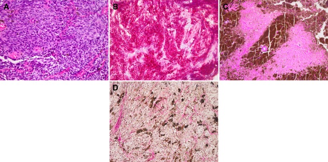
Background: Spinal melanocytoma is one of the most infrequent space-occupying lesions of the central nervous system. To the best of our knowledge, this is the first report of primary bifocal intradural melanocytoma of heterogeneous pathological grade to date.

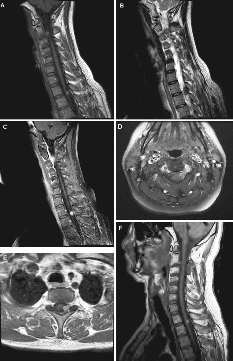
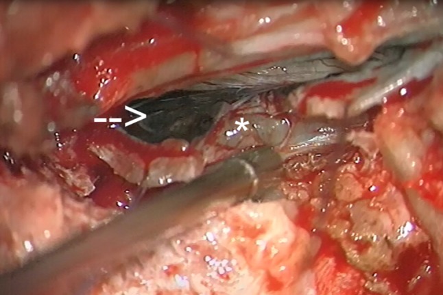
Case description: We report the case of a 43-year old patient with primary bifocal melanocytoma, clinically and radiologically resembling benign schwannoma. The patient presented with myeloradiculopathy of the left C3 dermatome. Magnetic resonance imaging of the upper spine revealed two space-occupying lesions with paraspinal extension, initially diagnosed as neurofibroma. Definitive histopathological classification of both lesions was melanocytoma. Both tumours were only partially removed due to adherence to surrounding structures. The patient underwent stereotactic external beam irradiation (EBR). Follow-up at 1 year after surgery revealed no recurrence and the patient remained free of symptoms. The clinical, radiological and pathological features of this rare tumour entity are presented and the available literature is reviewed.

Conclusions: Intradural melanocytoma, although exceedingly rare, requires a thorough work-up to exclude malignant melanoma. With only two previous reports of multifocal melanocytoma published in the literature, standard therapy has not yet been established and complete surgical removal remains the modality of choice. Patients should be closely monitored to detect local recurrence or malignant degeneration. EBR may be considered in cases where total excision is not achievable and reduces risk of local recurrences.


Figures

Fig. 1
Fig. 1
Magnetic resonance imaging. Sagittal MRI of the cervical/upper thoracic spine reveals two intradural, extra-axial lesions, extending through the neuroforamen of C2-C3 (a, b, d) and T1-T2 (a, b, e). Both enhance homogenously after gadolinium administration (d, e), the cervical lesion shows diffuse leptomeningeal enhancement (c). No progression is seen 12 months after initial surgery and external beam irradiation (f)
Fig. 2
Fig. 2
Intraoperative photography. Intraoperative photographs demonstrating the cervical lesion in the C2-3 segment as an intradural mass (arrow) with extradural extension (asterisk). Note the diffuse meningeal hyperpigmentation overlying the tumour
Fig. 3
Fig. 3
Histopathology. Haematoxylin–eosin (HE) staining of the cervical lesion displays variably pigmented, tightly packed tumour cells in a nested growth pattern. Cells possess oval to bean-shaped nuclei, with occasional grooves (a). Pan-melanoma immuno-histochemistry preparation (b) demonstrates diffuse cytoplasmatic labeling. HE-stained sections from the thoracic biopsy reveal monomorphic tumor cells surrounded by densely pigmented cells and extracellular melanin. Note the small focus of necrosis (c). Gomori reticulin stain fails to demonstrate reticulin fibers surrounding individual tumor cells (d)

References

    1. Ali Y, Rahme R, Moussa R, Abadjian G, Menassa-Moussa L, Samaha E. Multifocal meningeal melanocytoma: a new pathological entity or the result of leptomeningeal seeding? J Neurosurg. 2009;111(3):488–491. doi: 10.3171/2009.3.JNS081096. - DOI - PubMed
    1. Azarpira N, Torabineghad S, Sepidbakht S, Rakei M, Bagheri MH. Cytologic findings in pigmented melanotic schwannoma: a case report. Acta Cytol. 2009;53(1):113–115. doi: 10.1159/000325096. - DOI - PubMed
    1. Brat DJ, Giannini C, Scheithauer BW, Burger PC. Primary melanocytic neoplasms of the central nervous systems. Am J Surg Pathol. 1999;23(7):745–754. doi: 10.1097/00000478-199907000-00001. - DOI - PubMed
    1. Classen J, Hehr T, Paulus W, Plate K, Bamberg M. Suprasellar melanocytoma: a case of primary radiotherapy and review of the literature. J Neurooncol. 2002;58(1):39–46. doi: 10.1023/A:1015872207398. - DOI - PubMed
    1. El-Khashab M, Koral K, Bowers DC, Johnson-Welch S, Swift D, Nejat F. Intermediate grade meningeal melanocytoma of cervical spine. Child’s Nerv Syst. 2009;25:407–410. doi: 10.1007/s00381-008-0782-6. - DOI - PubMed