. 2013 Jun 21;113(1):52-61.
doi: 10.1161/CIRCRESAHA.112.300683. Epub 2013 Apr 12.

Macrophage ADAM17 deficiency augments CD36-dependent apoptotic cell uptake and the linked anti-inflammatory phenotype

Affiliations

Macrophage ADAM17 deficiency augments CD36-dependent apoptotic cell uptake and the linked anti-inflammatory phenotype

Will S Driscoll et al. Circ Res. .

Abstract

Rationale: Apoptotic cell phagocytosis (efferocytosis) is mediated by specific receptors and is essential for resolution of inflammation. In chronic inflammation, apoptotic cell clearance is dysfunctional and soluble levels of several apoptotic cell receptors are elevated. Reports have identified proteolytic cleavage as a mechanism capable of releasing soluble apoptotic cell receptors, but the functional implications of their proteolysis are unclear.

Objective: To test the hypothesis that ADAM17-mediated cleavage of apoptotic cell receptors limits efferocytosis in vivo.

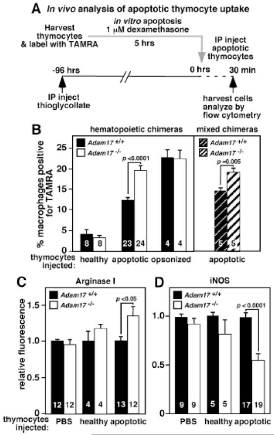
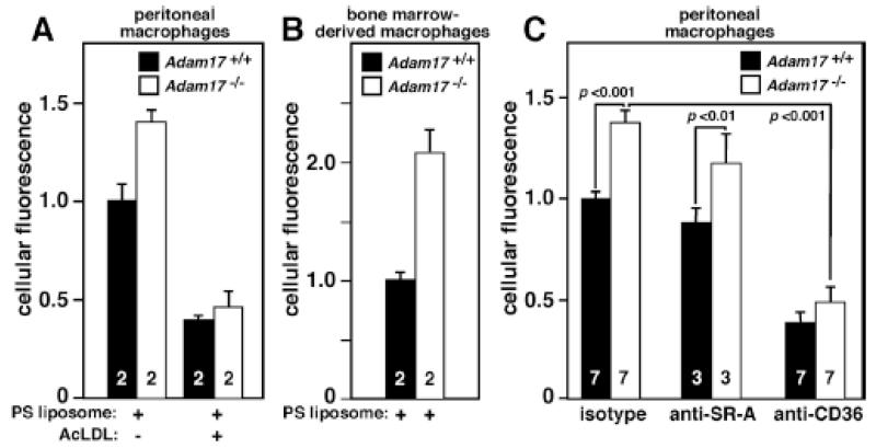
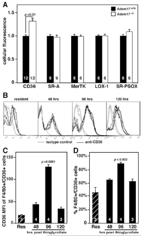
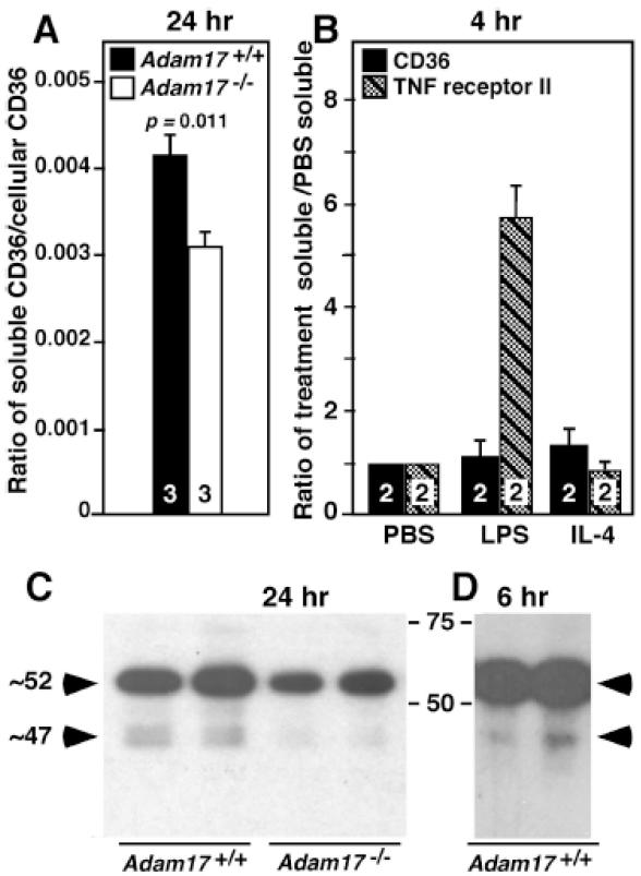
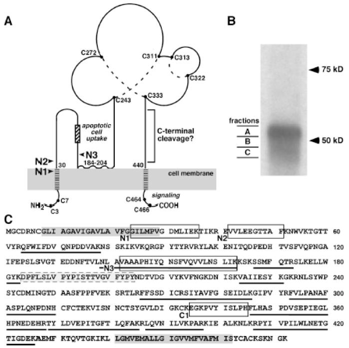
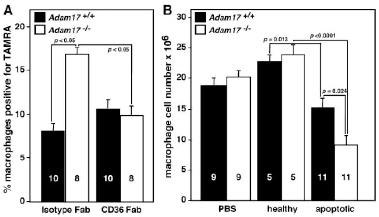
Methods and results: In vivo comparison of macrophage efferocytosis in wild-type and Adam17-null hematopoietic chimeras demonstrates that ADAM17 deficiency leads to a 60% increase in efferocytosis and an enhanced anti-inflammatory phenotype in a model of peritonitis. In vitro uptake of phosphatidylserine liposomes identifies the dual-pass apoptotic cell receptor CD36 as a major contributor to enhanced efferocytosis, and CD36 surface levels are elevated on macrophages from Adam17-null mice. Further, temporal elevation of CD36 expression with inflammation may also contribute to its impact. Soluble CD36 from macrophage-conditioned media comprises 2 species based on Western blotting, and mass spectrometry identifies 3 N-terminal peptides that represent probable cleavage sites. Levels of soluble CD36 are decreased in Adam17-null conditioned media, providing evidence for involvement of ADAM17 in CD36 cleavage. Importantly, enhanced efferocytosis in vivo by macrophages lacking ADAM17 is CD36 dependent and accelerates macrophage clearance from the peritoneum, thus promoting resolution of inflammation and highlighting the impact of increased apoptotic cell uptake.

Conclusions: Our studies demonstrate the importance of ADAM17-mediated proteolysis for in vivo efferocytosis regulation and suggest a possible mechanistic link between chronic inflammation and defective efferocytosis.

Keywords: apoptotic cells; inflammation; macrophage; metalloproteinase; proteolysis.


Figures

Figure 1
Figure 1. Increased in vivo efferocytosis of apoptotic thymocytes by Adam17-null macrophages is cell autonomous, and shifts them to a less inflammatory phenotype
A. The scheme for in vivo peritoneal macrophage uptake of TAMRA-labeled apoptotic thymocytes is shown. Healthy thymocytes and opsonized thymocytes were evaluated as controls. B. WT, Adam17-null, or mixed hematopoietic chimera mice that have ~50% WT and ~50% Adam17-null leukocytes were injected with the indicated thymocytes. The percent of TAMRA-positive macrophages was assessed by flow cytometry. C. Following in vivo exposure to PBS, healthy or apoptotic thymocytes, Adam17-null or WT macrophages were analyzed immediately following harvest for intracellular levels of arginase I, or D. cultured ex-vivo for 20 hours with lipopolysaccharide and IFNγ and analyzed for intracellular iNOS by flow cytometry. Numbers in the bars indicate the number of mice evaluated, and p values for significant differences are shown.
Figure 2
Figure 2. Adam17-null peritoneal and bone marrow-derived macrophages show increased phosphatidylserine liposome binding/uptake, and peritoneal macrophage uptake is CD36-dependent
A. Peritoneal macrophages from WT or Adam17-null hematopoietic chimera mice were harvested 96 hours after thioglycollate injection, and either plated and treated with or without acetylated-LDL at 4°C for 15 minutes followed by incubation with 40 μmol/L fluorescently labeled phosphatidylserine (PS) liposomes for 4 hours at 4°C. B. Bone-marrow-derived macrophages were isolated from whole bone marrow of WT or Adam17-null hematopoietic chimera mice and cultured for 7 days with M-CSF. Adherent cells were replated, and PS liposome binding was monitored as described in A. C. Peritoneal macrophages were plated and incubated with 160 μmol/L fluorescently labeled PS liposomes and 5 mg/L of either receptor-blocking or isotype control antibody for one hour at 37°C. Fluorescence was assessed by flow cytometry, numbers within the bars indicate the number of mice evaluated, and p values for significant differences are shown.
Figure 3
Figure 3. Cell surface levels of CD36 are elevated on peritoneal macrophages lacking ADAM17, and maximal expression of CD36 on WT macrophages is during the resolution phase of peritonitis
WT resident peritoneal macrophages (Res) or peritoneal macrophages from WT or Adam17-null hematopoietic chimera mice were harvested at different times after thioglycollate injection and immediately stained with antibodies to the indicated apoptotic cell receptors and evaluated by flow cytometry. A. Levels of different apoptotic cell receptors were evaluated 96 hours after thioglycollate. B. Histograms following staining of resident peritoneal macrophages or peritoneal macrophages 48, 96 and 120 hours after thioglycollate with anti-CD36 or isotype control (n=3-4 mice/time point). C. The mean fluorescent index (MFI) of CD36 is indicated for F4/80+ macrophages that are CD36+. D. The % of F4/80+ cells expressing CD36 is shown. Numbers within bars indicate the number of mice evaluated, and p values for significant differences are shown.
Figure 4
Figure 4. Adam17 deletion results in a 25% decrease in the ratio of soluble to cell- associated CD36
CD36 content and biochemical characteristics of microparticle-depleted conditioned medium and cell lysates from Adam17-null and WT peritoneal macrophages after the indicated incubation times were analyzed. A. CD36 levels in lysate and media were quantified by ELISA and expressed as the ratio of soluble to cellular CD36, and numbers within the bars indicate the number of mice of different genotypes whose macrophages were evaluated. These data are representative of 2 experiments. B. Soluble levels of CD36 and TNF receptor II were evaluated by ELISA following stimulation of WT peritoneal macrophages for 4 hours. Data is expressed relative to levels in PBS. Values are from two different experiments using macrophages from different mice determined in duplicate. C. and D. Western blot analysis of conditioned media from Adam17-null or WT peritoneal macrophages following deglycosylation with PNGase F. Each lane represents media collected from macrophages of different mice after the indicated time, and the collections for C are from a different experiment than shown in A.
Figure 5
Figure 5. Identification of novel proteolytic cleavage sites in soluble CD36
A. Topology and domains of CD36 and possible ADAM17 cleavage sites are indicated with arrows. Possible N-terminal ADAM17 cleavage sites are indicated with arrows. A hydrophobic region between amino acids 184-204 may interact with the plasma membrane, creating additional regions adjacent to the plasma membrane. All of the cysteine residues in CD36 are shown as a solid circle, and dotted lines indicate disulfide bonds determined from MS analysis. CD36 domain data particularly implicate the C-terminal cytoplasmic domain in assembly of signaling complexes that mediate its multiple functions., This diagram was adapted from Silverstein and Febbraio. B. Soluble CD36 immunoprecipitated from microparticle-depleted 24-hour-conditioned media was separated on SDS-PAGE following PNGase F treatment. Western showing three gel fractions (A, B, C) from the major regions of CD36 seen in Figure 4 and the area between were excised as indicated for mass spectrometric analysis. C. CD36 protein sequence is shown and peptides identified in the 3 gel slices are underlined, two putative transmembrane domains are in dark gray boxes, and a hydrophobic region that may interact with the plasma membrane is the gray box with dashed outline. The unique N-terminal peptides (N1, N2 and N3) and a C-terminal non-tryptic peptide (C1) are indicated by open black boxes. All of the indicated peptides were identified in at least 2 of 3 separate samples.
Figure 6
Figure 6. Enhanced efferocytosis by Adam17-null macrophages is abolished by CD36 blocking Fab and resolution of inflammation is accelerated in mice lacking leukocyte ADAM17
The in vivo apoptotic cell assay was performed as shown in Figure 1A. A. Fluorescently labeled apoptotic thymocytes, along with 80 μg of either isotype, or CD36-blocking Fab, were injected into the peritoneal cavity of Adam17-null and WT chimeric mice and TAMRA-positive macrophages assessed by flow cytometry. B. Total macrophage number in the peritoneal cavity was evaluated 30 min after injection of PBS, healthy thymocytes or apoptotic thymocytes to monitor the resolution of inflammation. The number of mice analyzed (within bars) and p values of significant differences are shown.

Comment in

References

    1. Devitt A, Marshall LJ. The innate immune system and the clearance of apoptotic cells. J Leukoc Biol. 2011;90:447–457. - PubMed
    1. Huynh M-L, Fadok V, Henson P. Phosphatidylserine-dependent ingestion of apoptotic cells promotes tgf-beta1 secretion and the resolution of inflammation. J Clin Invest. 2002;109:41–50. - PMC - PubMed
    1. Merched A, Ko K, Gotlinger K, Serhan C, Chan L. Atherosclerosis: Evidence for impairment of resolution of vascular inflammation governed by specific lipid mediators. Faseb J. 2008;22:3595–3606. - PMC - PubMed
    1. Khanna S, Biswas S, Shang Y, Collard E, Azad A, Kauh C, Bhasker V, Gordillo GM, Sen CK, Roy S. Macrophage dysfunction impairs resolution of inflammation in the wounds of diabetic mice. PLoS One. 2010;5:e9539. - PMC - PubMed
    1. Schrijvers DM, De Meyer GR, Kockx MM, Herman AG, Martinet W. Phagocytosis of apoptotic cells by macrophages is impaired in atherosclerosis. Arterioscler Thromb Vasc Biol. 2005;25:1256–1261. - PubMed

Publication types

MeSH terms