. 2013 May;31(5):426-33.
doi: 10.1038/nbt.2561. Epub 2013 Apr 14.

Transcription factor-mediated reprogramming of fibroblasts to expandable, myelinogenic oligodendrocyte progenitor cells

Affiliations

Transcription factor-mediated reprogramming of fibroblasts to expandable, myelinogenic oligodendrocyte progenitor cells

Fadi J Najm et al. Nat Biotechnol. 2013 May.

Abstract

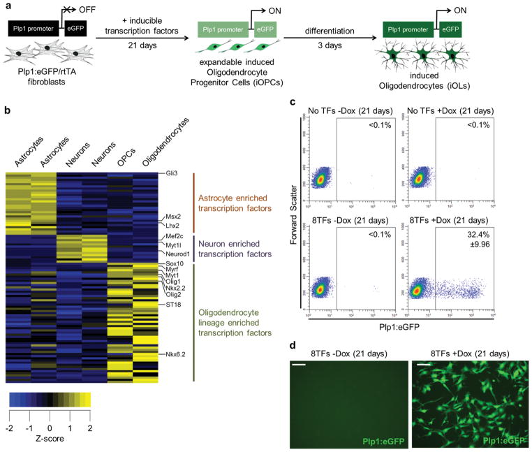
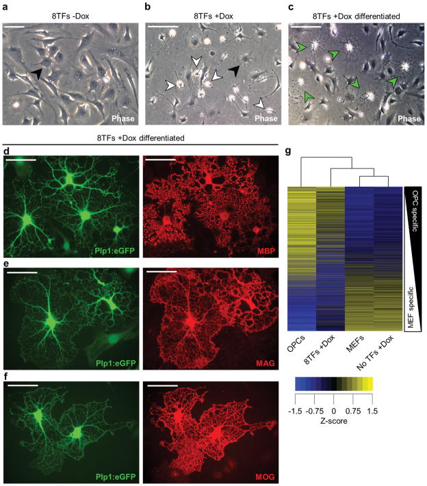
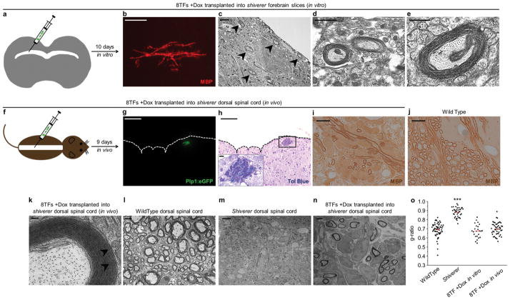
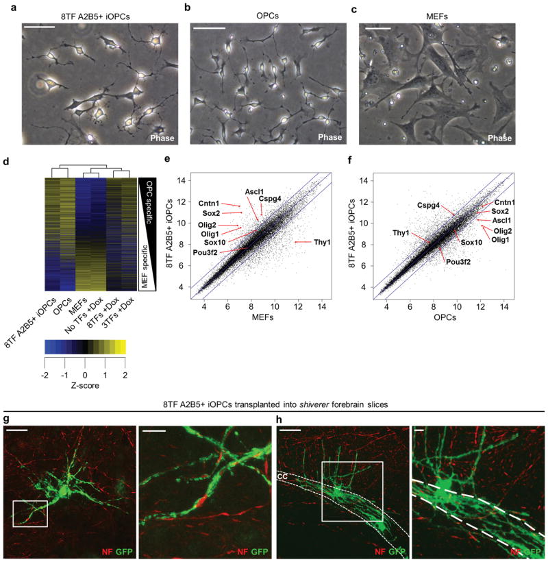
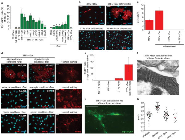
Cell-based therapies for myelin disorders, such as multiple sclerosis and leukodystrophies, require technologies to generate functional oligodendrocyte progenitor cells. Here we describe direct conversion of mouse embryonic and lung fibroblasts to induced oligodendrocyte progenitor cells (iOPCs) using sets of either eight or three defined transcription factors. iOPCs exhibit a bipolar morphology and global gene expression profile consistent with bona fide OPCs. They can be expanded in vitro for at least five passages while retaining the ability to differentiate into multiprocessed oligodendrocytes. When transplanted to hypomyelinated mice, iOPCs are capable of ensheathing host axons and generating compact myelin. Lineage conversion of somatic cells to expandable iOPCs provides a strategy to study the molecular control of oligodendrocyte lineage identity and may facilitate neurological disease modeling and autologous remyelinating therapies.


Conflict of interest statement

COMPETING FINANCIAL INTERESTS

P.J.T., R.H.M., and F.J.N. have a pending patent application for this technology.

Figures

Figure 1
Figure 1. Eight transcription factors can reprogram mouse embryonic fibroblasts to induced oligodendrocyte progenitor cells
a, Experimental design overview and timeline for transcription factor-mediated reprogramming of Plp1:eGFP/rtTA fibroblasts to induced oligodendrocyte progenitor cells (iOPCs) which are expandable and capable of differentiating to induced oligodendrocytes (iOLs). The modified Plp1:eGFP transgene is expressed in both OPCs and OLs in vivo. b, Gene expression heat map showing transcription factors (TFs) enriched (yellow) in each of the three major CNS lineages (see Supplementary Table 1 for complete list of genes). c, Representative flow cytometry plots and the average percentage of Plp1:eGFP+ cells after 21 days in OPC culture conditions for: non-infected and uninduced cells (No TFs −Dox); non-infected and induced cells (No TFs +Dox); infected with 8TFs but not induced (8TFs −Dox); and infected with 8TFs and induced (8TFs +Dox). Only infection of 8TFs and induction gave rise to Plp:eGFP+ cells at day 21 (8TFs +Dox = 32.4% +/− 9.9%; n = 19 independent biological replicates from 3 independent lots of lentivirus). All other conditions were devoid of Plp1:eGFP+ cells at day 21 (<0.1%). d, Representative live-cell fluorescent images of Plp:eGFP MEF cultures that were infected with inducible lentiviruses containing the 8TF pool and cultured for 21 days in the absence (−Dox) or presence (+Dox) of TF induction. Scale bar, 50μm (d).
Figure 2
Figure 2. Eight transcription factor induced MEFs exhibit properties of bona fide OPCs
a, b, Phase-contrast images highlighting the dramatic morphological differences between 21 day 8TF-uninduced (a; −Dox) and induced (b; +Dox) MEFs. Non-reprogrammed cells exhibit typical fibroblast morphology (black arrowheads) while a portion of the 8TF-induced cultures show the characteristic bipolar morphology of OPCs (white arrowheads). c, Phase-contrast image of day 21 8TF-induced MEFs passaged into oligodendrocyte differentiation media for 3 days showing the generation of induced oligodendrocytes (iOLs; green arrowheads) that exhibit the distinctive multiprocessed morphology of oligodendrocytes. d–f, Representative immunofluorescent images of iOLs differentiated from 8TF-induced MEFs, containing the Plp1:eGFP reporter and expressing the specific and defining markers of mature oligodendrocytes MBP (d), MAG (e), and MOG (f). g, Clustered heat map of z-scored global gene expression values comparing pluripotent stem cell-derived bona fide OPCs, 8TF-induced MEFs (8TFs +Dox), MEFs, and uninfected MEFs plus doxycycline (No TFs +Dox). Plot is rank ordered with OPC-specific genes at the top and MEF-specific genes at the bottom and includes >13,000 genes for which there was signal above background in at least one of the samples. Scale bars, 50μm (a), 100μm (b, c), 25μm (d–f).
Figure 3
Figure 3. Eight transcription factor induced MEFs function to generate compact myelin
a, Experimental scheme for testing the ability of 8TF-induced MEFs to myelinate axons in vitro. 8TF-induced MEFs were transplanted into P5 shiverer forebrain slices and cultured for 10 days. b, Representative immunofluorescent image of MBP+ myelin tracts generated from expanded (passage 3, day 32) 8TF-induced MEFs 10 days after transplantation into coronal forebrain slice cultures of shiverer mutant mice (lethally hypomyelinated due to a lack of Mbp). c–e, Electron micrograph images of multi-layered compact myelin (black arrowheads) generated by donor 8TF-induced MEFs 10 days after transplantation into coronal forebrain slice cultures of shiverer mutant mice. f, Experimental scheme for testing the ability of 8TF-induced MEFs to myelinate axons in vivo. 8TF-induced MEFs were transplanted into the dorsal spinal cord of P3-4 shiverer mutant mice. g, h, Representative immunofluorescent image (g) and matched toluidine blue (tol blue) stained section (h) showing localization of 8TF-induced cells (containing Plp1:eGFP transgene) 9 days after transplantation into the dorsal spinal cord of shiverer mutant mice (n=4 mice). The pia mater of the dorsal spinal cord is indicated by the dashed lines. (h, inset) Enlarged view of black box from (h) showing numerous myelinated axons generated from 8TF-induced cells. i–j, Representative immunostaining of MBP showing that the myelin produced in shiverer hosts 9 days after transplantation with 8TF-induced MEFs is of donor origin (i). Identically processed wild type control staining is shown in (j). k, Electron micrograph images of multi-layered compact myelin generated by 8TF-induced MEFs 9 days after transplantation into the dorsal spinal cord of shiverer mutant mice. Major dense lines are evident and indicated by black arrowheads. l–n, Electron micrographs of wild type dorsal column axons (l), untreated shiverer dorsal column axons (m), and shiverer dorsal column axons myelinated by transplanted 8TF-induced MEFs (n). o, g-ratios were calculated for wild type in vivo spinal cord (0.69±0.07), shiverer in vivo spinal cord (0.88±0.05) and 8TF-induced MEF (in vitro slices: 0.68±0.07 and in vivo spinal cord: 0.70±0.07) myelin. Differences between wild type and each of the other groups was compared using a two-tailed Student’s t-test (***p<2.2×10⁻¹⁶ wild type vs. shiverer. All others not significant). Scale bars, 25μm (b), 2μm (c, ln) 1μm (d), 500nm (e), 100μm (g, h), 100nm (k), and 10μm (i, j)
Figure 4
Figure 4. A2B5 immunosorting allows for the prospective enrichment of iOPCs
a–c, Phase contrast images highlighting the bipolar morphology of 8TF-induced MEFs (sorted at day 21 for both Plp1:eGFP and A2B5; denoted as 8TF A2B5+ iOPCs) (a) compared to bona fide OPCs (b), and MEFs (c). d, Clustered heat map of z-scored global gene expression values comparing 8TF A2B5+ iOPCs to the same three control cell samples in Figure 2g [MEFs, uninfected MEFs plus doxycycline (No TFs +Dox), and pluripotent stem cell-derived bona fide OPCs]. Plot is rank ordered with OPC-specific genes at the top and MEF-specific genes at the bottom and includes >13,000 genes for which there was signal above background in at least one of the samples. e, f, Pairwise comparison of log2-adjusted global gene expression values of 8TF-induced A2B5+ iOPCs with MEFs (e) and bona fide OPCs (f). Blue lines denote a 2-fold difference in gene expression. Characteristic MEF-enriched (Thy1) and OPC-enriched (all others cited) genes are indicated with red arrows. g, h, Confocal images collected 10 days after transplantation of 8TF A2B5+ iOPCs, containing the Plp1:eGFP reporter, at passage 3 into coronal forebrain slice cultures of shiverer mutant mice showing extensive ensheathment of neuron axons by 8TF A2B5+ iOLs. Neurons were visualized with anti-neurofilament (NF) and iOLs with anti-GFP antibodies. The corpus callosum (cc) is indicated with dashed white lines. Zoomed insets are presented to the right of each image. Scale bars, 50μm (a–c, g, h), and 10μm (g, h insets).
Figure 5
Figure 5. Sox10, Olig2, and Nkx6.2 are sufficient to reprogram fibroblasts to iOPCs
a, Summary graph quantifying the percentage of Plp1:eGFP+ cells induced by subsets of the original 8TF pool at day 21 (n = 3 independent biological replicates from 1 lot of lentivirus; see Supplementary Figure 4 for representative flow cytometry plots). A 3TF pool of Sox10, Olig2, and Nkx6.2 showed the robust ability to induce Plp1:eGFP+ cells from MEFs (20.8% +/− 1.5%). Differences between groups were compared using a two-tailed Student’s t-test (**p<0.02 and *p<0.05 versus 8TFs +Dox). b, c, Immunofluorescent (b) and quantification (c) data showing the capacity of 3TF-induced MEFs at day 21 (3TFs +Dox) to respond to differentiation signals (3TFs +Dox differentiated) and generate multiprocessed O4+ oligodendrocytes. Note that the undifferentiated cultures (3TFs +Dox) contain a population of O4+ cells which are largely bipolar. 3TF-uninduced MEFs (3TFs −Dox differentiated) and non-infected induced MEFs (No TFs +Dox differentiated) yielded no O4+ cells. O4+ cells were manually scored from triplicate wells and data are presented as mean ± S.E.M. (n = 3 independent biological replicates from 1 lot of lentivirus). d, Representative immunofluorescent images showing the differentiation potential of 3TF-induced MEFs (3TFs +Dox) when exposed to three different lineage inducing conditions. 3TF-induced MEFs differentiated in 3 days into iOLs that expressed MBP when exposed to oligodendrocyte differentiation conditions both in the presence (+Dox) or absence (−Dox) of doxycycline. 3TF-induced MEFs never gave rise to neurons (MAP2) or astrocytes (GFAP) either in the presence (+Dox) or absence (−Dox) of doxycycline when exposed to the respective neuron or astrocyte promoting culture conditions. Positive control cell types that were stained simultaneously to ensure function of each antibody: pluripotent stem cell derived oligodendrocytes (MBP), astrocytes (GFAP), and neurons (MAP2). e, Quantitative efficiency of 3TF-induced MEFs (3TF +Dox) to differentiate into MBP+ oligodendrocytes when exposed to oligodendrocyte differentiation conditions for 3 days. Data are presented as mean ± S.E.M. of MBP+ iOLs per 4×10⁴ cells seeded (n = 10 independent biological replicates from 3 lots of lentivirus). 3TF +Dox cells generated with a high viral titer showed a parallel increase in the efficiency of generating MBP+ iOLs (3TF +Dox high virus titer; n= 8 independent biological replicates from 2 lots of lentivirus). f, Electron micrograph image of multi-layered compact myelin generated from day 21 3TF-induced MEFs 10 days after transplantation into coronal forebrain slice cultures of shiverer mutant mice. g, Immunofluorescent images of engraftment and morphology of Plp1:eGFP+ 3TF-induced MEFs 10 days after transplantation into P5 coronal forebrain slice cultures of shiverer mutant mice. h, g-ratios were calculated from 3TF-induced MEFs transplanted into shiverer forebrain slices (0.63±0.09). Differences between groups and wild type (shown are the same control samples as in Figure 3n: wild type (0.69±0.07), shiverer (0.88±0.05) and 8TF-induced MEFs transplanted into shiverer forebrain slices and dorsal spinal cords (0.69±0.07)) were compared using a two-tailed Student’s t-test (***p<2.2×10⁻¹⁶ wild type vs. shiverer. All others not significant).. Scale bars, 25μm (b, d), 100μm (g), and 100nm (f).

Comment in

References

    1. Goldman SA, Nedergaard M, Windrem MS. Glial progenitor cell-based treatment and modeling of neurological disease. Science. 2012;338:491–495. - PMC - PubMed
    1. Franklin RJ, Ffrench-Constant C. Remyelination in the CNS: from biology to therapy. Nat Rev Neurosci. 2008;9:839–855. - PubMed
    1. Windrem MS, et al. Neonatal chimerization with human glial progenitor cells can both remyelinate and rescue the otherwise lethally hypomyelinated shiverer mouse. Cell Stem Cell. 2008;2:553–565. - PMC - PubMed
    1. Sim F, et al. CD140a identifies a population of highly myelinogenic, migration-competent, and efficiently engrafting human oligodendrocyte progenitor cells. Nature Biotechnol. 2011 - PMC - PubMed
    1. Najm FJ, et al. Rapid and robust generation of functional oligodendrocyte progenitor cells from epiblast stem cells. Nat Methods. 2011;8:957–962. - PMC - PubMed

Publication types

Substances

Associated data